Role of Other Ions in Action Potential
Introduction to Ion Involvement in Action Potentials
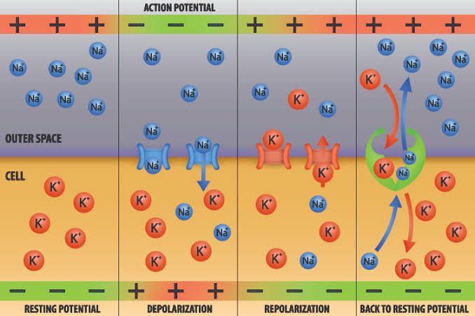
Action potentials are primarily driven by the movement of sodium (Na⁺) and potassium (K⁺) ions across the neuronal membrane. However, other ions also play significant roles in modulating the action potential process and influencing neuronal excitability. Understanding these roles requires a step-by-step analysis of how different ions contribute to the phases of an action potential.
1. Sodium Ions (Na⁺)
Sodium ions are crucial for the initiation and propagation of action potentials. When a neuron is stimulated, voltage-gated sodium channels open, allowing Na⁺ to flow into the cell. This influx causes depolarization, which is the rapid rise in membrane potential that characterizes the action potential. The rapid opening of these channels is what leads to the steep upward slope of the action potential curve.
2. Potassium Ions (K⁺)
After depolarization, potassium channels open in response to changes in membrane potential. The efflux of K⁺ out of the neuron helps repolarize the membrane back toward its resting state. This outward flow occurs after the peak of the action potential and is essential for returning the membrane potential to its negative resting value.
3. Calcium Ions (Ca²⁺)
Calcium ions have a dual role in action potentials, particularly in certain types of neurons and muscle cells. In neurons, calcium channels can open during depolarization, contributing to further depolarization and facilitating neurotransmitter release at synaptic terminals. In cardiac muscle cells, Ca²⁺ plays a critical role by prolonging depolarization through calcium-induced calcium release mechanisms, which are essential for muscle contraction.
4. Chloride Ions (Cl⁻)
Chloride ions generally have an inhibitory effect on neuronal excitability when they enter neurons through chloride channels or transporters. The influx of Cl⁻ can stabilize or hyperpolarize the membrane potential, making it less likely for an action potential to occur following stimulation. This inhibitory role is vital for balancing excitatory signals within neural circuits.
5. Role of Ion Concentration Gradients
The concentration gradients maintained by ion pumps such as the sodium-potassium pump (Na⁺/K⁺ ATPase) are fundamental for generating resting membrane potentials and facilitating action potentials. These pumps actively transport Na⁺ out of and K⁺ into cells against their concentration gradients, ensuring that sufficient ion concentrations are available for rapid changes during an action potential.
Conclusion
In summary, while sodium and potassium ions are central players in generating and propagating action potentials, other ions like calcium and chloride also significantly influence neuronal behavior and signal transmission through their modulatory effects on excitability and neurotransmitter release.
Effect of Hypocalcemia on Neuron Excitability
Introduction to Calcium’s Role in Neuronal Function
Calcium ions (Ca2+) play a crucial role in various physiological processes, particularly in neuronal function. They are essential for neurotransmitter release at synapses and for the generation of action potentials in neurons. The concentration of calcium in the extracellular fluid is tightly regulated, as deviations from normal levels can significantly impact neuronal excitability.
Mechanism of Hypocalcemia-Induced Neuronal Excitability
Hypocalcemia refers to abnormally low levels of calcium in the blood. When calcium levels drop, several physiological changes occur that enhance neuronal excitability:
- Voltage-Gated Sodium Channels: Under normal conditions, calcium ions inhibit sodium movement through voltage-gated sodium channels. This inhibition is critical for maintaining the resting membrane potential and preventing excessive neuronal firing. In hypocalcemia, the reduced availability of Ca2+ leads to diminished inhibition of these channels, allowing increased sodium influx into neurons.
- Increased Action Potential Generation: With enhanced sodium entry due to decreased calcium inhibition, neurons become more depolarized and more likely to reach the threshold needed to generate action potentials. This results in an increased frequency of action potentials, leading to heightened neuronal activity.
- Spontaneous Discharge: At plasma Ca2+ concentrations significantly below normal (approximately 50% lower), peripheral nerve fibers may become so excitable that they start discharging spontaneously. This spontaneous activity can lead to abnormal signaling patterns and contribute to symptoms such as muscle spasms or tetany.
- Clinical Manifestations: The hyper-excitability resulting from hypocalcemia can manifest clinically as various neurological symptoms:
- Tetany: Characterized by involuntary muscle contractions due to sustained muscle fiber activation.
- Chvostek’s Sign: A clinical test where tapping over the facial nerve causes twitching of facial muscles on the same side, indicating hyperexcitability.
- Seizures: In severe cases, hypocalcemia can lead to seizures due to excessive neuronal firing and excitatory neurotransmission.
- Pathophysiological Conditions: Certain medical conditions predispose individuals to hypocalcemic seizures or increased excitability. These include congenital disorders affecting parathyroid hormone production, renal insufficiency leading to poor calcium homeostasis, or drug-induced changes in calcium metabolism.
- Neurotransmitter Release Dynamics: While it might seem counterintuitive that low calcium levels could enhance excitability given its role in neurotransmitter release, the loss of inhibitory effects on sodium channels leads to a net increase in excitatory signaling within the nervous system.
- Conclusion on Hypocalcemia’s Impact: Overall, hypocalcemia enhances neuronal excitability through mechanisms involving voltage-gated sodium channels and altered neurotransmission dynamics. This paradoxical relationship highlights the complex interplay between ion concentrations and neuronal behavior.
In summary, hypocalcemia leads to increased neuronal excitability primarily by reducing calcium’s inhibitory effects on sodium channels, resulting in spontaneous discharges and heightened sensitivity of neurons.


