Course of the Radial Artery
The radial artery originates from the bifurcation of the brachial artery at the inferior portion of the cubital fossa. It appears as a direct continuation of the brachial artery and runs distally along the lateral aspect of the forearm. As it travels down, it crosses over the distal tendon of the biceps brachii muscle and lies deep to the brachioradialis muscle in its proximal part. Distally, it becomes more superficial, lying between the tendon of the brachioradialis and flexor carpi radialis muscles, and is covered only by fascia and skin.
Relations in the Forearm
In terms of anatomical relations, while in the forearm, the radial artery is closely associated with several structures:
- Muscles: It runs alongside important muscles such as the brachioradialis and flexor carpi radialis.
- Nerves: The radial nerve runs in proximity to this artery but does not directly accompany it throughout its course.
- Veins: The radial vein typically accompanies the radial artery, forming a paired structure that helps facilitate venous return from the forearm.
Termination in Hand
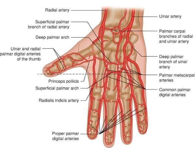
As it approaches the wrist, specifically at the anatomical snuffbox, which is formed by tendons of extensor muscles, it makes a lateral turn around to enter into the palm. Here, it divides into two significant branches:
- Superficial Palmar Branch: This branch contributes to forming part of the superficial palmar arch which primarily receives blood supply from ulnar artery.
- Deep Palmar Branch: This branch continues deeper into the hand to form part of the deep palmar arch. The deep palmar arch is crucial for supplying blood to metacarpal bones and digital arteries that supply fingers.
Clinical Significance
The clinical significance of the radial artery is multifaceted:
- Pulse Measurement: The radial artery’s location at its distal end makes it an accessible site for measuring pulse rate. This can provide vital information regarding heart rate and rhythm.
- Allen’s Test: This test assesses collateral circulation in hand by evaluating both radial and ulnar arteries’ patency. It is particularly important before procedures like arterial cannulation or harvesting for grafts.
- Surgical Considerations: Knowledge about its course is essential during surgeries involving wrist or hand to avoid inadvertent damage to this vessel which could lead to ischemia or necrosis in hand tissues.
- Variability: In less than 1% of individuals, variations exist where it takes a more superficial course through anatomical snuffbox; awareness of such variations can prevent misdiagnosis during vascular assessments.
In summary, understanding both its anatomical course and clinical relevance aids healthcare professionals in diagnostics and surgical interventions involving upper limb vascularity.
Course of the Ulnar Artery
The ulnar artery is a major vessel that supplies blood to the forearm and hand. It originates from the brachial artery in the cubital fossa, typically arising just below the radial head. The ulnar artery then travels obliquely downward along the medial side of the forearm. As it descends, it runs deep to several muscles, including the pronator teres and flexor carpi ulnaris.
In its course through the forearm, the ulnar artery lies between two significant muscles: flexor digitorum superficialis and flexor carpi ulnaris. This positioning is critical as it allows for adequate blood supply to these muscles while also providing a pathway for branches that supply other structures in the forearm.
Relations of the Ulnar Artery
As it courses downwards, the ulnar artery maintains close anatomical relationships with various structures:
- Muscles: It is covered by muscles such as flexor digitorum superficialis and flexor carpi ulnaris.
- Nerves: The median nerve runs near the ulnar artery but is separated by the ulnar head of pronator teres.
- Veins: Accompanying veins are present alongside the ulnar artery, forming a venous plexus that aids in venous return from the forearm.
These relations are clinically significant because they can influence surgical approaches or interventions involving vascular access or repair.
Termination in Hand
Upon reaching the wrist, the ulnar artery passes beneath the transverse carpal ligament at a point just radial to pisiform bone and superficial to flexor retinaculum. In this region, it divides into two terminal branches:
- Superficial Palmar Arch: This branch contributes significantly to blood supply in the palm of the hand.
- Deep Palmar Branch: This branch dives deeper into hand anatomy, contributing to deeper structures.
The superficial palmar arch is particularly important as it forms anastomoses with branches from the radial artery, ensuring collateral circulation within the hand.
Clinical Significance
The clinical significance of understanding the course, relations, and termination of the ulnar artery cannot be overstated:
- Surgical Access: Knowledge of its location is crucial for procedures such as creating arteriovenous fistulas for hemodialysis patients when other arteries are unsuitable.
- Vascular Injury Prevention: Awareness of its proximity to nerves and muscles helps prevent inadvertent injury during surgical procedures or invasive interventions.
- Diagnosis of Vascular Conditions: Understanding variations in anatomy can aid clinicians in diagnosing conditions related to compromised blood flow or vascular malformations.
In summary, detailed knowledge about the ulnar artery’s course, relations, termination points in hand anatomy, and their clinical implications enhances surgical precision and improves patient outcomes.
Formation of the Superficial Palmar Arch
The superficial palmar arch is primarily formed by the ulnar artery, which arises at the level of a horizontal line that passes through the distal margin of the base of the thumb. It courses anterolaterally towards the midshaft of the carpal bones and then takes a posterolateral course to form a distally convex arch across the palm. The arch also receives a contribution from the radial artery, specifically through its superficial palmar branch. In some individuals, variations may occur where either the radialis indicis or princeps pollicis arteries participate in forming this arch instead of the radial artery.
Branches of the Superficial Palmar Arch
The superficial palmar arch gives rise to three main branches known as common palmar digital arteries. These arteries further divide into proper palmar digital arteries that supply blood to digits 2-4. The common palmar digital arteries run distally across the second to fourth lumbricals and anastomose with corresponding branches from the deep palmar arch. Each common palmar digital artery subsequently divides into two proper palmar digital arteries, which travel along adjacent sides of digits 2-4, providing vascular supply to these areas.
Areas of Distribution for Superficial Palmar Arch
The primary function of the superficial palmar arch is to supply blood to various structures in digits 2-4, including:
- Proximal phalanges
- Metacarpophalangeal joints
- Proximal interphalangeal joints
Once reaching their respective digits, proper palmar digital arteries give off terminal branches that connect with similar branches on opposite sides, forming arterial arcades that ensure adequate blood flow to distal elements such as:
- Distal interphalangeal joints
- Middle and distal phalanges
- Surrounding soft tissues
Formation of the Deep Palmar Arch
The deep palmar arch is mainly formed by the radial artery, which serves as its primary source. It arises between two heads of the adductor pollicis muscle and courses medially across bases of metacarpal bones while receiving contributions from the ulnar artery via its deep branch. Occasionally, it may end before connecting with this deep ulnar branch, resulting in what is termed an ‘incomplete deep palmar arch.’
Branches of the Deep Palmar Arch
The deep palmar arch gives off several important branches:
- Palmar Metacarpal Arteries: These run distally across lumbricals and anastomose with common digital branches from the superficial palmar arch.
- Perforating Branches: These arise from the concavity of the deep arch and run through interosseous spaces 2-4, connecting with dorsal metacarpal arteries.
- Recurrent Branches: These ascend proximally to supply carpal bones and joints.
Areas of Distribution for Deep Palmar Arch
The deep palmar arch primarily supplies blood to deeper structures within the hand including:
- Bones (metacarpals)
- Joints (metacarpophalangeal and proximal interphalangeal)
- Muscles (adductor pollicis, dorsal interossei, lumbrical muscles)
Additionally, it contributes vascularization to radiocarpal joints through its recurrent branches.


