Different Forms of Transport of Oxygen in the Blood
Oxygen transport in the blood occurs primarily through two mechanisms: dissolved oxygen and hemoglobin-bound oxygen. Each mechanism plays a crucial role in ensuring that tissues throughout the body receive adequate oxygen for metabolic processes.
1. Dissolved Oxygen
A small fraction of oxygen is transported dissolved directly in the plasma, which is the liquid component of blood. This form accounts for approximately 1.5% of the total oxygen content in the blood. The amount of oxygen that can dissolve in plasma is influenced by several factors, including temperature, salinity, and partial pressure of oxygen (P(O2)). However, this method alone is insufficient to meet the body’s demands for oxygen, especially during physical exertion or high metabolic activity.
2. Hemoglobin-Bound Oxygen
The majority of oxygen transport—about 98.5%—occurs via hemoglobin (Hb), a specialized protein found within red blood cells (erythrocytes). Hemoglobin consists of four subunits: two alpha and two beta chains, each containing an iron-containing heme group capable of binding one molecule of oxygen. This allows a single hemoglobin molecule to carry up to four molecules of oxygen.
When we inhale, fresh oxygen enters the lungs and diffuses across the alveolar-capillary membrane into the bloodstream. Here’s how hemoglobin facilitates efficient oxygen transport:
- Binding Mechanism: As each heme group binds an oxygen molecule, hemoglobin undergoes a conformational change that increases its affinity for additional oxygen molecules. This cooperative binding means that once one molecule binds, it becomes easier for subsequent molecules to attach.
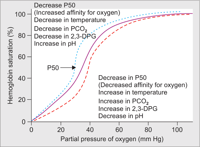
- Oxygen Saturation: The relationship between P(O2) and hemoglobin saturation can be graphically represented by an S-shaped curve known as the oxygen dissociation curve. At higher P(O2) levels (such as in the lungs), hemoglobin becomes saturated with oxygen; conversely, at lower P(O2) levels (such as in metabolically active tissues), hemoglobin releases its bound oxygen.
- Factors Affecting Binding: Several physiological factors influence hemoglobin’s affinity for oxygen:
- Carbon Dioxide Levels: Increased CO2 levels lead to a decrease in blood pH (more acidic conditions), which reduces hemoglobin’s affinity for oxygen—a phenomenon known as the Bohr effect.
- Temperature: Elevated body temperatures also decrease hemoglobin’s affinity for oxygen, facilitating greater release to active tissues.
- BPG Levels: 2,3-bisphosphoglycerate (BPG) produced during glycolysis can bind to hemoglobin and further reduce its affinity for oxygen under certain conditions.
In summary, while only a small percentage of oxygen is transported dissolved in plasma, most is carried by hemoglobin within red blood cells. This dual mechanism ensures that sufficient amounts of oxygen are delivered efficiently throughout the body to support cellular respiration and energy production.
Oxyhemoglobin Dissociation Curve
The oxyhemoglobin dissociation curve is a graphical representation that illustrates how hemoglobin in the blood binds to and releases oxygen. On this curve, the vertical axis represents the percentage of hemoglobin saturation with oxygen (SO2), while the horizontal axis represents the partial pressure of oxygen (PO2) in mmHg. The relationship depicted by this curve is crucial for understanding how effectively hemoglobin transports oxygen from the lungs to tissues throughout the body.
The shape of the curve is typically sigmoidal (S-shaped), which reflects the cooperative binding nature of hemoglobin. When one molecule of oxygen binds to a hemoglobin molecule, it induces a conformational change that makes it easier for additional oxygen molecules to bind. This means that at lower partial pressures of oxygen, hemoglobin has a lower saturation level, but as PO2 increases, saturation rises steeply until it approaches full saturation at high PO2 levels.
Key points about the oxyhemoglobin dissociation curve include:
- P50 Value: The P50 value is defined as the partial pressure of oxygen at which hemoglobin is 50% saturated. For healthy individuals, this value is typically around 26.6 mmHg.
- Steep and Plateau Regions: The curve has two distinct regions: a steep portion where small changes in PO2 result in significant changes in SO2 (typically found in systemic capillaries), and a plateau region where large increases in PO2 do not significantly increase SO2 (typically found in pulmonary capillaries).
- Shifts in the Curve: Factors such as pH, temperature, carbon dioxide concentration, and 2,3-bisphosphoglycerate levels can shift this curve to the right or left, affecting hemoglobin’s affinity for oxygen.
Bohr Effect
The Bohr effect describes how changes in carbon dioxide concentration and pH affect hemoglobin’s affinity for oxygen. Specifically, an increase in carbon dioxide concentration or a decrease in pH (more acidic conditions) results in a rightward shift of the oxyhemoglobin dissociation curve. This shift indicates that hemoglobin’s affinity for oxygen decreases; thus, it becomes easier for hemoglobin to release bound oxygen into tissues.
This physiological response is particularly beneficial during aerobic exercise or periods of increased metabolic activity when tissues produce more carbon dioxide and lactic acid. As these byproducts accumulate:
- Increased Carbon Dioxide: Higher levels of CO2 lead to increased formation of carbonic acid, which lowers blood pH.
- Enhanced Oxygen Delivery: The rightward shift allows more efficient unloading of oxygen from hemoglobin into actively respiring tissues that require it most urgently.
- Facilitated Oxygen Release: This mechanism ensures that during times of heightened demand—such as exercise—muscles receive adequate amounts of oxygen necessary for aerobic metabolism.
In summary, both the oxyhemoglobin dissociation curve and the Bohr effect are critical components of respiratory physiology that ensure effective delivery and utilization of oxygen throughout the body under varying conditions.
Factors Causing Rightward Shift of Oxyhemoglobin Dissociation Curve
A rightward shift in this curve indicates that hemoglobin has a decreased affinity for oxygen, allowing it to release oxygen more readily to tissues. Several physiological factors contribute to this rightward shift:
- Increased pCO2 Concentration: Elevated levels of carbon dioxide (pCO2) in the blood lead to increased production of carbonic acid, which lowers blood pH (acidosis). This effect is known as the Bohr effect, where higher pCO2 enhances oxygen unloading from hemoglobin.
- Acidosis: A decrease in blood pH (increased acidity) reduces hemoglobin’s affinity for oxygen. This can occur due to metabolic processes or respiratory issues that increase CO2 levels, further promoting oxygen release.
- Raised Temperature: An increase in body temperature can also cause a rightward shift in the curve. Higher temperatures enhance metabolic activity and promote the release of oxygen from hemoglobin, which is particularly important during exercise when tissues require more oxygen.
- High Concentrations of 2,3-Diphosphoglycerate (2,3 DPG): 2,3 DPG is a byproduct of glycolysis found in red blood cells. Increased levels of 2,3 DPG reduce hemoglobin’s affinity for oxygen, facilitating greater oxygen delivery to tissues under conditions such as chronic hypoxia or anemia.
- Increased Levels of Hydrogen Ions (H+): As hydrogen ion concentration increases (which occurs during acidosis), it can bind to hemoglobin and promote a conformational change that decreases its affinity for oxygen.
These factors collectively enable hemoglobin to release more oxygen into tissues that are metabolically active or under stress, ensuring adequate supply during times of increased demand.
Factors Causing Leftward Shift of Oxyhemoglobin Dissociation Curve
A leftward shift in this curve indicates an increased affinity of hemoglobin for oxygen, meaning that hemoglobin holds onto oxygen more tightly and releases it less readily to tissues. Several physiological and biochemical factors can cause this leftward shift:
- Decreased Carbon Dioxide Levels (Hypocapnia): Lower levels of carbon dioxide in the blood lead to a decrease in hydrogen ion concentration (increased pH), which enhances hemoglobin’s affinity for oxygen. This phenomenon is part of the Bohr effect, where decreased CO2 results in increased pH, shifting the curve to the left.
- Increased pH (Alkalosis): An increase in blood pH, often due to respiratory alkalosis or metabolic alkalosis, causes a leftward shift. Higher pH levels reduce the release of oxygen from hemoglobin, as it stabilizes the R state (relaxed state) of hemoglobin, which has a higher affinity for oxygen.
- Decreased Temperature: A lower body temperature reduces metabolic activity and decreases the release of 2,3-bisphosphoglycerate (2,3-BPG) from red blood cells. Since 2,3-BPG binds to deoxygenated hemoglobin and promotes oxygen release, its reduction leads to a higher affinity for oxygen and shifts the curve leftward.
- Decreased 2,3-BPG Levels: 2,3-BPG is produced during glycolysis and plays a crucial role in regulating hemoglobin’s affinity for oxygen. Lower levels of 2,3-BPG result in increased binding affinity for oxygen by stabilizing the R state of hemoglobin.
- Presence of Fetal Hemoglobin (HbF): Fetal hemoglobin has a higher affinity for oxygen compared to adult hemoglobin (HbA). The presence of HbF shifts the dissociation curve to the left because it does not bind 2,3-BPG as effectively as HbA does.
- Carbon Monoxide Binding: Carbon monoxide competes with oxygen for binding sites on hemoglobin but binds much more tightly than oxygen does. The presence of carbon monoxide increases hemoglobin’s affinity for whatever oxygen is still bound, effectively shifting the curve to the left.
- Certain Genetic Variants: Some genetic mutations can lead to alterations in hemoglobin structure that increase its affinity for oxygen without affecting its ability to transport it effectively.
In summary, these factors—decreased carbon dioxide levels, increased pH, decreased temperature, decreased 2,3-BPG levels, presence of fetal hemoglobin, carbon monoxide binding, and certain genetic variants—contribute to a leftward shift in the oxyhemoglobin dissociation curve by enhancing hemoglobin’s affinity for oxygen.
Definition of Cyanosis
Cyanosis is a medical condition characterized by a bluish or purplish discoloration of the skin, lips, and/or nails. This occurs due to a lack of oxygen in the blood, which can result from various underlying health issues. The bluish tone is most noticeable in areas where the skin is thinner, such as the lips, tongue, earlobes, and fingernails.
Types of Cyanosis
- Circumoral (Perioral) Cyanosis: This type affects only the mouth or lips and often occurs when blood vessels constrict in response to cold temperatures. It is common in newborns and may appear in older children during cold weather.
- Peripheral Cyanosis: This type involves the extremities such as hands, fingers, feet, and toes turning blue. It can occur due to exposure to cold weather but is rarely life-threatening. However, it is important to determine its cause to prevent potential injury.
- Central Cyanosis: This type affects other parts of the body beyond just the extremities, including the chest, cheeks, tongue, gums, and lips. Central cyanosis may indicate serious underlying conditions related to the heart or lungs and requires immediate medical attention.
Causes of Cyanosis
Cyanosis typically arises from conditions that lead to inadequate oxygen levels in the blood. Some common causes include:
- Lung Disorders: Conditions that impair oxygen absorption in the lungs can lead to cyanosis. Examples include chronic obstructive pulmonary disease (COPD), pneumonia, asthma exacerbations, or pulmonary embolism.
- Heart Conditions: Congenital heart defects or other heart-related issues can disrupt normal blood flow and oxygenation. Conditions such as tetralogy of Fallot or transposition of great vessels are examples that may cause central cyanosis.
- Blood Abnormalities: Certain blood disorders can affect how oxygen is carried in the bloodstream. For instance, methemoglobinemia results from abnormal hemoglobin that cannot effectively bind oxygen.
- Environmental Factors: Exposure to extreme cold can cause peripheral cyanosis as blood vessels constrict to preserve core body temperature.
- Other Medical Conditions: Various other health issues like anemia or carbon monoxide poisoning can also contribute to cyanotic symptoms by affecting oxygen transport or availability.
In summary, cyanosis indicates an underlying issue with oxygen delivery within the body and necessitates thorough evaluation for appropriate diagnosis and treatment.


