Definition of Lung Compliance
Lung compliance refers to the ability of the lungs to stretch and expand in response to changes in pressure. It is a measure of the lung’s expandability and is calculated by dividing the change in lung volume (V) by the change in pressure (P) applied to the lungs, expressed mathematically as C = ∆V ÷ ∆P. High lung compliance indicates that the lungs can easily expand with minimal pressure, while low compliance suggests that more pressure is required for expansion.
Factors Affecting Lung Compliance
Several factors influence lung compliance, including:
- Elastic Fibers: The presence of elastin in the connective tissue of the lungs plays a crucial role. More elastic fibers lead to greater ease of expansion, thereby increasing compliance. Conversely, a decrease in elastic fibers can reduce compliance.
- Surface Tension: Surface tension within the alveoli significantly affects lung compliance. The thin fluid lining inside alveoli creates surface tension that opposes expansion. Surfactant production decreases this surface tension, facilitating easier lung expansion.
- Transmural Pressure: This refers to the difference between alveolar pressure and intrapleural pressure. Positive transmural pressures are necessary for lung expansion; however, these pressures are achieved through negative pressures created within the intrapleural space rather than positive pressures within the alveoli.
- Lung Volume: Compliance varies with lung volume; it tends to be higher at lower volumes when alveoli are deflated and lower at higher volumes when they are inflated.
- Pathological Conditions: Various diseases can alter lung compliance. For example, restrictive lung diseases (like pulmonary fibrosis) decrease compliance due to stiffening of lung tissue, while obstructive conditions (like emphysema) may increase compliance but impair overall function.
- Age and Health Status: Age-related changes and health conditions can also affect elasticity and surface tension dynamics, impacting overall lung compliance.
In summary, understanding these factors is essential for assessing respiratory function and diagnosing potential pulmonary issues.
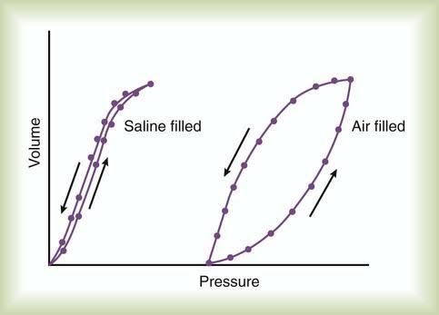
Compliance Diagram of Air-Filled and Saline-Filled Lungs
Introduction to Compliance Diagrams
The compliance diagram is a graphical representation that illustrates the relationship between lung volume and pressure during inflation and deflation. It helps in understanding how different factors, such as surface tension and elastic recoil, affect lung compliance. The compliance curves for air-filled and saline-filled lungs differ significantly due to the distinct properties of air and saline in relation to surface tension.
Air-Filled Lungs
In air-filled lungs, the compliance curve typically demonstrates a characteristic shape influenced by the presence of surfactant, which reduces surface tension within the alveoli. During inflation, as pressure increases, the volume of air in the lungs also increases. The initial slope of the curve indicates high compliance because small changes in pressure result in significant changes in volume. However, as the lungs approach their total capacity, the curve flattens out due to increased elastic recoil forces opposing further expansion.
- Initial Phase: At low lung volumes, there is a steep increase in volume with minimal pressure change due to surfactant action.
- Mid-Range Phase: As more air enters, the compliance decreases because of increased elastic resistance from stretched lung tissue.
- End-Phase: Near total lung capacity, further inflation requires disproportionately higher pressures due to significant elastic recoil.
Saline-Filled Lungs
In contrast, saline-filled lungs exhibit a different compliance curve primarily because saline does not have surfactant properties like air does. When lungs are filled with saline:
- Initial Phase: The compliance is lower than that of air-filled lungs at similar volumes because saline does not reduce surface tension effectively.
- Linear Increase: The curve shows a more linear relationship between pressure and volume throughout most of its range since there is less hysteresis (the difference between inflation and deflation curves) compared to air-filled lungs.
- Higher Pressure Requirement: More pressure is needed for inflation at all volumes compared to air-filled lungs due to lack of surfactant reducing surface tension.
Comparison Between Air-Filled and Saline-Filled Lungs
When comparing both diagrams:
- The air-filled lung curve starts off steeply sloped due to surfactant but flattens out as it approaches total lung capacity.
- The saline-filled lung curve remains relatively linear with a consistently lower compliance throughout its range.
This difference highlights how surfactant plays a crucial role in enhancing lung compliance by reducing surface tension within alveoli during normal breathing conditions.
Conclusion
Understanding these differences through compliance diagrams aids clinicians in assessing respiratory function and diagnosing various pulmonary conditions based on how well lungs can expand under different filling conditions.
Components of Surfactant
Pulmonary surfactant is primarily composed of lipids and proteins. The key components include:
- Phospholipids: These make up 80 to 90% of the surfactant’s molecular weight. The most abundant phospholipid is dipalmitoylphosphatidylcholine (DPPC), which constitutes approximately 70% of the lipid portion. Other phospholipids present in smaller amounts include phosphatidylglycerol, phosphatidylinositol, phosphatidylserine, and phosphatidylethanolamine.
- Surfactant Proteins: There are four main surfactant-associated proteins:
- SP-A (Surfactant Protein A): A hydrophilic protein involved in immune responses and pathogen clearance.
- SP-B (Surfactant Protein B): A hydrophobic protein that enhances the surface tension-reducing properties of surfactant and has some antimicrobial activity.
- SP-C (Surfactant Protein C): Another hydrophobic protein whose exact role is less clear but is believed to be essential for surfactant function.
- SP-D (Surfactant Protein D): Similar to SP-A, this hydrophilic protein plays a role in pulmonary host defense.
Role of Surfactant in Lung Compliance
Pulmonary surfactant significantly contributes to lung compliance, which is defined as the ability of the lungs and thorax to expand. The primary roles of surfactant in enhancing lung compliance include:
- Reduction of Surface Tension: Surfactant reduces the surface tension at the air-water interface within the alveoli. This reduction is crucial because high surface tension can lead to alveolar collapse (atelectasis) at the end of expiration. By lowering surface tension from approximately 70 dyn/cm (in pure water) to about 25 dyn/cm or even lower in the lungs, surfactant allows for easier inflation of alveoli during inhalation.
- Increased Compliance: By decreasing surface tension, surfactant increases lung compliance, meaning that a smaller pressure difference is required to inflate the lungs. This reduction in work associated with breathing allows for more efficient ventilation.
- Prevention of Alveolar Collapse: Surfactant helps maintain alveolar stability by preventing collapse during expiration, ensuring that all alveoli can expand uniformly during inhalation.
- Facilitation of Recruitment: Surfactant aids in recruiting collapsed airways by allowing them to reopen more easily due to reduced surface tension.
Overall, pulmonary surfactant plays a vital role in maintaining proper lung function and efficiency by enhancing compliance and preventing atelectasis.
Surfactant and Respiratory Distress Syndrome
Surfactant is a complex mixture of lipids and proteins that plays a crucial role in reducing surface tension within the alveoli of the lungs. In premature infants, particularly those born before 34 weeks of gestation, the lungs are often underdeveloped, leading to insufficient production of surfactant. This condition is known as Respiratory Distress Syndrome (RDS). The lack of adequate surfactant results in increased surface tension, which can cause the alveoli to collapse during expiration, thereby reducing the total surface area available for gas exchange.
Function of Surfactant in the Lungs
The primary function of surfactant is to lower the surface tension at the air-liquid interface within the alveoli. By doing so, it prevents alveolar collapse (atelectasis) and ensures that the lungs can expand effectively during inhalation. Surfactant also plays a role in stabilizing alveoli of different sizes; smaller alveoli tend to collapse into larger ones due to pressure differences, but surfactant helps maintain their structure and function.
In premature infants with RDS, insufficient surfactant leads to several physiological problems:
- Decreased Lung Compliance: The stiffness of the lungs increases due to high surface tension, making it more difficult for infants to breathe.
- Impaired Gas Exchange: With collapsed or poorly functioning alveoli, oxygen uptake is reduced and carbon dioxide elimination is impaired, leading to hypoxia (low oxygen levels) and hypercapnia (high carbon dioxide levels).
- Increased Work of Breathing: Infants must exert more effort to breathe against increased resistance in their lungs.
Surfactant Replacement Therapy
Given its critical role in lung function, surfactant replacement therapy has become a standard treatment for RDS in preterm infants. This therapy involves administering exogenous surfactant directly into the trachea via intubation or through less invasive methods such as thin catheters or nebulization.
- Types of Surfactants Used: There are two main types of surfactants used in clinical practice:
- Natural Surfactants: Derived from animal sources (e.g., porcine or bovine), these have been shown to be more effective than synthetic alternatives.
- Synthetic Surfactants: These have been developed to mimic natural surfactants and include protein-containing formulations like lucinactant.
- Benefits of SRT:
- Reduces mortality rates associated with RDS.
- Decreases the need for mechanical ventilation and reduces complications such as pulmonary air leaks.
- Improves overall lung function and oxygenation.
- Administration Techniques: Traditional methods involve intubating the infant, administering surfactant, and then extubating them shortly after (INSURE method). Newer non-invasive techniques are being explored to minimize trauma associated with intubation while still delivering effective treatment.
- Timing and Dosage: Early administration of surfactant has been shown to yield better outcomes compared to delayed treatment. Multiple doses may also improve efficacy.
- Future Directions: Research continues into aerosolized forms of surfactant that could allow for non-invasive delivery methods without compromising effectiveness.
Conclusion
In summary, surfactant plays an essential role in maintaining lung function in premature infants by reducing surface tension within the alveoli and preventing atelectasis. Surfactant replacement therapy has significantly improved outcomes for infants suffering from RDS by enhancing lung compliance and facilitating gas exchange.


