Origin, Course, Relations, Branches/Tributaries and Termination of Nerves and Vessels of Anterior, Lateral, and Posterior Compartments of Leg
1. Anterior Compartment of the Leg
- Origin: The anterior compartment is primarily innervated by the deep fibular nerve (L4-L5) and contains muscles that are responsible for dorsiflexion of the foot.
- Course: The deep fibular nerve travels down the leg alongside the anterior tibial artery. It runs in a plane between the tibialis anterior muscle and the extensor digitorum longus muscle.
- Relations: The anterior compartment is bounded laterally by the fibula and medially by the tibia. It lies deep to the fascia cruris.
- Branches/Tributaries: The main vessels in this compartment include:
- Anterior tibial artery: A branch of the popliteal artery that supplies blood to the anterior compartment.
- Dorsalis pedis artery: Continuation of the anterior tibial artery after it crosses the ankle joint.
- Termination: The deep fibular nerve terminates by supplying sensation to the skin between the first and second toes. The dorsalis pedis artery continues into the foot where it branches into various arteries supplying different regions.
2. Lateral Compartment of the Leg
- Origin: This compartment is innervated by the superficial fibular nerve (L5-S2) and contains muscles that are primarily responsible for eversion of the foot.
- Course: The superficial fibular nerve descends in a lateral direction beneath the fascia cruris, emerging at about mid-leg level to supply sensation to part of the dorsum of the foot.
- Relations: The lateral compartment is bordered medially by the anterior compartment and laterally by a layer of fascia. It is also adjacent to both peroneus longus and peroneus brevis muscles.
- Branches/Tributaries:
- Fibular artery (peroneal artery): A branch from posterior tibial artery that supplies blood to this compartment.
- Termination: The superficial fibular nerve terminates as it divides into cutaneous branches supplying sensation to parts of the lower leg and dorsum of foot. The fibular artery contributes branches that supply surrounding structures before terminating as well.
3. Posterior Compartment of the Leg
- Origin: Innervated by the tibial nerve (L4-S3), this compartment includes muscles responsible for plantarflexion at ankle joint.
- Course: The tibial nerve travels downwards through this compartment, running between two groups of muscles (superficial and deep).
- Relations: This compartment is divided into two groups:
- Superficial group includes gastrocnemius, soleus, and plantaris.
- Deep group includes popliteus, flexor hallucis longus, flexor digitorum longus, and tibialis posterior.
- Branches/Tributaries:
- Posterior tibial artery: A continuation from popliteal artery which supplies blood to both superficial and deep groups.
- Fibular artery branches off posterior tibial artery providing additional supply.
- Termination: The tibial nerve bifurcates into medial plantar nerve and lateral plantar nerve at around ankle level. The posterior tibial artery divides into medial and lateral plantar arteries upon reaching foot.
In summary, each compartment has distinct nerves (deep/superficial fibular nerves for lateral/anterior; tibial for posterior), arteries (anterior/posterior tibial arteries), with specific origins, courses through defined anatomical relations leading to their respective terminations in either sensory or muscular territories.
Cutaneous Nerves and Vessels of the Leg
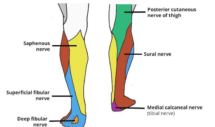
The cutaneous innervation of the leg is primarily provided by several key peripheral nerves, each responsible for specific areas of skin. Understanding these nerves is crucial for diagnosing and treating conditions related to sensory deficits or pain in the lower limb.
1. Posterior Cutaneous Nerve of Thigh
- The posterior cutaneous nerve of the thigh supplies the skin over the central superior and posterior aspects of the leg. It originates from the sacral plexus, specifically from spinal nerves S1 to S3. This nerve plays a significant role in providing sensation to the posterior thigh and proximal leg.
2. Saphenous Nerve
- The saphenous nerve is a sensory branch of the femoral nerve, which arises from the lumbar plexus (L2-L4). It supplies sensation to the anteromedial aspect of the leg and extends down to provide sensory innervation to the medial aspect of the foot. It is notable for its long course along with the great saphenous vein.
3. Superficial Fibular Nerve
- The superficial fibular nerve is a branch of the common fibular nerve (which itself branches from the sciatic nerve). This nerve supplies sensation to the anterolateral aspect of the leg and also covers most of the dorsum of the foot, except for a small area between the first and second toes.
4. Sural Nerve
- The sural nerve is formed by contributions from both tibial and common fibular nerves. It provides sensory innervation to the posterolateral aspect of the leg and lateral margin of both hindfoot and midfoot. This nerve is often used in clinical settings for nerve grafting due to its accessibility.
5. Deep Fibular Nerve
- The deep fibular nerve, another branch of the common fibular nerve, supplies sensation to a small area between the first and second toes on the dorsum of the foot. It also innervates muscles in this region, contributing to motor functions as well.
6. Medial Plantar Nerve
- The medial plantar nerve is a terminal branch of the tibial nerve that supplies sensation to most parts of the sole (medial two-thirds) as well as some digital branches that innervate toes 1 through 3.
7. Lateral Plantar Nerve
- Also a terminal branch of the tibial nerve, it supplies sensation to lateral third of sole and contributes sensory fibers to toes 4 and 5.
Vascular Supply
The vascular supply in conjunction with these nerves includes major arteries such as:
- Anterior Tibial Artery: Supplies blood to anterior compartment structures.
- Posterior Tibial Artery: Supplies blood to posterior compartment structures.
- Fibular Artery: A branch off posterior tibial artery that provides additional supply mainly to lateral compartment structures.
These arteries are accompanied by corresponding veins that drain deoxygenated blood back towards heart via larger venous structures like popliteal vein leading into femoral vein.
In summary, understanding both cutaneous nerves and vascular supply is essential for comprehending how sensory information is relayed from various regions within lower limb back towards central nervous system while ensuring adequate perfusion throughout tissues involved.
Dermatomes of the Leg
Dermatomes are specific areas of skin that are innervated by sensory fibers from a single spinal nerve root. In the case of the leg, dermatomes correspond to the lumbar and sacral regions of the spinal cord. Understanding these dermatomes is crucial for diagnosing and treating conditions related to nerve damage or dysfunction.
The dermatomes of the leg can be categorized based on their anatomical locations:
- L1 Dermatome: This dermatome covers the upper part of the groin area and extends downwards towards the lateral aspect of the thigh.
- L2 Dermatome: The L2 dermatome encompasses the anterior and lateral aspects of the thigh, extending down to approximately the mid-thigh region.
- L3 Dermatome: This area includes the medial aspect of the thigh and extends down to just above the knee. It is particularly important in assessing knee pain or dysfunction.
- L4 Dermatome: The L4 dermatome covers a significant portion of the medial side of the leg, including parts of the knee, and extends down to include part of the foot, specifically around the medial malleolus (the bony prominence on either side of your ankle).
- L5 Dermatome: This dermatome includes most of the lateral aspect of the leg and extends down into the dorsum (top) of the foot, covering areas such as between the first and second toes.
- S1 Dermatome: The S1 dermatome covers much of the posterior aspect of the leg and lateral side, extending down to include parts of both heel and plantar surface (the bottom) of foot.
- S2 Dermatome: This area encompasses parts of both posterior thighs and legs, extending down towards parts near but not including most areas on feet.
- S3-S5 Dermatomes: These dermatomes cover areas around perineum and buttocks but are less commonly referenced in clinical practice concerning lower limb issues.
The mapping out of these dermatomes is essential for clinicians when evaluating sensory loss or pain distribution in patients with potential radiculopathy or other neurological conditions affecting lower limbs. Testing these dermatomes can help pinpoint which spinal nerve root may be affected based on where a patient experiences numbness or pain.


