Assessing and Appropriately Resuscitating a Patient with Acute GI Haemorrhage
1. Initial Assessment
When a patient presents with acute gastrointestinal (GI) hemorrhage, the first step is to conduct a thorough assessment. This includes:
- History Taking: Gather information on the patient’s medical history, including any previous episodes of GI bleeding, liver disease, use of anticoagulants, or non-steroidal anti-inflammatory drugs (NSAIDs).
- Physical Examination: Look for signs of hypovolaemic shock such as tachycardia, hypotension, pallor, and altered mental status. Assess for abdominal tenderness or distension.
2. Risk Stratification
Utilize validated scoring systems like the Blatchford score or the Rockall score to stratify the risk of mortality and determine the urgency of intervention required.
3. Resuscitation Protocols
The principles of resuscitation follow the ABCs (Airway, Breathing, Circulation):
- Airway Management: Ensure that the airway is patent. If there are concerns about airway protection due to altered consciousness or significant vomiting, consider intubation.
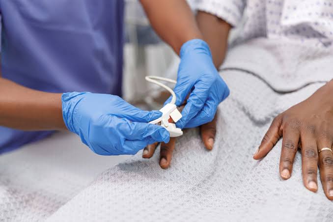
- Breathing Support: Administer supplemental oxygen via nasal cannula or mask to maintain adequate oxygen saturation levels.
- Circulation Support:
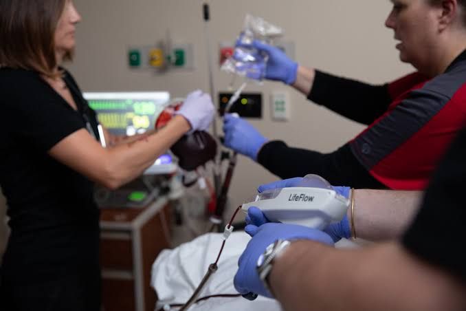
- Intravenous Access: Establish two large-bore intravenous (IV) lines (14–16G) for fluid resuscitation.
- Fluid Resuscitation: In cases of hypovolaemic shock (e.g., blood pressure <100 mmHg and pulse >100 bpm), administer 1–2 liters of crystalloid solution (e.g., 0.9% saline) rapidly while awaiting blood products.
- Be cautious in elderly patients or those with pre-existing cardiac conditions; a slower infusion rate may be necessary to prevent pulmonary edema.
4. Blood Transfusion
If the patient exhibits hemodynamic instability or has a hemoglobin level below 10 g/dl, initiate urgent blood transfusion following established protocols. Use O-negative blood if cross-matched blood is not immediately available.
- Monitor for potential complications associated with transfusion such as rebleeding rates and mortality risks.
5. Correction of Coagulopathy
Address any coagulopathy that may be present:
- For patients on warfarin who are actively bleeding, reverse anticoagulation using prothrombin complex concentrate along with intravenous vitamin K.
- In cases of thrombocytopenia where platelet counts are below 50 × 10^9/l and active bleeding is occurring, administer platelet transfusions.
6. Continuous Monitoring
Throughout the resuscitation process:
- Continuously monitor vital signs including heart rate, blood pressure, respiratory rate, and oxygen saturation.
- Assess urine output as an indicator of renal perfusion and overall fluid status.
7. Prepare for Further Intervention
Once initial stabilization is achieved:
- Consult gastroenterology for urgent endoscopy if indicated based on risk stratification.
- Be prepared for surgical intervention if endoscopic management fails or if there is ongoing massive hemorrhage.
In summary, effective management of acute GI hemorrhage involves rapid assessment and initiation of resuscitative measures focusing on stabilizing circulation through IV fluids and blood products while simultaneously addressing coagulopathies and preparing for potential therapeutic interventions.
Aetiopathology of Common Causes of Upper GI Bleeding
1. Duodenal Ulcer
Duodenal ulcers are primarily caused by an imbalance between aggressive factors (such as gastric acid and pepsin) and defensive factors (such as mucus and bicarbonate). The most common aetiological factor is infection with Helicobacter pylori, which leads to chronic inflammation of the gastric mucosa (chronic gastritis) and increased acid secretion. Nonsteroidal anti-inflammatory drugs (NSAIDs) also contribute to ulcer formation by inhibiting prostaglandin synthesis, which is essential for maintaining the protective mucosal barrier. The erosion of blood vessels in the submucosa can lead to bleeding.
2. Gastric Ulcer
Gastric ulcers share similar aetiological factors with duodenal ulcers but have distinct characteristics. They are often associated with H. pylori infection, NSAID use, and malignancy. Gastric ulcers occur when the protective mechanisms of the gastric mucosa are compromised, leading to damage from gastric acid. Unlike duodenal ulcers, which typically occur in younger individuals, gastric ulcers are more common in older adults. The ulceration can penetrate deeper into the stomach wall, potentially involving blood vessels and causing significant bleeding.
3. Gastric Erosions
Gastric erosions are superficial lesions that affect the gastric mucosa and can result from various factors including NSAID use, alcohol consumption, stress-related mucosal disease, or ischemia due to reduced blood flow. Unlike ulcers, erosions do not penetrate through the muscularis layer but can still cause bleeding if they affect underlying blood vessels. The acute nature of erosions often leads to sudden onset upper GI bleeding.
4. Oesophageal Varices
Oesophageal varices are dilated veins in the esophagus that develop due to portal hypertension, commonly resulting from liver cirrhosis or hepatic venous obstruction. Increased pressure in the portal vein causes collateral circulation to develop, leading to variceal formation. These varices are fragile and prone to rupture under increased pressure or trauma, resulting in significant upper GI bleeding.
5. Mallory-Weiss Tear
A Mallory-Weiss tear is a longitudinal laceration at the gastroesophageal junction caused by severe vomiting or retching that increases intra-abdominal pressure abruptly. This condition is often seen in individuals with alcohol use disorder or those experiencing severe gastrointestinal distress. The tear can lead to bleeding from the esophagus or proximal stomach due to disruption of blood vessels at this junction.
6. Oesophagogastric Cancer
Oesophagogastric cancer encompasses malignancies arising from either the esophagus or stomach and can lead to upper GI bleeding through several mechanisms: direct invasion into surrounding tissues causing erosion of blood vessels; obstruction leading to increased pressure; or ulceration associated with tumor growth. Risk factors include chronic gastroesophageal reflux disease (GERD), smoking, obesity, and dietary factors such as low fruit and vegetable intake.
In summary, upper GI bleeding can arise from various conditions characterized by different aetiopathological mechanisms ranging from infections and lifestyle factors to malignancies and anatomical changes due to increased pressures.
Role of Oesophago-Gastro-Duodenoscopy (OGD) in GI Bleeding Management
Oesophago-gastro-duodenoscopy (OGD), commonly referred to as upper gastrointestinal endoscopy, plays a crucial role in the management of upper gastrointestinal bleeding (UGIB). The procedure involves the use of a flexible tube equipped with a camera and light source, which allows healthcare providers to visualize the esophagus, stomach, and duodenum.
- Diagnosis: OGD is primarily utilized for diagnosing the source of UGIB. Common causes include peptic ulcers, esophageal varices, gastritis, and malignancies. By directly visualizing these areas, clinicians can identify lesions or abnormalities that may be causing bleeding.
- Therapeutic Intervention: Beyond diagnosis, OGD also serves therapeutic purposes. It enables interventions such as:
- Hemostasis: Techniques like thermal coagulation, band ligation for varices, or clipping can be employed to control active bleeding.
- Biopsy: Tissue samples can be taken for histological examination if malignancy is suspected.
- Removal of Obstructions: In cases where food or foreign bodies are obstructing the upper GI tract, OGD can facilitate their removal.
- Timing and Urgency: The timing of OGD is critical in managing UGIB. For patients with suspected variceal bleeding due to liver cirrhosis, guidelines recommend performing endoscopy within 12 hours after stabilization. For non-variceal UGIB, intervention should ideally occur within 24 hours for high-risk patients based on scoring systems like the Glasgow-Blatchford Score.
Role of Colonoscopy in GI Bleeding Management
Colonoscopy is essential for evaluating lower gastrointestinal bleeding (LGIB). This procedure allows direct visualization of the colon and rectum using a flexible endoscope.
- Diagnosis: Colonoscopy is effective in identifying sources of LGIB such as diverticulosis, colorectal cancer, inflammatory bowel disease (IBD), and angiodysplasia. It provides real-time imaging that aids in pinpointing the exact location and cause of bleeding.
- Therapeutic Measures: Similar to OGD, colonoscopy offers therapeutic options:
- Hemostasis Techniques: Methods such as argon plasma coagulation or clip application can be used to manage identified bleeding sites.
- Polypectomy: If polyps are found during colonoscopy that could potentially bleed or become malignant, they can be removed during the same procedure.
- Marking Lesions: In cases where lesions are detected but cannot be treated immediately due to other factors (e.g., patient stability), they can be marked for future surgical intervention.
- Preparation and Timing: Effective bowel preparation is vital prior to colonoscopy to ensure clear visualization of the colonic mucosa. The timing of colonoscopy following an episode of LGIB may vary; however, it is generally performed once patients are stabilized and adequately prepared.
In summary, both OGD and colonoscopy serve pivotal roles in diagnosing and managing gastrointestinal bleeding through their diagnostic capabilities and therapeutic interventions tailored to specific types of bleeding—upper versus lower GI tract.
Risk Factors for Upper GI Bleeding
Upper gastrointestinal (GI) bleeding can arise from various conditions and risk factors. The primary risk factors include:
- Medications:
- Non-steroidal anti-inflammatory drugs (NSAIDs) such as aspirin, ibuprofen, and naproxen are significant contributors to upper GI bleeding due to their effect on the gastric mucosa.
- Anticoagulants can increase the risk of bleeding by affecting blood clotting mechanisms.
- Helicobacter pylori Infection:
- This bacterium is known to cause peptic ulcers, which can lead to bleeding if not treated effectively.
- Chronic Alcohol Use:
- Excessive alcohol consumption can irritate the stomach lining and contribute to conditions like gastritis or ulcers that may bleed.
- History of Peptic Ulcers:
- Individuals with a history of peptic ulcers are at an increased risk for rebleeding.
- Liver Disease:
- Conditions such as cirrhosis can lead to variceal bleeding due to increased pressure in the portal vein.
- Age:
- Older adults are generally at a higher risk due to the cumulative effects of medications, comorbidities, and physiological changes in the GI tract.
- Chronic Vomiting:
- Conditions that cause chronic vomiting can lead to tears in the esophagus or stomach lining (e.g., Mallory-Weiss tears).
- Inflammatory Bowel Disease:
- Diseases such as Crohn’s disease and ulcerative colitis can cause inflammation and ulceration leading to bleeding.
- Cancer:
- Tumors in the GI tract can erode blood vessels or cause other complications leading to bleeding.
- Diverticulosis:
- This condition involves pouches forming in the colon wall that can become inflamed or bleed.
Role of the GP in Prevention
General practitioners (GPs) play a crucial role in preventing upper GI bleeding through several strategies:
- Risk Assessment:
- GPs should evaluate patients’ medical histories for risk factors such as previous ulcers, current medication use (especially NSAIDs and anticoagulants), alcohol consumption, and any existing liver disease.
- Patient Education:
- Educating patients about the risks associated with certain medications, particularly NSAIDs and their potential gastrointestinal side effects, is essential.
- GPs should inform patients about lifestyle modifications that could reduce their risk, such as moderating alcohol intake and avoiding smoking.
- Screening for Helicobacter pylori:
- GPs should consider testing for H. pylori infection in patients with a history of peptic ulcers or those presenting with dyspepsia symptoms.
- If detected, appropriate eradication therapy should be initiated.
- Medication Management:
- For patients requiring NSAIDs or anticoagulants, GPs should assess whether gastroprotective agents like proton pump inhibitors (PPIs) are necessary.
- Regular review of medication regimens is important to minimize risks while managing pain or cardiovascular conditions effectively.
- Monitoring High-Risk Patients:
- GPs should closely monitor patients with chronic conditions that predispose them to upper GI bleeding.
- Regular follow-ups may be necessary for those on long-term NSAID therapy or those with a history of gastrointestinal issues.
- Referral for Specialist Care:
- When indicated, GPs should refer patients for endoscopic evaluation or specialist care if they exhibit signs of significant gastrointestinal distress or have complex medical histories that increase their risk of bleeding.
By implementing these preventive measures, GPs can significantly reduce the incidence of upper GI bleeding among at-risk populations.
Role and Indications for Investigations
- Initial Assessment: The initial evaluation of patients with GIB begins with a thorough history and physical examination. This assessment helps determine whether the bleeding is from an upper or lower gastrointestinal source. Signs such as tachycardia, orthostatic hypotension, and chronic anemia may indicate significant blood loss.
- Laboratory Tests: Laboratory tests including complete blood count (CBC), liver function tests, and coagulation profiles are essential to assess the severity of bleeding and guide treatment decisions.
- Imaging Studies:
- Endoscopy: Upper endoscopy (esophagogastroduodenoscopy) is often performed for suspected upper GIB to visualize the source directly and potentially treat it via cauterization or band ligation.
- Colonoscopy: For lower GIB, colonoscopy allows for direct visualization of the colon and can also be therapeutic.
- Radiological Imaging: In cases where endoscopic intervention fails or is not possible, imaging studies such as computed tomography (CT) angiography can be utilized to localize the source of bleeding. Nuclear scintigraphy may also be employed in specific scenarios to detect active bleeding.
Role of Interventional Radiology
Interventional radiology plays a crucial role in managing GIB when conventional medical management and endoscopic therapies are inadequate:
- Angiography: This technique allows for direct visualization of blood vessels supplying the gastrointestinal tract. It can help identify areas of active bleeding that may not be visible through other means.
- Transcatheter Embolization: Once a bleeding vessel is identified via angiography, interventional radiologists can perform embolization to occlude the vessel, effectively stopping the hemorrhage. This method has become increasingly safe due to advancements in catheter technology.
- Indications for Interventional Radiology:
- Persistent or recurrent GIB despite medical therapy.
- Patients who are hemodynamically unstable where rapid control of bleeding is necessary.
- Cases where endoscopic intervention has failed or is deemed inappropriate due to anatomical considerations.
Role of Surgery
Surgical intervention remains an important option in managing GIB:
- Indications for Surgery:
- Surgical intervention may be required when there is massive hemorrhage that cannot be controlled by less invasive means.
- Patients with specific conditions such as perforated ulcers or malignancies causing significant bleeding may necessitate surgical resection.
- In cases where interventional radiology fails to control the bleed or when there are complications from embolization (e.g., ischemia), surgery becomes necessary.
- Types of Surgical Procedures:
- Resection of affected bowel segments in cases involving malignancy or diverticular disease.
- Surgical repair of vascular anomalies contributing to GIB.
- Laparotomy may be performed in emergency situations where rapid access to the abdominal cavity is needed for control of hemorrhage.
In summary, the management of gastrointestinal bleeding requires a multidisciplinary approach involving careful investigation to ascertain the source and nature of the bleed, followed by appropriate use of interventional radiology techniques when indicated, and surgical intervention when necessary.


