Parts of the Diaphragm
The diaphragm is a complex musculotendinous structure that consists of three main parts, each with specific attachments and neurovascular supply. These parts are:
1. Sternal Part
- Attachments: The sternal part originates from the xiphoid process of the sternum.
- Neurovascular Supply: It receives innervation from the phrenic nerve (C3-C5) and blood supply primarily from the superior phrenic artery.
2. Costal Part
- Attachments: The costal part attaches to the inner surfaces of the lower six ribs (ribs 7-12) and their costal cartilages.
- Neurovascular Supply: This part is also innervated by the phrenic nerve and receives blood from both the inferior phrenic arteries and intercostal arteries.
3. Lumbar Part
- Attachments: The lumbar part arises from the lumbar vertebrae (L1-L3) via tendinous structures known as crura, specifically the right crus and left crus. The right crus arises from L1-L3 and their intervertebral discs, while the left crus arises from L1-L2.
- Neurovascular Supply: Like other parts, it is innervated by the phrenic nerve, with additional sensory innervation provided by the lower intercostal nerves (6th to 11th). Blood supply comes mainly from the inferior phrenic arteries.
In summary, each part of the diaphragm has distinct origins that contribute to its overall function in respiration, with a consistent pattern of neurovascular supply ensuring proper muscle function and sensation.
Role of the Diaphragm in Respiration
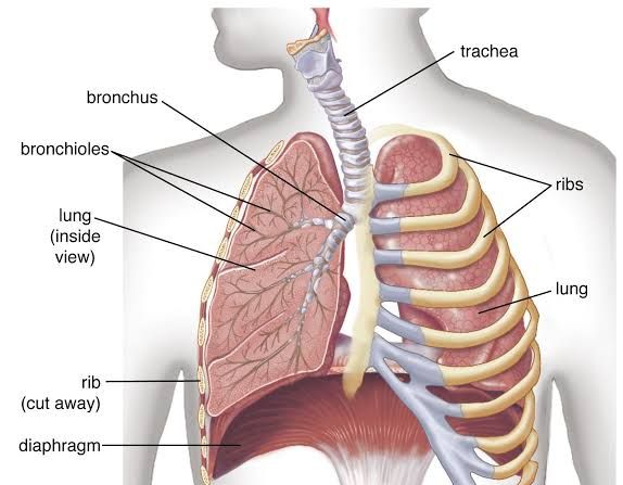
The diaphragm is a crucial muscle located beneath the lungs that plays a significant role in the process of respiration. Its primary function is to facilitate breathing by contracting and relaxing rhythmically, which allows for the inhalation and exhalation of air.
Inhalation Process
- Diaphragm Contraction: When you inhale, the diaphragm contracts and moves downward. This contraction flattens the dome-shaped muscle, increasing the volume of the thoracic (chest) cavity.
- Vacuum Creation: As the diaphragm moves downwards, it creates a negative pressure (vacuum) within the chest cavity. This negative pressure causes air to be drawn into the lungs through the trachea.
- Lung Expansion: The expansion of the chest cavity allows the lungs to expand as well, filling with air that travels through bronchial tubes to reach alveoli, where gas exchange occurs.
Exhalation Process
- Diaphragm Relaxation: During exhalation, the diaphragm relaxes and returns to its original dome shape. This relaxation decreases the volume of the thoracic cavity.
- Air Expulsion: As the chest cavity’s volume decreases, pressure inside increases, forcing air out of the lungs and back through the trachea to exit through the nose or mouth.
- Role of Other Muscles: While exhalation is typically a passive process during rest, it can become active during physical exertion when abdominal muscles contract to push more air out forcefully.
Additional Functions of the Diaphragm
- Pressure Regulation: The diaphragm also helps regulate pressure in both thoracic and abdominal cavities. This regulation aids in functions such as urination and defecation by increasing intra-abdominal pressure.
- Prevention of Acid Reflux: By maintaining pressure on adjacent structures like the esophagus, it assists in preventing acid reflux.
In summary, the diaphragm is essential for respiration as it facilitates inhalation by contracting and creating negative pressure in the thoracic cavity while also aiding in exhalation by relaxing and allowing for air expulsion from the lungs.
Diaphragmatic Apertures and Their Structures
The diaphragm has three major apertures that allow structures to pass between the thoracic and abdominal cavities. Each aperture corresponds to a specific vertebral level and is associated with particular anatomical structures. Below are the details of these apertures:
- Caval Opening (Vena Caval Foramen)
- Vertebral Level: T8
- Structures Traversing:
- Inferior vena cava (IVC)
- Phrenic nerve (phrenicN)
- Esophageal Hiatus
- Vertebral Level: T10
- Structures Traversing:
- Esophagus
- Vagus nerves (CN X)
- Aortic Hiatus
- Vertebral Level: T12
- Structures Traversing:
- Aorta
- Thoracic duct
- Azygos vein
These apertures are crucial for the passage of blood vessels, nerves, and other structures between the thorax and abdomen, facilitating essential physiological functions.


