Histological Features of Plasma Membrane and Cellular Organelles Correlated with Their Function
Plasma Membrane
The plasma membrane is a critical structure that surrounds the cell, acting as a barrier between the cytosol and the extracellular environment. Its histological features include:
- Lipid Bilayer: The plasma membrane is primarily composed of phospholipids arranged in a bilayer formation, which creates an amphipathic structure. This bilayer allows for selective permeability, enabling the passage of certain molecules while restricting others.
- Proteins: Integral and peripheral proteins are embedded within or associated with the lipid bilayer. Integral proteins can function as channels or pumps to facilitate transport across the membrane, while peripheral proteins often play roles in signaling and maintaining the cell’s shape.
- Cholesterol: Cholesterol molecules interspersed within the phospholipid bilayer contribute to membrane fluidity and stability. They help maintain structural integrity and influence permeability to small water-soluble molecules.
- Glycolipids: These are found on the extracellular side of the membrane and play a role in cell recognition and signaling processes.
The functions of these features include:
- Selective Permeability: The lipid bilayer allows for controlled entry and exit of substances.
- Cell Communication: Membrane proteins act as receptors that bind ligands, facilitating communication with external signals.
- Structural Support: The presence of cholesterol and cytoskeletal linkers provides mechanical support to maintain cell shape.
Cellular Organelles
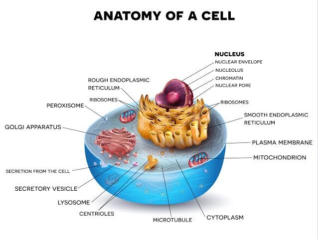
- Endoplasmic Reticulum (ER):
- Rough ER (rER): Characterized by ribosomes on its surface, it is involved in protein synthesis and folding. Proteins synthesized here are often destined for secretion or for use in lysosomes.
- Smooth ER (sER): Lacks ribosomes and is involved in lipid synthesis, detoxification processes, and calcium ion storage.
- Golgi Apparatus:
- Composed of flattened membranous sacs (cisternae), it modifies, sorts, and packages proteins received from the rER for transport to their final destinations (e.g., secretion outside the cell or delivery to lysosomes).
- Mitochondria:
- Known as the powerhouse of the cell, mitochondria have a double-membrane structure that facilitates ATP production through oxidative phosphorylation. Their inner membrane contains folds called cristae that increase surface area for energy production.
- Lysosomes:
- These organelles contain hydrolytic enzymes necessary for breaking down waste materials and cellular debris. Their acidic interior allows them to effectively degrade macromolecules.
- Peroxisomes:
- Contain enzymes that oxidize fatty acids and amino acids; they also detoxify harmful substances like hydrogen peroxide through catalase activity.
- Secretory Vesicles:
- These vesicles transport materials such as hormones or neurotransmitters from one part of the cell to another or out of the cell via exocytosis.
Each organelle’s structure correlates directly with its function—membranous organelles compartmentalize various biochemical processes essential for cellular homeostasis, while non-membranous structures like ribosomes facilitate protein synthesis directly within the cytoplasm.
In summary, both plasma membranes and cellular organelles exhibit specific histological features that are intricately linked to their respective functions within eukaryotic cells.
Membranous Organelles
Membranous organelles are cellular structures that are surrounded by a lipid bilayer membrane, which separates their internal environment from the cytoplasm. This compartmentalization allows for specialized functions to occur within these organelles. The main membranous organelles include:
- Endoplasmic Reticulum (ER): The ER is a network of membranes involved in protein and lipid synthesis. It is divided into two types:
- Rough Endoplasmic Reticulum (rER): Studded with ribosomes, the rER is primarily responsible for the synthesis of proteins that are either secreted from the cell or incorporated into cellular membranes.
- Smooth Endoplasmic Reticulum (sER): Lacking ribosomes, the sER is involved in lipid synthesis, metabolism, and detoxification processes.
- Golgi Apparatus: This organelle consists of stacked membranous sacs known as cisternae. It modifies, sorts, and packages proteins received from the rER for transport to their final destinations within or outside the cell.
- Mitochondria: Often referred to as the powerhouses of the cell, mitochondria have a double membrane structure. They are essential for energy production through cellular respiration, converting nutrients into adenosine triphosphate (ATP).
- Peroxisomes: These single-membrane-bound organelles contain enzymes that oxidize fatty acids and amino acids, producing hydrogen peroxide as a byproduct, which is then broken down by catalase.
- Lysosomes: Membranous sacs filled with hydrolytic enzymes that digest macromolecules and recycle cellular components through processes such as autophagy and phagocytosis.
Non-Membranous Organelles
Non-membranous organelles lack a surrounding membrane and are typically involved in structural support or cellular processes that do not require compartmentalization. Key non-membranous organelles include:
- Ribosomes: These are complexes made up of ribosomal RNA (rRNA) and proteins that synthesize proteins by translating messenger RNA (mRNA). Ribosomes can be found free-floating in the cytoplasm or attached to the rER.
- Cytoskeleton: This network provides structural support to the cell and facilitates movement. It consists of three main components:
- Microfilaments: Composed mainly of actin filaments; they play roles in muscle contraction and cell motility.
- Intermediate Filaments: Provide mechanical strength to cells.
- Microtubules: Hollow tubes made of tubulin; they help maintain cell shape and are involved in intracellular transport as well as mitotic spindle formation during cell division.
- Centrioles: Cylindrical structures composed of microtubules that play a crucial role in organizing microtubules during cell division.
In summary, membranous organelles are characterized by their lipid bilayer membranes allowing compartmentalized functions essential for various cellular activities, while non-membranous organelles contribute primarily to structural integrity and protein synthesis without such compartmentalization.
Histological Characteristics of an Apoptotic Cell
Apoptosis, or programmed cell death, is characterized by a series of distinct histological features that can be observed under a microscope. These features are crucial for identifying apoptotic cells in tissue samples. The following are the main histological characteristics of an apoptotic cell:
1. Cell Shrinkage:
One of the earliest morphological changes during apoptosis is cell shrinkage. This occurs due to the loss of intracellular water and ions, leading to a reduction in cell volume. The cytoplasm becomes denser as organelles condense.
2. Chromatin Condensation:
During apoptosis, chromatin undergoes significant changes. It condenses and aggregates along the nuclear membrane, resulting in a characteristic appearance known as pyknosis. This condensation is often accompanied by nuclear fragmentation.
3. Nuclear Fragmentation:
As apoptosis progresses, the nucleus may break apart into smaller fragments, a process referred to as karyorrhexis. This fragmentation contributes to the overall breakdown of cellular structure.
4. Formation of Apoptotic Bodies:
The dying cell begins to form small membrane-bound vesicles known as apoptotic bodies. These bodies contain cellular components and are formed as the cell’s cytoplasm and organelles are packaged into these vesicles.
5. Membrane Blebbing:
The plasma membrane exhibits blebbing, where small protrusions (blebs) form on its surface due to cytoskeletal reorganization and loss of structural integrity. This blebbing is a hallmark feature of apoptotic cells.
6. Loss of Membrane Integrity:
While apoptotic cells maintain their membrane integrity longer than necrotic cells, they eventually lose it as apoptosis progresses, allowing for the release of signals that attract phagocytes for clearance.
7. DNA Fragmentation:
A key biochemical event in apoptosis is the cleavage of DNA into oligonucleosomal fragments, which can be detected using specific staining techniques such as TUNEL assay or agarose gel electrophoresis.
These histological characteristics collectively distinguish apoptotic cells from necrotic cells and provide insight into the underlying mechanisms governing programmed cell death.


