Trachea
The trachea, commonly referred to as the windpipe, is a crucial component of the respiratory system. It serves as a conduit for air to travel from the larynx (voice box) down to the bronchi, which then direct air into the lungs. The trachea is approximately 4 inches (10 centimeters) long and about 1 inch (2.5 centimeters) in diameter, making it roughly the width of an adult’s finger.
Structure and Relations of the Trachea
The trachea is composed of 16 to 20 C-shaped rings of cartilage that provide structural support while allowing flexibility. These rings are connected by smooth muscle known as the trachealis muscle, which can contract or relax to adjust airflow during breathing. The inner lining of the trachea consists of mucosa, which contains goblet cells that secrete mucus to trap dust and debris, along with cilia that help move this mucus out of the airway.
In terms of anatomical relations, the trachea is located in the lower neck and upper chest region. It sits anteriorly to the esophagus and extends from the larynx at approximately the level of C6 vertebra down to T5 vertebra where it bifurcates into the right and left main bronchi. The trachea is positioned behind the sternum and between the two lobes of the lungs.
Subdivision of Trachea
The trachea does not have formal subdivisions like other organs; however, it can be described in terms of its proximal and distal ends:
- Proximal End: This end connects with the larynx.
- Distal End: This end bifurcates into two main bronchi (right and left), leading into each lung.
Pleura and Pleural Cavity
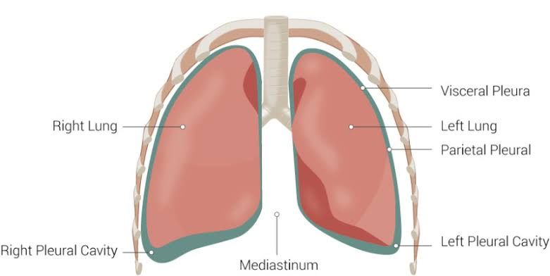
The pleura refers to a set of serous membranes that envelop each lung and line the thoracic cavity. These membranes play a vital role in facilitating respiration by allowing smooth movement between lung surfaces during inhalation and exhalation.
Parts of Pleura:
- Visceral Pleura: This layer directly covers the surface of each lung, extending into fissures between lobes.
- Parietal Pleura: This thicker layer lines the internal surface of the thoracic cavity and can be further subdivided based on its location:
- Mediastinal Pleura: Covers parts adjacent to mediastinum.
- Cervical Pleura: Extends into the neck region.
- Costal Pleura: Lines inner aspects of ribs.
- Diaphragmatic Pleura: Covers superior surface of diaphragm.
Pleural Cavity: The pleural cavity is a potential space between these two layers (visceral and parietal pleura). It contains a small amount of serous fluid that serves two primary functions:
- Lubrication: Allows pleural surfaces to slide over one another smoothly.
- Surface Tension: Helps keep both layers together so that when thoracic volume changes during breathing, lung expansion occurs simultaneously.
Pleural Recesses: There are specific recesses within this cavity where opposing pleural surfaces come close together:
- Costodiaphragmatic Recess: Located between costal pleurae and diaphragmatic pleurae.
- Costomediastinal Recess: Found between costal pleurae and mediastinal pleurae behind sternum.
These recesses are clinically significant because they can serve as sites for fluid accumulation in conditions such as pleural effusion.
Pleural Nerve Supply
The pleura is a serous membrane that envelops the lungs and lines the thoracic cavity. It consists of two layers: the visceral pleura, which covers the lungs, and the parietal pleura, which lines the thoracic wall and diaphragm. The nerve supply to the pleura is crucial for its sensory functions and is derived from different sources depending on which layer of the pleura is being considered.
- Visceral Pleura: The visceral pleura is innervated by autonomic nerves, specifically from the pulmonary plexus, which contains both sympathetic and parasympathetic fibers. These fibers are responsible for reflex actions such as coughing and bronchoconstriction but do not convey pain sensations.
- Parietal Pleura: In contrast, the parietal pleura has a rich sensory nerve supply primarily from somatic nerves. The intercostal nerves provide sensation to the costal part of the parietal pleura, while the phrenic nerve supplies sensation to the mediastinal and diaphragmatic parts. This somatic innervation allows for pain perception in conditions affecting the parietal pleura, such as pleuritis or pneumothorax.
The distinction between these two types of innervation is important clinically; visceral pain is often referred and poorly localized, while pain from irritation of the parietal pleura can be sharp and well-localized due to its somatic nerve supply.
Lungs: Lobes, Fissures, Surfaces
The human lungs are paired organs located in the thoracic cavity that facilitate gas exchange through respiration. Each lung has distinct anatomical features including lobes, fissures, surfaces, and variations between right and left lungs.
- Lobes:
- Right Lung: The right lung consists of three lobes:
- Superior lobe
- Middle lobe
- Inferior lobe
- Left Lung: The left lung has two lobes:
- Superior lobe
- Inferior lobe
- Right Lung: The right lung consists of three lobes:
- Fissures:
- Right Lung: It has two fissures:
- Horizontal fissure (separates superior from middle lobe)
- Oblique fissure (separates middle from inferior lobe)
- Left Lung: It has one oblique fissure that separates its superior and inferior lobes.
- Right Lung: It has two fissures:
- Surfaces:
- Both lungs have three main surfaces:
- Costal surface (facing the ribs)
- Mediastinal surface (facing the heart and other mediastinal structures)
- Diaphragmatic surface (facing down towards the diaphragm)
- Both lungs have three main surfaces:
- Comparison Between Right and Left Lungs:
- The right lung is larger than the left lung due to anatomical constraints imposed by the heart’s position in the thoracic cavity.
- The right main bronchus is wider and more vertically oriented compared to the left main bronchus, making it more susceptible to foreign body aspiration.
- The left lung features a cardiac notch on its anterior border to accommodate space for the heart.
In summary, while both lungs serve similar functions in respiration, they exhibit significant differences in structure due to their anatomical positioning within the thoracic cavity.
Bronchopulmonary Segments
The bronchopulmonary segments are the functional divisions of the lungs, each receiving its own air and blood supply. Here is a detailed list of the bronchopulmonary segments for both the right and left lungs:
Right Lung (10 Segments):
- Apical segment (B1)
- Posterior segment (B2)
- Anterior segment (B3)
- Lateral segment (B4) – part of the middle lobe
- Medial segment (B5) – part of the middle lobe
- Superior segment (B6) – part of the lower lobe
- Medial basal segment (B7)
- Anterior basal segment (B8)
- Lateral basal segment (B9)
- Posterior basal segment (B10)
Left Lung (8 Segments):
- Apicoposterior segment (B1/2)
- Anterior segment (B3)
- Superior segment (B6) – part of the lower lobe
- Anteromedial segment (B7/8)
- Lateral segment (B9)
- Posterior segment (B10)
Innervations, Blood Supply, and Lymphatic Drainage of the Lungs
1. Innervations:
The lungs receive autonomic innervation from the pulmonary plexus, which is formed by branches from both the sympathetic and parasympathetic nervous systems:
- Sympathetic Innervation: This comes from thoracic spinal nerves T1-T4, leading to bronchodilation and reduced glandular secretion.
- Parasympathetic Innervation: This is primarily through the vagus nerve, which causes bronchoconstriction and increased glandular secretion.
2. Blood Supply:
The lungs receive dual blood supply:
- Pulmonary Circulation: The pulmonary arteries carry deoxygenated blood from the right ventricle to the lungs for oxygenation.
- Bronchial Circulation: The bronchial arteries supply oxygenated blood to lung tissue itself; these arise from either the intercostobronchial trunk or directly from the descending thoracic aorta.
3. Lymphatic Drainage:
Lymphatic drainage in the lungs is crucial for maintaining fluid balance and immune function:
- Superficial Lymphatic Plexus: Located beneath the pleura, it drains lymph from lung surfaces into regional lymph nodes.
- Deep Lymphatic Plexus: Found within the bronchial walls, it drains lymph from deeper structures like bronchi and pulmonary vessels into hilum-associated lymph nodes.
Parts and Contents of the Mediastinum
The mediastinum is a central compartment in the thoracic cavity, situated between the lungs. It is divided into four main parts: the superior mediastinum, anterior mediastinum, middle mediastinum, and posterior mediastinum. Each part contains specific structures and organs.
- Superior Mediastinum:
- Location: Extends from the thoracic inlet to the horizontal plane at the level of T4-T5 vertebrae.
- Contents:
- Thymus gland (in children)
- Great vessels: aorta (ascending, arch, descending), brachiocephalic trunk, left common carotid artery, left subclavian artery
- Trachea
- Esophagus
- Thoracic duct
- Vagus nerves and phrenic nerves
- Anterior Mediastinum:
- Location: Lies between the sternum and pericardium.
- Contents:
- Thymus gland (in adults it may be small or absent)
- Fat tissue
- Lymph nodes
- Middle Mediastinum:
- Location: Contains structures that are centrally located within the thorax.
- Contents:
- Heart and pericardium
- Ascending aorta
- Pulmonary arteries and veins
- Mainstem bronchi
- Internal thoracic arteries and veins
- Posterior Mediastinum:
- Location: Located behind the heart and pericardium.
- Contents:
- Descending aorta
- Esophagus
- Thoracic duct
- Azygos vein system (azygos vein, hemiazygos vein)
- Sympathetic trunks
Internal Thoracic Artery
The internal thoracic artery (ITA), also known as the internal mammary artery, is an important vessel in the thorax that supplies blood to various structures in the chest wall and breasts.
- Origin: The internal thoracic artery arises from the first part of the subclavian artery, typically just distal to its origin from the brachiocephalic trunk on the right side or directly from the subclavian on the left side.
- Location: The ITA descends vertically along each side of the sternum within the thorax.
- Course: After its origin, it travels downwards along the inner surface of the rib cage, running parallel to but slightly lateral to the sternum.
- Branches: The internal thoracic artery gives rise to several branches as it descends:
- Anterior intercostal arteries (branches to supply intercostal spaces 1-6)
- Perforating branches that supply skin and muscles of anterior chest wall
- Musculophrenic artery (supplies diaphragm and lower intercostal spaces)
- Superior epigastric artery (continues downward into abdominal wall)
The ITA plays a crucial role in surgical procedures such as coronary artery bypass grafting due to its robust blood supply.
Surface Markings of the Trachea, Lungs, and Pleura
The surface markings of the trachea, lungs, and pleura are critical for understanding normal anatomy as well as identifying pathological conditions on imaging studies such as chest X-rays and CT scans.
Trachea Surface Markings
The trachea extends from the cricoid cartilage at the level of C6 to the carina at T5-T6. On a chest X-ray, it is typically visualized in the midline of the neck and thorax. The trachea is approximately 10-12 cm long and is composed of C-shaped cartilaginous rings that maintain its patency. The surface marking can be traced downwards from the suprasternal notch (approximately at T2) to its bifurcation into the right and left main bronchi at the carina.
Lung Surface Markings
The lungs are divided into lobes: three on the right (upper, middle, lower) and two on the left (upper and lower). On a chest X-ray, lung markings should extend to the thoracic wall. The upper lobe typically reaches up to about T1-T2 posteriorly, while the lower lobe extends down to about T10-T12 during expiration. The costophrenic angles should be sharp and well-defined; blunting may indicate fluid accumulation (pleural effusion).
Pleura Surface Markings
The pleura consists of two layers: visceral pleura covering the lungs and parietal pleura lining the thoracic cavity. In normal circumstances, these layers are not distinctly visible on imaging unless there are abnormalities present. On a chest X-ray or CT scan, signs of pleural disease such as thickening or effusion can be assessed by examining areas around each lung’s edge.
Typical Appearance on Chest X-Ray
A standard chest X-ray provides a two-dimensional view of thoracic structures. Key features include:
- Clear visualization of lung fields with vascular markings extending to the edges.
- Presence or absence of pleural effusions indicated by blunted costophrenic angles.
- Identification of pneumothorax if there is a visible visceral pleura with air surrounding it.
- Assessment for any abnormal opacities that may suggest masses or infiltrates.
Typical Appearance on CT Scan
CT scans offer cross-sectional imaging that provides detailed views of thoracic structures:
- The trachea appears as a tubular structure with clear delineation against surrounding tissues.
- Lung parenchyma can be evaluated for nodules, consolidations, or ground-glass opacities.
- Pleural spaces can be assessed for effusions or thickening more accurately than on X-rays due to higher resolution.
- CT also allows for evaluation in multiple planes (axial, coronal) which aids in assessing complex pathologies.
In summary, understanding these surface markings helps radiologists identify normal anatomy and detect abnormalities effectively using both chest X-rays and CT scans.


