Relationship Between Pressure, Flow, and Resistance
The relationship between pressure, flow, and resistance in the circulatory system can be understood through principles derived from physics, particularly Ohm’s Law. In hemodynamics, blood flow (F) is analogous to electrical current (I), the pressure difference (ΔP) across a vessel is akin to voltage difference (ΔV), and vascular resistance (R) corresponds to electrical resistance.
- Pressure Difference (ΔP): This is the driving force for blood flow and is defined as the difference in pressure between two points in the circulatory system. For instance, when considering blood flow through an organ like the kidney, ΔP can be expressed as the difference between arterial pressure (PA) and venous pressure (PV). Thus, ΔP = PA – PV.
- Blood Flow (F): Blood flow refers to the volume of blood that passes through a given point in the circulation over a specific period. It can be influenced by various factors including heart rate and stroke volume.
- Resistance (R): Resistance to blood flow arises from several factors including the diameter of blood vessels, viscosity of the blood, and length of the vessel. The relationship can be summarized mathematically as: F = ΔP ÷ R. This equation indicates that for a given pressure difference, an increase in resistance will lead to a decrease in blood flow.
- Regulation of Blood Flow: The body regulates blood flow primarily by altering vascular resistance rather than changing perfusion pressures significantly. When resistance increases due to constriction of blood vessels or other factors, it leads to reduced flow at any given ΔP.
- Impact of Pathological Conditions: Under normal conditions with laminar flow, there is a linear relationship between perfusion pressure and flow; however, this relationship changes under pathological conditions leading to turbulent flow where increased resistance results in decreased flow despite higher pressures.
Laminar and Turbulent Blood Flow
Blood flow can be categorized into two primary types: laminar and turbulent.
- Laminar Flow:
- Laminar flow occurs when blood moves smoothly in parallel layers without disruption. This type of flow is characteristic of healthy arteries where there are no obstructions.
- In laminar conditions, each layer flows at different velocities—typically faster in the center than near the vessel walls—but maintains an orderly pattern.
- Laminar flow is generally associated with lower Reynolds numbers (<2300), indicating that viscous forces dominate over inertial forces.
- Turbulent Flow:
- Turbulent flow arises when there are disturbances within the bloodstream that disrupt this orderly movement. Factors such as arterial narrowing due to atherosclerosis or branching vessels contribute to turbulence.
- In turbulent conditions, eddies and vortices form within the fluid leading to chaotic movement which can generate sounds such as murmurs or bruits detectable during clinical examinations.
- Turbulent flow typically occurs at higher Reynolds numbers (>4000), where inertial forces dominate over viscous forces.
- Clinical Implications:
- The transition from laminar to turbulent flow has significant clinical implications since turbulence increases vascular resistance which can elevate blood pressure and reduce overall perfusion efficiency.
- Understanding these dynamics helps clinicians assess cardiovascular health and identify potential issues related to arterial health.
In summary, understanding how pressure influences blood flow through varying levels of resistance provides crucial insights into cardiovascular physiology while recognizing how laminar versus turbulent flows impact overall vascular health is essential for diagnosing and managing cardiovascular diseases effectively.
Understanding Methods for Measurement of Blood Flow
Blood flow can be measured using several methods, each with its own principles and applications. The most common methods include:
- Doppler Ultrasound: This non-invasive technique uses sound waves to measure the velocity of blood flow in vessels. By emitting ultrasound waves and analyzing their reflection from moving red blood cells, it provides information about the speed and direction of blood flow.
- Magnetic Resonance Imaging (MRI): MRI can visualize blood flow through the use of magnetic fields and radio waves. It is particularly useful for assessing large vessels and complex vascular structures.
- Electromagnetic Flowmetry: This method involves placing an electromagnetic sensor around a blood vessel. As blood flows through the magnetic field, it induces a voltage proportional to the flow rate, allowing for real-time measurement.
- Laser Doppler Flowmetry: This technique uses laser light to measure microvascular blood flow. It is often used in research settings to assess skin perfusion or other localized areas.
- Thermal Dilution: Commonly used in critical care settings, this method involves injecting a known quantity of cold saline into the bloodstream and measuring changes in temperature downstream to calculate cardiac output.
Each of these methods has specific indications based on clinical needs, patient conditions, and the type of vascular assessment required.
Definition of Blood Pressure and Its Standard Unit
Blood pressure is defined as the force exerted by circulating blood against the walls of blood vessels, primarily arteries. It is measured as two values:
- Systolic Blood Pressure (SBP): The pressure in the arteries when the heart beats.
- Diastolic Blood Pressure (DBP): The pressure in the arteries when the heart rests between beats.
The standard unit for measuring blood pressure is millimeters of mercury (mmHg). A typical reading might be expressed as 120/80 mmHg, where 120 represents systolic pressure and 80 represents diastolic pressure.
Resistance to Blood Flow
Blood flow resistance is a critical factor in the circulatory system, influencing how easily blood can move through vessels. Resistance is primarily determined by three factors: the viscosity of the blood, the length of the blood vessels, and the radius of the vessels. According to Poiseuille’s law, resistance (R) can be expressed mathematically as:
R = 8ηL ÷ πr4
where η is the viscosity of the blood, L is the length of the vessel, and r is the radius of the vessel. This equation highlights that even small changes in vessel radius can lead to significant changes in resistance due to its fourth power relationship.
Peripheral Resistance
Peripheral resistance refers specifically to the resistance encountered by blood as it flows through systemic circulation, particularly in smaller arteries and arterioles. These vessels are highly muscular and capable of vasoconstriction and vasodilation, which allows for regulation of blood flow and pressure. Factors such as blood viscosity (which can be influenced by hematocrit levels), vessel diameter, and overall vascular health play crucial roles in determining peripheral resistance.
Increased peripheral resistance can lead to higher blood pressure as the heart must work harder to pump against this resistance. Conditions such as hypertension are often associated with elevated peripheral resistance due to factors like increased vascular tone or changes in blood composition.
Pulmonary Resistance
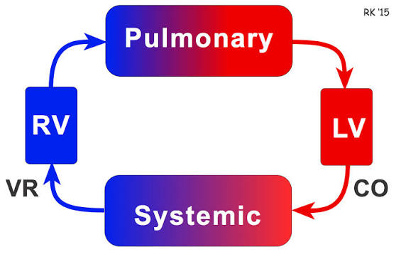
Pulmonary resistance refers to the opposition encountered by blood flow within the pulmonary circulation—the network of vessels that carry deoxygenated blood from the right side of the heart to the lungs for oxygenation and then return oxygenated blood back to the left side of the heart. The pulmonary vasculature is generally more compliant than systemic circulation; however, it still experiences significant influences from various factors including lung volume, vascular tone, and hematocrit levels.
The relationship between pulmonary arterial pressure and flow rate is essential for understanding pulmonary vascular dynamics. Increased pulmonary vascular resistance can lead to conditions such as pulmonary hypertension, which may arise from various pathologies affecting lung function or structure.
Effect of Hematocrit on Vascular Resistance
Hematocrit—the proportion of blood volume occupied by red blood cells—has a direct impact on both peripheral and pulmonary vascular resistance. Changes in hematocrit affect blood viscosity; higher hematocrit levels increase viscosity, leading to greater flow resistance. Conversely, lower hematocrit levels decrease viscosity but may not significantly reduce flow resistance under certain conditions.
Research indicates that at constant pulmonary blood volume, reducing hematocrit through hemodilution has only minor effects on pulmonary flow resistance. In contrast, increasing hematocrit through hemoconcentration markedly increases flow resistance up to 130%. This suggests that while lower hematocrit may not drastically improve flow dynamics under controlled conditions, higher levels can significantly impede it.
Moreover, variations in pulmonary blood volume also influence pressure-flow relationships across different hematocrit levels. Incremental increases in pulmonary blood volume tend to shift these relationships downward for all investigated hematocrits (0-50%), indicating that maintaining optimal pulmonary blood volume is crucial for effective gas exchange and overall cardiovascular health.
In summary, both peripheral and pulmonary resistances are influenced by hematocrit levels through their effects on blood viscosity and vessel dynamics. Understanding these interactions helps elucidate mechanisms underlying various cardiovascular conditions.
Vascular Distensibility
Vascular distensibility refers to the ability of blood vessels to expand in response to changes in pressure. It is a critical characteristic of the vascular system that allows blood vessels to accommodate the pulsatile output from the heart and maintain a smooth, continuous flow of blood through smaller vessels. Distensibility is typically quantified as the fractional increase in volume per unit increase in pressure, expressed mathematically as:
Distensibility = ΔV ÷ V0 ⋅ ΔP
where ΔV is the change in volume, V0 is the original volume, and ΔP is the change in pressure (in mm Hg). For example, if an increase of 1 mm Hg causes a vessel originally containing 10 ml of blood to increase its volume by 1 ml, then the distensibility would be calculated as 0.1 per mm Hg or 10% per mm Hg.
Differences Between Arteries and Veins
The primary difference between arteries and veins regarding distensibility lies in their structural characteristics and functional roles within the circulatory system:
- Structural Differences:
- Arteries have thicker and stronger walls compared to veins. This structural integrity is necessary because arteries must withstand higher pressures generated by the heart’s pumping action.
- Veins, on the other hand, are more compliant due to their thinner walls and larger luminal diameters.
- Distensibility Comparison:
- On average, veins are about eight times more distensible than arteries. This means that for a given increase in pressure, veins can accommodate significantly more blood than arteries can.
- The greater distensibility of veins allows them to serve as reservoirs for storing large quantities of blood (approximately 0.5 to 1.0 liter extra), which can be mobilized when needed elsewhere in circulation.
- Compliance:
- Compliance differs from distensibility; it takes into account both the distensibility of a vessel and its volume. Compliance is defined as:
Compliance = Distensibility × V
where V represents the volume of blood contained within the vessel.
- Systemic veins have about 24 times greater compliance compared to their corresponding arteries due to their higher distensibility and larger volumes.
Laplace’s Law
Laplace’s law describes the relationship between tension in a vessel wall (T), internal pressure (P), and radius (r). It states that:
T = P ⋅ r
This law implies that for a given internal pressure, larger vessels will experience greater wall tension than smaller vessels due to their larger radius. In practical terms:
- In arteries: As they expand with each heartbeat, they must withstand high internal pressures while maintaining structural integrity.
- In veins: Although they are more distensible, they operate under lower pressures; thus, their wall tension remains manageable even with significant volume changes.
Understanding Laplace’s law helps explain why arterial walls need to be thicker and stronger compared to venous walls despite veins being able to hold more blood at lower pressures.
In summary, vascular distensibility plays a crucial role in how arteries and veins function differently within the circulatory system—arteries manage high-pressure situations while maintaining structural integrity, whereas veins provide flexibility and storage capacity for blood.
Vascular Compliance and Delay Compliance
Vascular compliance refers to the ability of blood vessels, particularly veins and arteries, to expand and contract in response to changes in pressure. It is defined as the change in volume (ΔV) per unit change in pressure (ΔP), mathematically expressed as:
C = ΔV ÷ ΔP
Where:
- C is compliance,
- ΔV is the change in volume,
- ΔP is the change in pressure.
High compliance indicates that a vessel can accommodate a large volume of blood with only a small increase in pressure, which is characteristic of veins. Conversely, arteries have lower compliance due to their thicker walls and higher elastic properties, allowing them to withstand higher pressures without significant expansion.
Delay compliance, on the other hand, refers to the time-dependent aspect of vascular compliance. It describes how blood vessels respond over time to changes in volume and pressure. When a sudden increase in blood volume occurs (for example, during systole), there may be an immediate but temporary increase in pressure followed by a gradual adjustment as the vessel walls stretch and accommodate this new volume. This phenomenon is particularly relevant for larger arteries where elastic recoil plays a significant role.
Arterial Pressure Pulsation and Transmission of Pressure Pulses to Peripheral Arteries
Arterial pressure pulsation is primarily driven by the rhythmic contraction of the heart during systole and relaxation during diastole. When the left ventricle contracts, it ejects blood into the aorta, creating a pressure wave that travels through the arterial system. This wave consists of both forward-moving components (the actual pulse wave) and reflected waves from peripheral sites.
The characteristics of this pulsatile flow are influenced by several factors including:
- Elasticity of Arteries: The elasticity allows arteries to expand during systole when they receive blood from the heart.
- Diameter of Vessels: Larger diameter vessels can transmit pressure waves more effectively than smaller ones.
- Distance from Heart: As one moves further away from the heart, such as into peripheral arteries, there are changes in both amplitude and shape of the pressure wave due to resistance offered by smaller arterioles.
The transmission of these pressure pulses involves several key processes:
- Wave Propagation: The speed at which these pulses travel depends on arterial stiffness; stiffer arteries transmit waves faster than compliant ones.
- Reflection: As pressure waves encounter branching points or changes in vessel diameter (such as moving from large arteries into smaller arterioles), some energy is reflected back towards the heart.
- Damping: The pulsatile nature diminishes as it travels through smaller vessels due to frictional losses and increased resistance.
In summary, while arterial compliance allows for accommodation of varying volumes under different pressures, delay compliance reflects how quickly these adjustments occur over time. The transmission of arterial pressure pulses illustrates how these dynamics play out within our circulatory system—highlighting both immediate responses and longer-term adaptations.
Function of the Veins
Veins are blood vessels that carry deoxygenated blood back to the heart, with the exception of pulmonary veins, which carry oxygenated blood from the lungs to the heart. The primary functions of veins include:
- Transporting Blood: Veins collect blood from various parts of the body and return it to the heart. They have a larger lumen compared to arteries, allowing them to accommodate varying volumes of blood.
- Storage Capacity: Veins can act as reservoirs for blood due to their ability to stretch and hold more blood than arteries. This is particularly important during times of increased demand for blood flow, such as during exercise.
- Regulating Blood Flow: By constricting or dilating, veins help regulate venous return and maintain adequate circulation throughout the body.
Venous Pressure
Venous pressure refers to the pressure within the venous system. It is generally lower than arterial pressure and varies depending on several factors:
- Gravity: Venous pressure in standing individuals is influenced by gravity; thus, it is higher in veins located below the heart compared to those above it.
- Muscle Contraction: During physical activity, skeletal muscles contract and compress nearby veins, increasing venous pressure and aiding in venous return.
- Respiratory Activity: The act of breathing creates a negative pressure in the thoracic cavity during inhalation, which helps draw blood into the thoracic veins and ultimately towards the heart.
Venous Resistance
Venous resistance is primarily determined by factors such as vein diameter and compliance (the ability of a vessel to expand). While veins have lower resistance compared to arteries due to their larger diameter, several factors can influence venous resistance:
- Vasoconstriction/Dilation: Smooth muscle contraction or relaxation within vein walls can alter their diameter, affecting resistance.
- Valvular Function: The presence of valves in veins prevents backflow and contributes to maintaining unidirectional flow towards the heart, thereby influencing overall resistance.
- Pathological Conditions: Conditions like varicose veins can increase resistance due to impaired function or structural changes in vein walls.
Venous Valves
Venous valves are one-way structures located within many veins that prevent backflow of blood as it returns to the heart. Their functions include:
- Maintaining Unidirectional Flow: Valves ensure that blood moves toward the heart rather than pooling in extremities or flowing backward due to gravity.
- Facilitating Venous Return: By preventing backflow during muscle relaxation phases, valves assist in maintaining efficient venous return even when skeletal muscles are not actively contracting.
- Reducing Venous Pressure Fluctuations: Valves help stabilize venous pressure by preventing sudden drops caused by gravitational effects when standing still.
Venous Pump
The venous pump refers to mechanisms that facilitate venous return against gravity:
- Muscle Pump Mechanism: As skeletal muscles contract during movement or exercise, they compress adjacent veins, pushing blood toward the heart while simultaneously closing valves that prevent backflow.
- Respiratory Pump Mechanism: Changes in thoracic pressure during breathing create a suction effect that aids in drawing blood into thoracic veins from peripheral areas.
- Postural Changes: Shifting positions (e.g., standing up) activates these pumps more effectively as they adaptively respond to changes in body posture and gravity’s effects on venous return.
Blood Venous Reservoir
The term “blood venous reservoir” refers to a portion of total blood volume stored within systemic veins at any given time:
- Functionality as a Reservoir: Approximately 60-70% of total blood volume resides within veins at rest; this capacity allows for rapid adjustments in circulating volume based on physiological demands (e.g., during hemorrhage or intense physical activity).
- Regulation of Blood Volume Distribution: The reservoir function enables redistribution of blood volume between different vascular compartments (e.g., from storage sites in veins into active circulation) depending on immediate needs for oxygen delivery or metabolic support across tissues.
- Homeostatic Role During Stressors: In response to stressors such as dehydration or significant bleeding, this reservoir can be mobilized quickly into circulation through vasoconstriction mechanisms mediated by sympathetic nervous system activation.
In summary, understanding these components—veins’ structure and function, pressures involved, resistance factors, valve roles, pumping mechanisms, and reservoir capabilities—provides insight into how our circulatory system maintains homeostasis under varying conditions.


