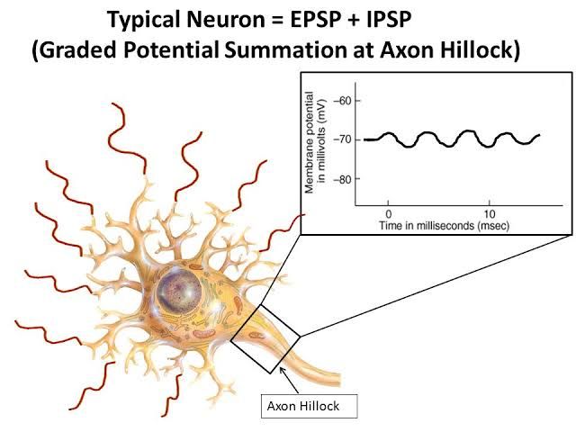
Graded potentials are essential physiological phenomena that occur in neurons and muscle cells, representing localized changes in membrane potential. Unlike action potentials, which are all-or-nothing responses, graded potentials vary in magnitude depending on the strength of the stimulus. This variability allows for a nuanced response to different levels of input.
Physiological Basis of Graded Potentials
- Nature of Graded Potentials: Graded potentials arise from the opening or closing of ligand-gated ion channels in response to stimuli such as neurotransmitter binding or sensory input. These changes can either depolarize (make the inside of the cell more positive) or hyperpolarize (make it more negative) the membrane potential.
- Mechanism of Action: When a stimulus occurs, it causes specific ions (such as Na+, K+, Ca2+, or Cl-) to flow across the cell membrane through these channels. For example, when sodium channels open, Na+ ions enter the neuron, leading to depolarization. Conversely, if chloride channels open or potassium channels close, hyperpolarization occurs.
- Local Changes: Graded potentials are localized events that occur primarily in dendrites and cell bodies where voltage-gated ion channels are absent. They do not propagate along the axon like action potentials but instead diminish over time and distance due to leakage and resistance within the cellular membrane.
- Types of Graded Potentials:
- Excitatory Postsynaptic Potentials (EPSPs): These are depolarizing graded potentials that increase the likelihood of an action potential occurring by making the membrane potential less negative.
- Inhibitory Postsynaptic Potentials (IPSPs): These are hyperpolarizing graded potentials that decrease the likelihood of an action potential by making the membrane potential more negative.
- Summation: The overall effect on a neuron is determined by summation processes:
- Temporal Summation: Occurs when multiple graded potentials arrive at a neuron in quick succession, building upon each other before they fade.
- Spatial Summation: Involves simultaneous inputs from multiple synapses that combine their effects on a single neuron.
- Threshold for Action Potential: If graded potentials reach a certain threshold (typically around -55 mV), they can trigger an action potential at the axon hillock region of a neuron.
Properties of Graded Potentials
- Variable Strength: The amplitude of graded potentials is proportional to the strength of the stimulus; stronger stimuli produce larger changes in membrane potential.
- Decremental Nature: Graded potentials decrease in amplitude with distance from their point of origin due to passive electrical properties of neurons.
- Reversible Changes: If a stimulus ceases, graded potentials will return to resting membrane potential without triggering an action potential if they do not reach threshold.
- Localized Events: They occur primarily at dendrites and cell bodies rather than along axons where action potentials propagate.
Physiological Basis and Properties of Compound Action Potential
Introduction to Compound Action Potential (CAP)
The compound action potential (CAP) is a measure of the electrical activity generated by a group of nerve fibers in response to stimulation. It represents the summed electrical responses of multiple individual axons within a nerve bundle, allowing researchers to assess the overall function and characteristics of the nerve.
Physiological Basis of CAP
The physiological basis for the CAP lies in the properties of individual action potentials generated by neurons. When a nerve is stimulated, it can produce an action potential if the stimulus exceeds a certain threshold. This threshold is typically around -50 to -55 mV, which must be reached for depolarization to occur. The all-or-none law applies here; once the threshold is met, an action potential will propagate along the axon without diminishing in amplitude.
In a nerve bundle, different types of fibers (e.g., A-alpha, A-beta) have varying diameters and conduction velocities. Larger diameter fibers tend to conduct impulses faster than smaller ones. As stimulus strength increases, more fibers are recruited into action, leading to a larger CAP due to the additive effect of individual action potentials from these fibers.
Properties of CAP
- Latency: The latency refers to the time delay between the application of a stimulus and the onset of the CAP. This can be measured from two points:
- From the onset of stimulus artifact to the beginning of CAP.
- From stimulus artifact to peak amplitude of CAP. The latency reflects how quickly signals travel through different fiber types within the nerve.
- Threshold Voltage: The threshold voltage is defined as the minimum stimulus voltage required to elicit at least one action potential in any fiber within the nerve bundle. This value can vary depending on factors such as temperature and health status but generally indicates how excitable a nerve is.
- Peak Amplitude: The peak amplitude represents the maximum voltage reached during the CAP response. It provides insight into how many fibers are actively conducting during stimulation; higher amplitudes indicate greater recruitment.
- Duration: The duration measures how long it takes for the CAP signal to return to baseline after reaching its peak. This duration can increase with stronger stimuli as more fibers contribute their action potentials over time.
- Shape: The shape of the CAP waveform can provide information about fiber recruitment patterns and conduction velocities within different populations of axons in response to varying stimulus strengths.
- Maximal Stimulus Voltage: Beyond a certain point, increasing stimulus voltage does not lead to further increases in CAP amplitude because all fast-conducting A-alpha fibers have been activated. However, additional slower fibers may still be recruited at even higher voltages.
Conclusion
Understanding these properties allows researchers and clinicians to evaluate nerve function effectively and diagnose various neurological conditions based on changes observed in CAP characteristics under different experimental conditions or pathologies.
Contrast between Action Potential and Graded Potential
Definition and Nature
- Action Potentials are rapid, all-or-nothing electrical signals that propagate along the axon of a neuron. They occur when a neuron’s membrane potential reaches a certain threshold due to depolarization.
- Graded Potentials, on the other hand, are changes in membrane potential that vary in size (amplitude) and can be either depolarizing or hyperpolarizing. They occur in response to stimuli and do not follow an all-or-nothing principle.
Amplitude and Strength
- The amplitude of graded potentials is proportional to the strength of the stimulus; stronger stimuli produce larger graded potentials. This means that graded potentials can vary widely in their magnitude.
- In contrast, action potentials have a fixed amplitude (approximately 100 mV) regardless of the strength of the stimulus once the threshold is reached. The frequency of action potentials encodes information about stimulus intensity rather than their amplitude.
Duration
- Graded potentials can last from milliseconds to seconds, depending on the type of stimulus and its duration.
- Action potentials, however, are relatively short-lived events, typically lasting only about 3-5 milliseconds.
Ion Channels Involved
- Graded potentials are primarily mediated by ligand-gated ion channels or other types of channels sensitive to mechanical or thermal changes. These channels open in response to neurotransmitters or sensory stimuli.
- Conversely, action potentials are generated by voltage-gated sodium (Na+) and potassium (K+) channels. When a neuron depolarizes past threshold, these channels open rapidly, leading to a swift influx of Na+ ions followed by K+ efflux.
Propagation Mechanism
- The propagation of graded potentials occurs through passive spread (electrotonic spread), meaning they diminish in amplitude as they travel away from their point of origin (decremental conduction).
- In contrast, action potentials propagate without losing strength due to regeneration at each segment along the axon. This non-decremental propagation ensures that action potentials maintain their full amplitude over long distances.
Refractory Periods
- There is no refractory period associated with graded potentials, allowing them to summate over time (temporal summation) or across different locations (spatial summation).
- However, action potentials have both absolute and relative refractory periods which prevent immediate re-firing after an action potential has occurred. This characteristic ensures unidirectional propagation along the axon.
In summary, while both graded and action potentials are essential for neuronal communication, they differ fundamentally in their mechanisms, characteristics, and roles within the nervous system.
Ionic Basis of Excitatory Post Synaptic Potential (EPSP), Inhibitory Post Synaptic Potential (IPSP), and End Plate Potential (EPP)
1. Excitatory Post Synaptic Potential (EPSP)
The EPSP is a transient depolarization of the postsynaptic membrane potential that occurs when excitatory neurotransmitters bind to their receptors on the postsynaptic neuron. The ionic basis of EPSPs primarily involves the influx of positively charged ions, particularly sodium ions (Na+).
- Mechanism: When an excitatory neurotransmitter, such as glutamate, binds to its receptor (e.g., AMPA or NMDA receptors), it causes the opening of ion channels that are permeable to Na+. This leads to a rapid influx of Na+ ions into the postsynaptic cell.
- Resulting Change in Membrane Potential: The influx of Na+ causes the inside of the neuron to become more positive relative to the outside, resulting in depolarization. If this depolarization reaches a certain threshold, it can trigger an action potential in the postsynaptic neuron.
- Duration and Summation: EPSPs are graded potentials, meaning their amplitude can vary depending on the amount of neurotransmitter released and the number of receptors activated. They can summate temporally or spatially, leading to a greater overall change in membrane potential.
2. Inhibitory Post Synaptic Potential (IPSP)
In contrast to EPSPs, IPSPs result in hyperpolarization of the postsynaptic membrane potential. This is primarily mediated by inhibitory neurotransmitters such as gamma-aminobutyric acid (GABA) and glycine.
- Mechanism: When GABA binds to its receptors (e.g., GABA_A receptors), it opens chloride channels that allow Cl- ions to flow into the cell. In some cases, K+ channels may also open, allowing K+ ions to exit.
- Resulting Change in Membrane Potential: The influx of Cl- or efflux of K+ makes the inside of the neuron more negative compared to its resting state, leading to hyperpolarization. This makes it less likely for an action potential to occur because it moves further away from the threshold needed for depolarization.
- Functionality: IPSPs serve as a critical mechanism for regulating neuronal excitability and preventing excessive firing. They can also summate with EPSPs; if an IPSP occurs simultaneously with an EPSP, they can counteract each other’s effects.
3. End Plate Potential (EPP)
The EPP is a specific type of synaptic potential that occurs at neuromuscular junctions where motor neurons communicate with muscle fibers.
- Mechanism: When an action potential reaches the presynaptic terminal at a neuromuscular junction, it triggers voltage-gated calcium channels to open. The influx of Ca2+ ions promotes vesicle fusion and release of acetylcholine (ACh) into the synaptic cleft.
- Receptor Activation: ACh binds to nicotinic acetylcholine receptors on the postsynaptic muscle fiber membrane. These receptors are ligand-gated ion channels that allow Na+ ions to enter and K+ ions to exit.
- Resulting Change in Membrane Potential: The net effect is a significant depolarization known as EPP due to more Na+ entering than K+ exiting. If this depolarization reaches threshold levels, it will initiate an action potential in muscle fibers leading to contraction.
- Importance in Muscle Contraction: EPPs are crucial for converting neural signals into mechanical responses in muscles; they ensure that muscle fibers contract effectively upon stimulation by motor neurons.
In summary, while EPSPs lead to depolarization through Na+ influx via excitatory neurotransmitters, IPSPs cause hyperpolarization through Cl- influx or K+ efflux via inhibitory neurotransmitters. EPPs represent a specialized form occurring at neuromuscular junctions involving ACh-induced depolarization necessary for muscle contraction.


