Histological Features of Different Types of Cartilages in the Body
Cartilage is a specialized connective tissue that provides support and flexibility to various structures in the human body. There are three main types of cartilage: hyaline cartilage, elastic cartilage, and fibrocartilage. Each type has distinct histological features and specific locations within the body.
1. Hyaline Cartilage
Histological Features: Hyaline cartilage is characterized by a glassy, translucent appearance due to its high content of collagen fibers (type II) and proteoglycans. The chondrocytes (cartilage cells) are located within lacunae (small cavities) and are often found in clusters called isogenous groups. The extracellular matrix is rich in water, which contributes to its resilience and ability to withstand compressive forces. The matrix appears homogeneous with a bluish tint when viewed under a microscope.
Location:
- Articular surfaces of bones (joints)
- Costal cartilages connecting ribs to the sternum
- Nose
- Trachea and bronchi
- Larynx
- Embryonic skeleton (initially formed as hyaline cartilage before ossification)
2. Elastic Cartilage
Histological Features: Elastic cartilage contains a dense network of elastic fibers in addition to collagen fibers, giving it greater flexibility compared to hyaline cartilage. Chondrocytes are also present in lacunae but are more numerous than in hyaline cartilage. The extracellular matrix has a yellowish tint due to the presence of elastic fibers, which can be visualized using special staining techniques.
Location:
- External ear (auricle or pinna)
- Epiglottis (the flap that covers the trachea during swallowing)
- Eustachian tube (auditory tube connecting the middle ear to the nasopharynx)
3. Fibrocartilage
Histological Features: Fibrocartilage is distinguished by its dense arrangement of collagen fibers (type I), which provide tensile strength and resistance to compression. The chondrocytes are arranged in rows between the bundles of collagen fibers, often appearing elongated or flattened. The extracellular matrix is less homogeneous than that of hyaline cartilage due to the presence of more fibrous components.
Location:
- Intervertebral discs (between vertebrae)
- Pubic symphysis (joint between pelvic bones)
- Menisci of the knee joint
- Temporomandibular joint (TMJ)
- Tendon insertions into bone
In summary, each type of cartilage serves unique functions based on its histological characteristics and locations throughout the body, contributing significantly to structural integrity and flexibility.
Features of Cartilage Development and Growth
Cartilage development and growth are characterized by several unique features that distinguish it from other types of connective tissues. The primary process through which cartilage is formed is known as chondrogenesis, which occurs from the mesoderm germ layer during embryonic development. Here are the key features:
- Cell Types: The main cells involved in cartilage are chondroblasts, which secrete the extracellular matrix components necessary for cartilage formation. As these cells mature, they become chondrocytes, which reside in small cavities called lacunae within the matrix.
- Matrix Composition: The extracellular matrix of cartilage is rich in proteoglycans, particularly aggrecan, and type II collagen fibers. This composition provides cartilage with its characteristic strength and flexibility.
- Avascular Nature: Cartilage is avascular, meaning it lacks blood vessels. Nutrients are supplied to chondrocytes through diffusion from surrounding tissues, which significantly affects its healing capacity.
- Slow Growth Rate: Cartilage grows primarily through interstitial growth (expansion from within) and appositional growth (growth at the surface). However, due to minimal cell division and the avascular nature of cartilage, its growth is slow compared to other tissues.
- Limited Regenerative Capacity: Once formed, cartilage has a limited ability to regenerate after injury because of its low cellularity and lack of a direct blood supply. Healing occurs slowly due to reliance on diffusion for nutrient supply.
Relation to Injury and Repair
The unique characteristics of cartilage development and growth have significant implications for injury and repair:
- Injury Susceptibility: Due to its avascular nature and low metabolic activity, articular cartilage injuries—common in athletes or individuals engaged in high-impact activities—are challenging to heal effectively. Damage can lead to pain, swelling, stiffness, and reduced joint function.
- Healing Challenges: When cartilage is injured, the limited regenerative capacity means that healing often does not occur adequately or may result in scar tissue formation rather than functional tissue restoration. This leads to long-term complications such as osteoarthritis.
- Repair Strategies: Various treatment strategies aim to promote repair or regeneration of damaged cartilage include microfracture surgery (creating small fractures in the underlying bone), autologous chondrocyte implantation (using cultured chondrocytes), and osteochondral grafting (transplanting healthy cartilage). These methods attempt to enhance blood supply or introduce new cells into the damaged area but still face challenges due to the inherent properties of cartilage.
In summary, while cartilage plays a crucial role in joint function due to its unique structural properties developed during embryogenesis, its limited capacity for repair following injury poses significant challenges for effective treatment options.
Cartilage Features and Clinical Aspects of Knee Joint Disorders
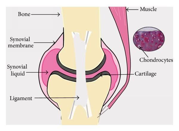
The primary types of cartilage present in the knee are hyaline cartilage, which covers the articular surfaces of bones, and fibrocartilage, which is found in structures like the menisci.
Features of Cartilage
- Composition: Cartilage consists mainly of water (about 70-80%), collagen fibers (primarily type II), proteoglycans, and chondrocytes. This unique composition allows it to withstand compressive forces while maintaining flexibility.
- Avascular Nature: Cartilage lacks blood vessels, which limits its ability to heal after injury. Nutrients are supplied through diffusion from surrounding synovial fluid.
- Degeneration: Over time or due to injury, cartilage can undergo degeneration characterized by changes such as fibrillation, loss of proteoglycans, and thinning.
Clinical Aspects Related to Cartilage Features
- Disc Prolapse:
- The intervertebral discs consist of an outer annulus fibrosus made up of fibrocartilage and an inner nucleus pulposus that contains a gel-like substance.
- Disc prolapse occurs when there is a rupture or herniation of the disc material, often leading to nerve compression.
- While primarily associated with spinal health, similar degenerative processes can affect knee cartilage integrity through altered biomechanics due to pain or instability from disc issues.
- Fibrosis:
- Fibrosis refers to the formation of excess fibrous connective tissue in response to injury or inflammation.
- In the context of knee joints, fibrosis can occur following trauma or surgery leading to conditions such as arthrofibrosis.
- This condition can restrict joint mobility and lead to further degeneration of cartilage due to abnormal loading patterns.
- Calcifications of Menisci:
- The menisci are C-shaped cartilaginous structures that provide stability and distribute load across the knee joint.
- Calcifications within meniscal tissue can occur due to chronic stress or degeneration over time.
- These calcifications may contribute to pain and dysfunction by limiting meniscal movement during knee flexion and extension.
Correlation Between Cartilage Features and Clinical Conditions
- The health status of cartilage directly influences clinical outcomes related to disc prolapse, fibrosis, and calcifications:
- Biomechanical Changes: Degenerative changes in cartilage can alter load distribution across the knee joint leading to increased stress on menisci and potentially resulting in tears or calcification.
- Inflammatory Responses: Damage to cartilage may trigger inflammatory responses that promote fibrosis around the joint capsule or within soft tissues adjacent to the knee.
- Pain Mechanisms: Degenerative changes in cartilage can lead to pain syndromes that mimic symptoms associated with disc prolapse; thus complicating diagnosis and treatment strategies.
- Understanding these correlations is essential for developing effective treatment plans aimed at preserving cartilage integrity while addressing associated clinical conditions like disc prolapse and meniscal calcifications.
In conclusion, there exists a complex interplay between cartilage features and various clinical aspects affecting knee health such as disc prolapse, fibrosis, and calcifications within menisci. Addressing these factors holistically is vital for optimal management strategies.


