Metabolic Bone Diseases: An Overview
Metabolic bone diseases are a group of disorders that affect the strength and structure of bones due to imbalances in minerals essential for bone health, such as calcium, phosphorus, and vitamin D. These conditions can lead to weakened bones, increased risk of fractures, and other complications. Understanding these diseases requires a thorough history-taking approach that considers various factors including age, dietary habits, medical history, and lifestyle choices.
Types of Metabolic Bone Diseases
- Osteoporosis
- Osteoporosis is characterized by a significant loss of bone mass and density, leading to fragile bones that are prone to fractures. It primarily affects older adults, especially postmenopausal women due to decreased estrogen levels. Risk factors include a family history of osteoporosis, small body size, poor nutrition (especially low calcium intake), excessive alcohol consumption, smoking, and sedentary lifestyle.
- Osteomalacia
- Osteomalacia refers to the softening of bones due to inadequate mineralization caused by vitamin D deficiency or insufficient calcium or phosphorus. In children, this condition is known as rickets. Symptoms may include bone pain and tenderness, muscle weakness, and skeletal deformities. Causes can include poor diet, lack of sunlight exposure (which is necessary for vitamin D synthesis), malabsorption syndromes (like celiac disease), and certain medications.
- Hyperparathyroidism
- This condition occurs when the parathyroid glands produce too much parathyroid hormone (PTH), leading to elevated calcium levels in the blood and subsequent bone resorption. Primary hyperparathyroidism often results from benign tumors on the parathyroid glands or gland hyperplasia. Secondary hyperparathyroidism can occur in response to chronic kidney disease or vitamin D deficiency.
- Paget’s Disease
- Paget’s disease involves abnormal bone remodeling where there is excessive breakdown and formation of bone tissue leading to enlarged and weakened bones. The exact cause is unknown but may involve genetic factors or viral infections. Symptoms can include pain in affected bones, deformities (such as bowing of the legs), and an increased risk of osteosarcoma (bone cancer).
- Osteogenesis Imperfecta (OI)
- Often referred to as “brittle bone disease,” OI is a genetic disorder characterized by fragile bones that break easily due to defects in collagen production. It varies in severity; some individuals may experience frequent fractures while others have only a few throughout their lives.
- Hypophosphatasia
- This rare genetic disorder affects the development of bones and teeth due to low levels of alkaline phosphatase enzyme which is crucial for mineralization processes. Symptoms can vary widely but often include weak bones leading to fractures or deformities.
- Other Conditions
- Other metabolic bone diseases may include conditions like renal osteodystrophy associated with chronic kidney disease or certain inherited disorders affecting mineral metabolism.
Conclusion
In summary, metabolic bone diseases encompass a variety of conditions that result from imbalances in essential minerals required for maintaining healthy bones. Each type has distinct causes, symptoms, risk factors, and treatment approaches that necessitate careful evaluation by healthcare professionals.
Clinical Examination and Identification of Deformities Associated with Metabolic Bone Diseases
The examination typically involves a thorough patient history, physical assessment, and specific tests aimed at revealing characteristic deformities.
Common Deformities Associated with Metabolic Bone Diseases
- Osteoporosis:
- Osteoporosis is characterized by decreased bone density, leading to fragile bones that are prone to fractures. During a clinical examination, signs may include:
- Loss of height due to vertebral compression fractures.
- Kyphosis (a forward curvature of the spine), often referred to as “dowager’s hump.”
- Increased susceptibility to fractures from minimal trauma.
- Osteoporosis is characterized by decreased bone density, leading to fragile bones that are prone to fractures. During a clinical examination, signs may include:
- Osteomalacia/Rickets:
- Osteomalacia in adults and rickets in children result from inadequate mineralization of bone tissue. Key deformities include:
- Bowing of the legs (genu varum) or knock knees (genu valgum) in children.
- Delayed growth and development in children due to weakened bones.
- Painful bone tenderness, particularly in the pelvis and lower back.
- Osteomalacia in adults and rickets in children result from inadequate mineralization of bone tissue. Key deformities include:
- Hyperparathyroidism:
- This condition leads to increased levels of parathyroid hormone (PTH), which can cause bone resorption. Clinical findings may include:
- Osteitis fibrosa cystica, characterized by brown tumors or cystic lesions on the bones.
- Bone pain and deformities resulting from weakened structural integrity.
- This condition leads to increased levels of parathyroid hormone (PTH), which can cause bone resorption. Clinical findings may include:
- Other Symptoms:
- Patients may also present with generalized symptoms such as:
- Aches or pains in the bones, particularly in the back, hips, and legs.
- Tooth problems due to poor mineralization affecting dental health.
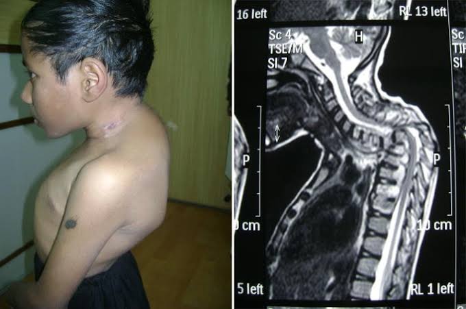
- Curvature of the spine or other skeletal deformities that develop over time.
- Patients may also present with generalized symptoms such as:
Physical Assessment Techniques
During a clinical examination for metabolic bone diseases, healthcare providers should employ several techniques:
- Visual Inspection: Look for asymmetry or abnormal postures that may indicate underlying skeletal issues.
- Palpation: Assess areas for tenderness or swelling that could suggest inflammation or structural changes.
- Range of Motion Tests: Evaluate joint mobility which may be limited due to pain or structural deformity.
- Functional Tests: Observe how patients perform daily activities; limitations may indicate significant skeletal weakness.
Diagnostic Imaging
In addition to clinical examination, imaging studies play a crucial role in diagnosing metabolic bone diseases:
- DEXA Scan: Measures bone mineral density and helps identify osteoporosis risk.
- X-rays: Can reveal fractures, deformations like bowing limbs, or other structural abnormalities indicative of osteomalacia/rickets.
Conclusion
Identifying deformities associated with metabolic bone diseases requires a comprehensive approach involving clinical examination techniques alongside diagnostic imaging. Recognizing these conditions early can lead to timely interventions that improve patient outcomes.
Comprehensive Overview of Basic Investigations for Metabolic Bone Disease
Metabolic bone diseases encompass a variety of conditions that affect bone density and structure, leading to increased risk of fractures and other complications. The diagnosis and differentiation of these diseases from hematological disorders require a systematic approach involving clinical evaluation, laboratory tests, and imaging studies.
1. Clinical Evaluation
The initial step in investigating metabolic bone disease is a thorough clinical evaluation. This includes:
- Patient History: Assessing for risk factors such as age, gender, family history of osteoporosis or fractures, dietary habits (calcium and vitamin D intake), physical activity level, and any history of endocrine disorders.
- Physical Examination: Checking for signs of bone fragility, deformities, tenderness over bones, and any evidence of previous fractures.
2. Laboratory Investigations
Laboratory tests are crucial in identifying metabolic abnormalities:
- Serum Calcium Levels: Both total and ionized calcium levels should be measured. Hypocalcemia or hypercalcemia can indicate various metabolic disorders.
- Phosphate Levels: Serum phosphate levels help differentiate between conditions like osteomalacia (low phosphate) and primary hyperparathyroidism (high phosphate).
- Alkaline Phosphatase (ALP): Elevated ALP levels may indicate increased bone turnover seen in conditions like Paget’s disease or osteoblastic metastases.
- Parathyroid Hormone (PTH): Measuring PTH helps distinguish between primary hyperparathyroidism (high PTH) and secondary causes due to low calcium levels (e.g., renal failure).
- Vitamin D Levels: 25-hydroxyvitamin D is the primary form assessed to evaluate vitamin D status; deficiency can lead to osteomalacia or osteoporosis.
- Bone Turnover Markers: These include markers such as N-telopeptide (NTx) or C-telopeptide (CTx) for resorption and osteocalcin for formation. They provide insight into the rate of bone remodeling.
3. Radiological Investigations
Radiological imaging plays an essential role in diagnosing metabolic bone diseases:
- Dual-Energy X-ray Absorptiometry (DEXA): This is the gold standard for measuring bone mineral density (BMD). It helps diagnose osteoporosis by comparing BMD against normative data.
- X-rays: Standard radiographs can reveal changes associated with metabolic bone diseases such as osteopenia, fractures, or specific patterns indicative of certain conditions like rickets or osteomalacia.
- CT Scans or MRI: These advanced imaging techniques may be used when more detailed visualization is required, particularly in complex cases where there is suspicion of malignancy or other pathologies affecting the bones.
Differentiating Metabolic Problems from Hematological Disorders
To differentiate metabolic bone diseases from hematological issues through investigations:
- Complete Blood Count (CBC): Anemia may suggest hematological disorders rather than purely metabolic issues. For instance, normocytic anemia could accompany chronic kidney disease affecting calcium metabolism.
- Bone Marrow Biopsy: In cases where hematological malignancies are suspected (e.g., multiple myeloma), a biopsy can clarify whether abnormal cells are present versus changes due to metabolic processes.
- Specific Markers:
- In multiple myeloma, serum protein electrophoresis would show monoclonal proteins.
- In contrast, elevated alkaline phosphatase with normal protein levels might suggest a primary metabolic issue rather than hematologic pathology.
- Imaging Findings:
- Osteoporosis typically presents with generalized low-density areas on X-rays without focal lesions.
- Hematological malignancies often show lytic lesions on imaging studies that are not characteristic of typical metabolic bone disease.
In summary, a combination of clinical evaluation, laboratory tests focusing on calcium-phosphate metabolism and hormone levels, along with appropriate imaging studies allows for accurate diagnosis and differentiation between various metabolic problems and hematological disorders related to the bones.
Treatment of Important Metabolic Bone Problems
1. Osteoporosis Treatment
Osteoporosis is the most common metabolic bone disease, characterized by decreased bone mass and increased fracture risk. Treatment options include:
- Medications:
- Bisphosphonates (e.g., alendronate, risedronate) are commonly prescribed to inhibit bone resorption and reduce fracture risk.
- Selective Estrogen Receptor Modulators (SERMs) (e.g., raloxifene) can help maintain bone density in postmenopausal women.
- Hormone Replacement Therapy (HRT) may be considered for women experiencing menopause to help maintain estrogen levels.
- Denosumab, a monoclonal antibody that inhibits RANKL, is used for patients at high risk of fractures.
- Teriparatide, a form of parathyroid hormone, stimulates new bone formation and is often used in severe cases.
- Lifestyle Modifications:
- Increasing calcium and vitamin D intake through diet or supplements.
- Engaging in weight-bearing exercises to strengthen bones.
- Avoiding smoking and limiting alcohol consumption.
- Fall Prevention Strategies:
- Implementing home safety measures to reduce the risk of falls that could lead to fractures.
2. Osteomalacia Treatment
Osteomalacia, often referred to as rickets in children, results from inadequate mineralization of bone due to deficiencies in calcium, phosphorus, or vitamin D. Treatment includes:
- Nutritional Supplements:
- High doses of vitamin D are typically prescribed to correct deficiencies. This may involve both oral supplements and dietary changes.
- Calcium and phosphorus supplements may also be necessary depending on blood levels.
- Dietary Changes:
- Encouraging a balanced diet rich in calcium (dairy products, leafy greens) and vitamin D (fatty fish, fortified foods).
- Addressing Underlying Conditions:
- If osteomalacia is caused by malabsorption syndromes (like celiac disease), treating the underlying condition is crucial for effective management.
3. Hyperparathyroidism Treatment
Hyperparathyroidism can lead to elevated calcium levels in the blood, resulting in weakened bones. Treatment options include:
- Surgical Intervention:
- Parathyroidectomy is often recommended for primary hyperparathyroidism when there are significant symptoms or complications such as osteoporosis or kidney stones.
- Medications:
- In cases where surgery isn’t an option, medications such as bisphosphonates can help manage bone density loss.
- Monitoring and Lifestyle Adjustments:
- Regular monitoring of calcium levels and kidney function is essential.
- Patients may need dietary adjustments to manage calcium intake effectively.
4. General Management Strategies
For all metabolic bone diseases:
- Regular monitoring through blood tests for calcium, phosphorus, and vitamin D levels helps guide treatment decisions.
- Bone density tests (DEXA scans) assess the effectiveness of treatment over time.
- Multidisciplinary approaches involving endocrinologists, nutritionists, physical therapists, and primary care providers ensure comprehensive care tailored to individual needs.
In summary, treatment strategies for metabolic bone diseases focus on correcting mineral deficiencies through medication and lifestyle changes while addressing any underlying health issues contributing to these conditions.


