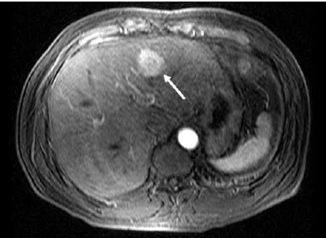
Space occupying lesions in the liver refer to abnormal growths or masses within the liver that can displace normal liver tissue.
Most Likely Causes for a Space Occupying Lesion of the Liver
Space occupying lesions in the liver can arise from various conditions, both benign and malignant. Understanding these causes is crucial for diagnosis and treatment. Below are the most likely causes categorized into benign and malignant lesions.
1. Benign Lesions:
- Hepatic Adenomas: These are benign tumors that may develop due to long-term use of birth control pills or anabolic steroids.
- Cavernous Hemangiomas: Usually present at birth, these vascular lesions develop from genetic mutations and are typically asymptomatic.
- Focal Nodular Hyperplasia (FNH): This lesion is caused by a malformation of blood vessels in the liver and is generally benign.
- Liver Cysts: While often of unknown origin, some cysts can be congenital or result from infections such as echinococcus.
2. Malignant Lesions:
- Hepatocellular Carcinoma (HCC): The most common type of primary liver cancer, often linked to chronic hepatitis B or C infections, cirrhosis, and other risk factors like heavy alcohol use.
- Metastatic Liver Disease: This occurs when cancer from other parts of the body spreads to the liver. Common primary cancers include colorectal, breast, and lung cancers.
- Cholangiocarcinoma: A rare cancer that originates in the bile ducts within or outside the liver.
3. Other Possible Causes:
- Liver Abscesses: These can be caused by bacterial or parasitic infections leading to localized collections of pus within the liver.
- Granulomatous Diseases: Conditions such as sarcoidosis or tuberculosis can lead to granuloma formation in the liver.
In summary, space occupying lesions in the liver can arise from a variety of benign conditions like hepatic adenomas and hemangiomas, as well as malignant conditions including hepatocellular carcinoma and metastatic disease. Accurate diagnosis through imaging studies and clinical evaluation is essential for determining appropriate management strategies.
Diagnosis for a Liver Abscess Based on History and Physical Examination
1. Patient History
The initial step in justifying a working diagnosis for a liver abscess is to gather a comprehensive patient history. Key components include:
- Symptoms: Patients may present with fever, chills, right upper quadrant pain, weight loss, and malaise. The presence of these symptoms can indicate an infectious process.
- Risk Factors: A history of underlying conditions such as diabetes, cirrhosis, or immunosuppression increases the likelihood of developing a liver abscess. Additionally, recent travel to endemic areas (e.g., Southeast Asia for melioidosis) or exposure to contaminated water may be relevant.
- Previous Medical History: Prior episodes of biliary disease, liver trauma, or surgery can provide insight into potential sources of infection.
- Medication Use: Immunosuppressive therapy or chemotherapy history should be noted as these can predispose patients to opportunistic infections.
2. Physical Examination
A thorough physical examination is crucial in supporting the diagnosis:
- Vital Signs: Fever and tachycardia may indicate systemic infection. Hypotension could suggest sepsis.
- Abdominal Examination: Tenderness in the right upper quadrant is often observed in patients with liver abscesses. Palpation may reveal hepatomegaly or a palpable mass if the abscess is large enough.
- Signs of Jaundice: Yellowing of the skin and eyes can indicate biliary obstruction or hepatic dysfunction associated with an abscess.
- Other Findings: Assessing for signs of dehydration or septic shock is important, especially if there are systemic symptoms.
3. Correlation Between History and Physical Findings
The combination of historical data and physical examination findings helps establish a working diagnosis:
- If the patient presents with fever, right upper quadrant pain, and has risk factors such as diabetes or recent travel to endemic regions, this raises suspicion for a liver abscess.
- The physical examination findings—particularly tenderness in the right upper quadrant—support this suspicion.
4. Next Steps
While history and physical examination are critical for forming a working diagnosis, further diagnostic imaging (such as ultrasound or CT scan) and laboratory tests (including blood cultures) are necessary to confirm the presence of an abscess and identify its etiology.
In summary, by carefully evaluating the patient’s history alongside physical examination findings—especially focusing on symptoms indicative of infection and any underlying risk factors—a strong justification for a working diagnosis of a liver abscess can be established.
Management Strategy for a Patient Suffering from a Malignant Space Occupying Lesion of the Liver
1. Initial Assessment and Diagnosis
The management of a patient with a malignant space-occupying lesion in the liver begins with a thorough assessment. This includes obtaining a detailed medical history, performing a physical examination, and conducting imaging studies such as ultrasound, computed tomography (CT), or magnetic resonance imaging (MRI) to characterize the lesion. Blood tests, including liver function tests and tumor markers (e.g., alpha-fetoprotein for hepatocellular carcinoma), are also essential.
2. Multidisciplinary Team Approach
Management should involve a multidisciplinary team comprising hepatologists, oncologists, interventional radiologists, and surgeons. This collaborative approach ensures comprehensive care tailored to the patient’s specific condition.
3. Staging of the Disease
Staging is crucial in determining the appropriate treatment strategy. The Barcelona Clinic Liver Cancer (BCLC) staging system is commonly used to classify hepatocellular carcinoma based on tumor size, number of lesions, presence of vascular invasion or metastasis, liver function status (Child-Pugh score), and performance status.
4. Treatment Options Based on Stage
- Early-Stage Disease:
- For patients with small tumors (<5 cm) without cirrhosis or with compensated cirrhosis, surgical resection may be considered if there are no significant comorbidities.
- Liver transplantation is an option for selected patients meeting criteria such as those outlined by the Milan criteria (single tumor ≤5 cm or up to three tumors each ≤3 cm without vascular invasion).
- Intermediate-Stage Disease:
- Transcatheter arterial chemoembolization (TACE) is often employed for patients who are not candidates for surgery but have preserved liver function. TACE delivers chemotherapy directly to the tumor while occluding its blood supply.
- Advanced-Stage Disease:
- Systemic therapies may be indicated for advanced disease where curative options are not feasible. Options include targeted therapies like sorafenib or immunotherapy combinations such as atezolizumab plus bevacizumab.
- Palliative care should also be integrated into management to address symptoms and improve quality of life.
5. Monitoring and Follow-Up
Regular follow-up through imaging studies and clinical assessments is vital to monitor treatment response and detect any recurrence early. Adjustments in therapy may be necessary based on disease progression or changes in liver function.
6. Management of Complications
Patients with malignant liver lesions often face complications due to underlying liver disease or treatment side effects. Management strategies should include:
- Addressing ascites with sodium restriction and diuretics.
- Managing variceal bleeding through endoscopic interventions.
- Treating hepatic encephalopathy using lactulose.
In summary, managing a malignant space-occupying lesion in the liver requires an individualized approach that considers the stage of cancer, underlying liver function, patient performance status, and potential treatment options available within a multidisciplinary framework.
Management Plan for a Patient Suffering from a Complicated Hydatid Cyst
1. Initial Assessment and Diagnosis
- Clinical Evaluation: Conduct a thorough history and physical examination to assess symptoms such as pain, dyspnea, or any signs of infection.
- Imaging Studies: Utilize ultrasound, CT scan, or MRI to determine the size, location, and characteristics of the hydatid cyst. Look for complications such as rupture or communication with other organs (e.g., biliary tree).
- Laboratory Tests: Perform serological tests (e.g., Echinococcus IgG antibodies) to confirm the diagnosis of echinococcosis.
2. Preoperative Preparation
- Anthelmintic Treatment: Consider administering albendazole or mebendazole preoperatively to reduce the risk of intraoperative spillage and recurrence.
- Assess Comorbidities: Evaluate the patient’s overall health status and manage any comorbid conditions that may affect surgical outcomes.
- Informed Consent: Discuss the risks, benefits, and potential complications of surgery with the patient and obtain informed consent.
3. Surgical Intervention
- Type of Surgery:
- For complicated cysts (e.g., those with biliary communication or significant compression of adjacent structures), consider procedures such as:
- Cystotomy (opening the cyst) with closure of any communicating structures.
- Capitonnage (suturing the cyst wall to prevent recurrence).
- Resection if necessary (segmentectomy or lobectomy in cases involving lung cysts).
- For complicated cysts (e.g., those with biliary communication or significant compression of adjacent structures), consider procedures such as:
- Surgical Approach: Choose an appropriate surgical approach based on cyst location (laparoscopic vs. open surgery).
4. Postoperative Care
- Monitoring: Closely monitor vital signs and watch for signs of complications such as infection, bleeding, or recurrence.
- Pain Management: Provide adequate analgesia to manage postoperative pain.
- Follow-Up Imaging: Schedule follow-up imaging studies to assess for any residual cysts or complications.
5. Long-Term Management
- Regular Follow-Up: Establish a follow-up schedule for monitoring potential recurrence through clinical evaluations and imaging studies.
- Continued Anthelmintic Therapy: If indicated, continue anthelmintic treatment postoperatively based on serological results and clinical findings.
- Patient Education: Educate the patient about hydatid disease prevention strategies, including proper hygiene practices to avoid reinfection.
6. Multidisciplinary Approach
- Involve specialists such as infectious disease experts, radiologists, and hepatobiliary surgeons in managing complex cases to ensure comprehensive care.
The management plan should be tailored based on individual patient factors including age, comorbidities, cyst characteristics, and available healthcare resources.
Informed Consent for Percutaneous Drainage of Echinococcal Cysts (PAIR)
Introduction to the Procedure
You are being asked to provide your consent for a medical procedure known as Percutaneous Aspiration, Injection, and Reaspiration (PAIR) for the treatment of echinococcal cysts. This procedure involves puncturing the cyst, aspirating its contents, injecting a sclerosing agent, and then reaspirating the cyst. The goal is to treat the cysts caused by Echinococcus granulosus, which can lead to complications if left untreated.
Purpose of the Procedure
The primary purpose of this procedure is to reduce or eliminate the size of echinococcal cysts in order to alleviate symptoms and prevent further complications such as rupture or infection. PAIR is considered less invasive than traditional surgical methods and can be performed under ultrasound or CT guidance.
Description of the Procedure
- Preparation: You will be positioned comfortably, and local anesthesia will be administered to minimize discomfort.
- Puncture: A thin needle will be inserted through your skin into the echinococcal cyst.
- Aspiration: The fluid within the cyst will be carefully aspirated (removed).
- Injection: A sclerosing agent may be injected into the cyst cavity to promote closure and prevent recurrence.
- Reaspiration: After a specified period, a follow-up aspiration may be performed to ensure that no residual fluid remains.
This procedure typically takes about 30 minutes to an hour.
Potential Risks and Complications
While PAIR is generally safe, there are potential risks associated with this procedure that you should be aware of:
- Infection: There is a risk of introducing bacteria during the puncture.
- Bleeding: Minor bleeding at the puncture site may occur.
- Cyst Rupture: If not performed correctly, there is a risk that the cyst could rupture, leading to spillage of its contents into surrounding tissues.
- Allergic Reaction: There may be an allergic reaction to medications used during the procedure.
- Need for Emergency Surgery: In rare cases where complications arise (such as severe infection or significant bleeding), emergency surgery may be required.
It is important that you discuss any concerns regarding these risks with your healthcare provider.
Alternatives to PAIR
You have options other than undergoing PAIR:
- Watchful Waiting: If your symptoms are mild or absent, monitoring without immediate intervention might be appropriate.
- Surgical Resection: Traditional surgery can remove larger or complicated cysts but comes with higher risks compared to PAIR.
- Medical Management: Medications such as albendazole may help manage symptoms but do not eliminate cysts.
Your healthcare provider will discuss these alternatives with you in detail.
Questions and Concerns
Please feel free to ask any questions about this procedure or express any concerns you might have before signing this consent form. It is crucial that you fully understand what this procedure entails and what it means for your health.
By signing below, you acknowledge that you have read this informed consent document, understand its contents, have had all your questions answered satisfactorily, and voluntarily agree to undergo PAIR for echinococcal cysts.
Patient Name: __
Signature: _
Date: _
I believe this informed consent process ensures that you are well-informed about your treatment options and their associated risks.


