Principles of Nerve Conduction Velocities (NCVs) and Electromyography (EMGs)
Nerve Conduction Velocities (NCVs)
Nerve conduction velocity (NCV) is a diagnostic test that measures the speed at which electrical impulses travel through a nerve. The principles underlying NCVs involve the following key concepts:
- Electrical Impulse Transmission: Nerves transmit signals through electrical impulses generated by the movement of ions across the nerve membrane. This process is essential for communication between the nervous system and muscles.
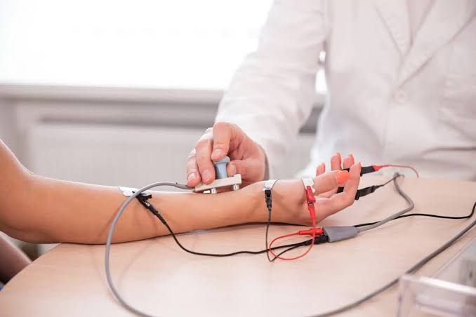
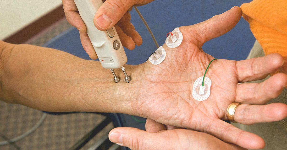
- Stimulation and Recording: During an NCV test, stimulating electrodes are placed on the skin over a nerve, delivering a small electrical pulse to activate the nerve. Recording electrodes are positioned on the skin over the muscle innervated by that nerve to capture its response.
- Conduction Velocity Calculation: The time it takes for the electrical impulse to travel from the stimulation site to the recording site is measured. The conduction velocity is calculated using the formula: Conduction Velocity = Distance ÷ Time, where distance is measured in centimeters and time in milliseconds.
- Assessment of Nerve Function: A normal conduction velocity indicates healthy nerve function, while slower velocities can suggest nerve damage or dysfunction, such as demyelination or neuropathy.
Electromyography (EMGs)
Electromyography (EMG) assesses muscle response to electrical stimulation from nerves, providing insights into neuromuscular health. The principles of EMG include:
- Muscle Electrical Activity: Muscles generate electrical signals when activated by motor neurons. These signals can be detected using electrodes inserted into or placed on the surface of muscles.
- Recording Techniques: During an EMG test, fine needles or surface electrodes are used to record electrical activity during different states—at rest, during slight contraction, and during maximal contraction. This helps evaluate how well muscles respond to nerve stimulation.
- Action Potentials Measurement: The electrical activity recorded reflects action potentials generated by muscle fibers when they contract in response to nerve signals. The size and shape of these action potentials provide information about muscle health and functionality.
- Differentiation Between Muscle and Nerve Disorders: By analyzing EMG results alongside NCV data, healthcare providers can differentiate between muscle disorders (myopathies) and nerve disorders (neuropathies), aiding in accurate diagnosis and treatment planning.
In summary, both NCVs and EMGs are critical tools in diagnosing neuromuscular disorders by measuring how well nerves conduct impulses and how effectively muscles respond to those impulses.
Purpose of Advising these Tests
The primary purpose of advising Nerve Conduction Velocities (NCVs) and Electromyography (EMG) tests is to evaluate the functionality of nerves and muscles in the body. These tests are essential diagnostic tools used by healthcare providers to identify various neuromuscular disorders.
1. Assessing Nerve Functionality:
- NCV tests measure how quickly electrical impulses travel through a nerve, which helps determine if there is any nerve damage or dysfunction. A slower conduction velocity can indicate conditions such as neuropathy, where nerves are damaged due to various factors like diabetes or autoimmune diseases.
2. Evaluating Muscle Response:
- EMG tests assess the electrical activity produced by muscles when they are at rest and during contraction. This helps in determining whether the muscles are responding appropriately to nerve signals. Abnormal electrical activity can indicate muscle disorders or issues with the neuromuscular junction.
3. Differentiating Between Muscle and Nerve Disorders:
- When performed together, NCV and EMG provide complementary information that helps differentiate between muscle disorders (myopathies) and nerve disorders (neuropathies). For instance, if NCV results show normal conduction but EMG shows abnormal muscle activity, it suggests a muscle disorder rather than a nerve issue.
4. Diagnosing Specific Conditions:
- These tests are used to diagnose specific conditions such as:
- Guillain-Barré syndrome
- Carpal tunnel syndrome
- Charcot-Marie-Tooth disease
- Herniated disk disease
- Sciatic nerve problems
5. Monitoring Disease Progression:
- In addition to diagnosis, NCV and EMG can be used to monitor the progression of neuromuscular diseases over time, helping healthcare providers adjust treatment plans accordingly.
In summary, advising NCVs and EMGs is crucial for accurately diagnosing neuromuscular conditions, assessing nerve and muscle function, differentiating between types of disorders, diagnosing specific diseases, and monitoring ongoing health issues.
How NCVs and EMG are Carried Out
Nerve Conduction Studies (NCS)
- Preparation: Before the test, the patient is typically asked to wear loose clothing for easy access to the area being tested. It is important that the skin is clean, so lotions, creams, or perfumes should not be applied for a day or two prior to the test.
- Positioning: The patient will sit or lie down comfortably on a table or bed.
- Electrode Placement:
- The healthcare provider will place stimulating electrodes on the skin over the nerve that is being tested.
- These electrodes deliver a mild electrical pulse to stimulate the nerve.
- Recording Electrodes: Recording electrodes are attached to the muscles that are controlled by those nerves. These electrodes will capture the muscle’s response to the electrical stimulation.
- Stimulation and Measurement:
- A small pulse of electricity is sent through the stimulating electrodes.
- This pulse stimulates the nerve, prompting it to send a signal to the muscle.
- The healthcare provider measures how long it takes for the muscle to respond, which is known as conduction velocity.
- Duration: The NCS may take anywhere from 15 minutes to over an hour, depending on how many nerves and muscles are being tested.
Electromyography (EMG)
- Preparation: Similar to NCS, patients should wear comfortable clothing and avoid lotions or creams before testing.
- Positioning: The patient will also sit or lie down on a table or bed during this procedure.
- Skin Cleaning: The provider cleans the skin over the muscle that will be tested.
- Electrode Insertion:
- A small needle electrode is inserted through the skin into the muscle.
- This electrode records electrical activity in response to nerve stimulation.
- Recording Muscle Activity:
- The machine records electrical signals from the muscle at rest and during contraction.
- Patients may be asked to contract their muscles by performing specific movements (e.g., lifting or bending a limb) while recordings are taken.
- Data Display and Analysis:
- The electrical activity appears as wavy and spiky lines on a monitor (oscilloscope) and can also be heard through an audio amplifier.
- The size and shape of these waves provide information about how well muscles respond when stimulated by nerves.
- Duration: An EMG test typically lasts between 30 minutes to an hour.
- Post-Test Care: After both tests, patients can usually resume normal activities unless otherwise advised by their healthcare provider.


