Sensory Pathways and Receptors
Sensory pathways are the routes through which sensory information travels from the peripheral nervous system to the central nervous system (CNS). These pathways are essential for processing sensory stimuli and enabling appropriate responses. Sensory receptors, located throughout the body, detect various types of stimuli, including touch, temperature, pain, and proprioception.
1. Types of Sensory Receptors
Sensory receptors can be categorized based on the type of stimulus they detect:
- Mechanoreceptors: These receptors respond to mechanical pressure or distortion. They are responsible for sensations such as touch, vibration, and proprioception. Mechanoreceptors include Merkel cells (for light touch), Meissner’s corpuscles (for light touch and vibration), Pacinian corpuscles (for deep pressure and vibration), and Ruffini endings (for skin stretch).
- Thermoreceptors: These receptors detect changes in temperature. They are classified into two main types: cold receptors that respond to lower temperatures and warm receptors that respond to higher temperatures.
- Nociceptors: These receptors are responsible for detecting pain. They can be activated by extreme mechanical, thermal, or chemical stimuli.
- Photoreceptors: Located in the retina of the eye, these receptors respond to light. There are two main types: rods (sensitive to low light levels) and cones (responsible for color vision).
- Chemoreceptors: These receptors respond to chemical stimuli. They play a crucial role in taste (gustatory receptors) and smell (olfactory receptors).
2. Sensory Pathways
The sensory pathways consist of a series of neurons that relay information from sensory receptors to specific areas in the brain where it is processed:
- First Order Neurons: The pathway begins with first-order neurons that have their cell bodies located in the dorsal root ganglion for somatosensory information from below the neck or in cranial nerve ganglia for sensations from the head and neck. These neurons detect stimuli through their sensory endings.
- Second Order Neurons: The axons of first-order neurons enter the spinal cord or brainstem where they synapse with second-order neurons. In the case of somatosensory pathways from below the neck, these second-order neurons reside in either the spinal cord or brainstem nuclei.
- Third Order Neurons: Second-order neurons then project their axons to third-order neurons located in the thalamus. The thalamus acts as a relay station for sensory information before it reaches its final destination in the cerebral cortex.
3. Major Sensory Pathways
There are two primary ascending pathways that carry somatosensory information:
- Dorsal Column-Medial Lemniscus Pathway: This pathway transmits fine touch, vibration, and proprioceptive information. First-order neurons enter through the dorsal column of white matter in the spinal cord; they ascend ipsilaterally until they synapse at nuclei in the medulla oblongata (nucleus gracilis for lower body sensations and nucleus cuneatus for upper body sensations). Second-order neurons then decussate (cross over) at this level before projecting to the thalamus.
- Spinothalamic Tract: This pathway carries pain and temperature sensations as well as crude touch. First-order neurons synapse with second-order neurons within the spinal cord shortly after entering it; these second-order neurons decussate immediately before ascending through this tract to reach the thalamus.
4. Topographical Representation
Sensory information is organized topographically within both pathways so that adjacent areas on the body correspond to adjacent areas in the CNS representation—this organization allows precise localization of sensory input.
In summary, sensory pathways involve complex networks of specialized receptor cells that convert various forms of environmental stimuli into electrical signals transmitted via a series of interconnected neurons leading ultimately to perception within specific regions of the brain.
Spinothalamic Pathway
The spinothalamic pathway is a crucial ascending sensory pathway in the central nervous system that transmits information regarding pain, temperature, and crude touch from the body to the brain. It is part of the anterolateral system and consists of several components:
- Anatomy: The spinothalamic tract is located anteriolaterally in the spinal cord and comprises three sets of neurons:
- First Order Neurons: These are nociceptive neurons that detect pain, thermal sensations, and crude touch. Their cell bodies reside in the dorsal root ganglia, and they enter the spinal cord to synapse with second-order neurons in the posterior grey horn.
- Second Order Neurons: Located within the posterior grey horn, these neurons decussate (cross over) at their respective segmental levels before ascending through the spinal cord as the spinothalamic tract. They then synapse with third-order neurons in the ventral posterior nucleus of the thalamus.
- Third Order Neurons: These neurons project from the thalamus to the primary somatosensory cortex (Brodmann areas 3, 1, and 2), where sensory information is processed.
- Function: The primary function of the spinothalamic tract is to relay sensory information about pain (nociception), temperature changes, and crude touch to higher brain centers for interpretation. The lateral spinothalamic tract specifically transmits pain and temperature sensations, while the anterior spinothalamic tract conveys crude touch and pressure.
- Clinical Relevance: Damage or dysfunction of this pathway can lead to significant sensory deficits or altered perception of stimuli.
Spinothalamic Damage
Damage to the spinothalamic tract can occur due to various conditions such as spinal cord injuries, tumors, or diseases affecting nerve pathways. The clinical manifestations depend on where along the pathway damage occurs:
- Symptoms:
- Patients may experience contralateral loss of pain and temperature sensation below the level of injury due to decussation occurring at or near their segmental level.
- Crude touch may remain intact because it is transmitted via other pathways (e.g., dorsal column-medial lemniscal system).
- Conditions Associated with Damage:
- Brown-Séquard Syndrome: This condition results from hemisection of the spinal cord leading to ipsilateral loss of proprioception and vibration sense (due to dorsal column involvement) while causing contralateral loss of pain and temperature sensation (due to spinothalamic damage).
- Peripheral Neuropathy: In conditions like diabetes mellitus, symmetrical damage can affect long peripheral nerves first, leading to a “stocking-glove” distribution pattern where patients lose sensation in distal extremities.
- Diagnosis and Assessment:
- Clinical assessment often involves testing for pinprick sensation (pain) and temperature discrimination using cold stimuli.
- Imaging studies such as MRI may be utilized for identifying structural causes leading to damage.
In summary, understanding both normal function and potential damage within the spinothalamic pathway is essential for diagnosing sensory disorders related to neurological conditions.
Dorsal Column Pathway
The dorsal column pathway, also known as the dorsal column-medial lemniscus (DCML) pathway, is a crucial sensory pathway in the central nervous system responsible for transmitting fine touch, vibration, two-point discrimination, and proprioceptive information from the body to the brain. This pathway consists of three orders of neurons:
- First-Order Neurons: These neurons are located in the dorsal root ganglia and are characterized as pseudo-unipolar neurons. Their peripheral axons receive sensory input from various receptors in the skin and joints. The types of receptors involved include:
- Tactile Mechanoreceptors: Such as Meissner’s corpuscles (fine touch and two-point discrimination), free nerve endings on hair follicles (fine touch), and Pacinian corpuscles (pressure and vibration).
- Proprioceptors: Including muscle spindles and Golgi tendon organs that detect changes in muscle length and tension.
After receiving sensory input, the central axons of these first-order neurons enter the spinal cord through the medial dorsal root entry zone. They ascend ipsilaterally within the dorsal columns of the spinal cord, forming two main tracts:
- Fasciculus Gracilis: Carries sensory information from the lower extremities.
- Fasciculus Cuneatus: Carries sensory information from the upper extremities.
- Second-Order Neurons: Located in the nucleus gracilis and nucleus cuneatus within the medulla oblongata, these neurons receive synaptic input from first-order neurons. The axons of second-order neurons cross over at this level (decussate) to form the medial lemniscus, which ascends through the brainstem while maintaining a somatotopic arrangement.
- Third-Order Neurons: These neurons reside in the ventral posterolateral (VPL) nucleus of the thalamus. Their axons project to the primary somatosensory cortex located in the postcentral gyrus of the parietal lobe, where sensory information is processed.
The primary function of this pathway is to convey detailed sensory information regarding fine touch, proprioception, and vibration sensations from various parts of the body to enable conscious perception.
Dorsal Column Damage
Damage to the dorsal column pathway can lead to significant neurological deficits due to its role in transmitting important sensory information. Conditions that may cause damage include trauma, vascular accidents (such as posterior spinal artery syndrome), or diseases like tabes dorsalis associated with untreated syphilis.
When there is damage to this pathway, individuals may experience symptoms such as:
- Loss of Fine Touch Sensation: Patients may find it difficult to perceive light touches or differentiate between two closely spaced points on their skin.
- Impaired Proprioception: Individuals may struggle with body awareness; for instance, they might not be able to accurately sense limb position without visual cues.
- Vibration Sense Loss: Patients often cannot feel vibrations applied to their skin or joints.
These deficits can lead to unsteady gait, increased risk of falls due to lack of proprioceptive feedback, diminished reflexes, and coordination problems. In severe cases, patients may also experience pain syndromes due to abnormal processing of nociceptive signals.
Overall, damage to this critical sensory pathway significantly impacts an individual’s quality of life by impairing their ability to interact safely with their environment.
Spinocerebellar Pathway
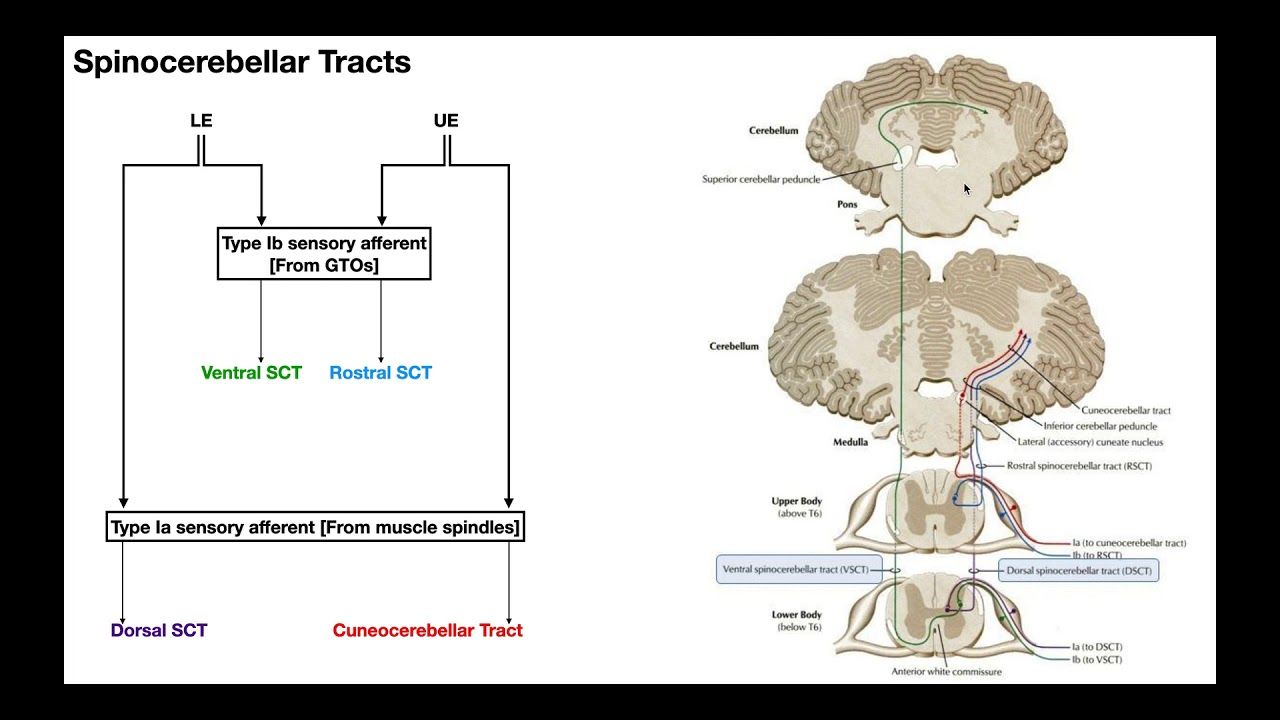
The spinocerebellar pathway is a critical neural pathway that transmits proprioceptive information from the body to the cerebellum, which plays a vital role in coordinating movement and maintaining balance. This pathway consists of several tracts, primarily the dorsal (posterior) spinocerebellar tract and the ventral (anterior) spinocerebellar tract.
- Dorsal Spinocerebellar Tract: This tract carries proprioceptive information from muscle spindles and Golgi tendon organs located in the lower limbs and trunk. The first-order neurons originate in the dorsal root ganglia and enter the spinal cord, where they synapse with second-order neurons in Clarke’s nucleus (located in the spinal cord). The axons of these second-order neurons ascend ipsilaterally through the lateral funiculus to reach the cerebellum via the inferior cerebellar peduncle.
- Ventral Spinocerebellar Tract: In contrast, this tract conveys proprioceptive information from lower limbs but involves a crossing over (decussation) at two points. First, it crosses to the opposite side of the spinal cord before ascending through the anterior white commissure. It then ascends to join the superior cerebellar peduncle, where it enters the cerebellum after crossing back to its original side.
- Cuneocerebellar Tract: This is analogous to the dorsal spinocerebellar tract but serves the upper limbs and neck. It originates from proprioceptors in these regions and travels through cuneate nuclei before reaching the cerebellum via inferior cerebellar peduncle.
- Rostral Spinocerebellar Tract: This tract transmits proprioceptive information from cranial parts of the body to the cerebellum, traveling ipsilaterally and terminating bilaterally within specific regions of the cerebellum.
Overall, these pathways are essential for providing continuous feedback about body position and movement, allowing for smooth coordination of motor activities.
Spinocerebellar Tract Damage
Damage to any part of the spinocerebellar tracts can lead to significant deficits in proprioception and coordination:
- Symptoms of Damage: Individuals may experience ataxia (lack of voluntary coordination), dysmetria (inability to control distance or scale of movements), and other motor dysfunctions due to impaired feedback regarding limb position and movement.
- Dorsal Spinocerebellar Tract Damage: Lesions here can result in loss of proprioceptive input from lower limbs leading to difficulties with balance and coordination while standing or walking. Patients may have difficulty performing tasks that require precise limb positioning.
- Ventral Spinocerebellar Tract Damage: Since this tract carries internally generated motor information about movement, damage can lead to issues with executing planned movements accurately despite intact sensory input from other pathways.
- Cuneocerebellar Tract Damage: Injury affecting this pathway may impair proprioception specifically related to upper limb movements, resulting in clumsiness or poor hand-eye coordination.
- Rostral Spinocerebellar Tract Damage: Damage here can affect coordination involving cranial structures, potentially impacting facial movements or upper limb actions as well.
In summary, damage along any segment of these tracts disrupts normal communication between peripheral receptors and central processing areas within the cerebellum, leading to various degrees of motor dysfunction depending on which specific tract is affected.


