Important Components of a Typical Bacterial Cell
1. Cell Envelope
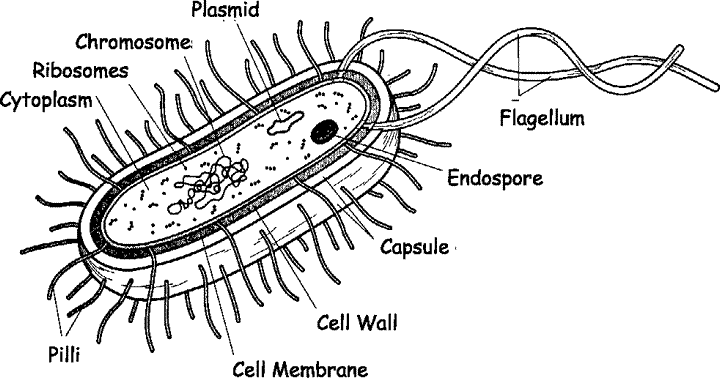
The cell envelope is a critical structure that surrounds the bacterial cell and consists of multiple layers. It typically includes:
- Cell Membrane: This is the innermost layer, composed of a phospholipid bilayer that regulates the movement of substances in and out of the cell. It plays a vital role in maintaining homeostasis and facilitating cellular processes.
- Cell Wall: The cell wall provides structural support and shape to the bacterium. It is primarily made up of peptidoglycan, which is a polymer consisting of sugars and amino acids. The composition of the cell wall can vary significantly among different bacterial species, which is crucial for classification and identification.
- Capsule (if present): Some bacteria have an additional outer layer known as a capsule, made up of polysaccharides. This structure helps protect the bacterium from desiccation and phagocytosis by larger microorganisms, enhancing its virulence.
2. Cytoplasm
The cytoplasm is the gel-like substance within the bacterial cell membrane that contains all cellular components. It houses various organelles, enzymes, nutrients, and genetic material necessary for metabolic processes.
3. Nucleoid Region
Bacteria lack a defined nucleus; instead, their genetic material (DNA) is located in a region called the nucleoid. The DNA is typically organized as a single circular chromosome that contains essential genes for survival and reproduction.
4. Ribosomes
Ribosomes are molecular machines found within the cytoplasm responsible for protein synthesis. Bacterial ribosomes are smaller than those found in eukaryotic cells (70S vs 80S), allowing them to efficiently translate mRNA into proteins.
5. Plasmids (if present)
Plasmids are small, circular DNA molecules separate from chromosomal DNA that can carry genes beneficial for survival, such as antibiotic resistance genes. They can replicate independently within the bacterial cell.
6. Appendages
Bacteria may possess appendages that aid in movement or attachment:
- Flagella: These are long, whip-like structures that enable motility by rotating like propellers.
- Pili (or fimbriae): These hair-like structures facilitate adhesion to surfaces or other cells and can play roles in conjugation (the transfer of genetic material between bacteria).
7. Inclusion Bodies (if present)
Inclusion bodies are granules found in some bacteria that store nutrients or other substances such as glycogen or polyhydroxybutyrate (PHB). They serve as energy reserves or building blocks for cellular functions.
In summary, a typical bacterial cell comprises several key components: the cell envelope (including membrane, wall, and possibly capsule), cytoplasm containing ribosomes and nucleoid region with DNA, plasmids for extra genetic information, appendages like flagella and pili for movement and attachment, and inclusion bodies for storage purposes.
Differentiating Characteristics of Gram Positive and Gram Negative Bacteria
1. Cell Wall Structure
The primary differentiating characteristic between gram-positive and gram-negative bacteria lies in their cell wall structure.
- Gram-Positive Bacteria: These bacteria have a thick peptidoglycan layer, which can be up to 90% of the cell wall composition. This thick layer retains the crystal violet stain used in the Gram staining procedure, resulting in a purple appearance under a microscope. Additionally, gram-positive bacteria may contain teichoic acids, which contribute to the rigidity of the cell wall and play roles in cell growth and division.
- Gram-Negative Bacteria: In contrast, gram-negative bacteria possess a much thinner peptidoglycan layer (about 10% of the cell wall) located between an inner cytoplasmic membrane and an outer membrane. The outer membrane contains lipopolysaccharides (LPS), which are significant for their role in immune response and pathogenicity. During the Gram staining process, these bacteria do not retain the crystal violet stain but take up the counterstain (safranin), appearing pink or red.
2. Staining Properties
The Gram staining technique is fundamental for distinguishing between these two groups:
- Gram-Positive: Retain crystal violet dye due to their thick peptidoglycan layer.
- Gram-Negative: Do not retain crystal violet; instead, they take up safranin due to their thinner peptidoglycan layer and presence of an outer membrane.
3. Sensitivity to Antibiotics
The differences in cell wall structure also influence antibiotic susceptibility:
- Gram-Positive Bacteria: Generally more susceptible to antibiotics that target peptidoglycan synthesis (e.g., penicillin) because their thick peptidoglycan layer is exposed directly to these agents.
- Gram-Negative Bacteria: Often more resistant to antibiotics due to their outer membrane acting as a barrier that prevents many drugs from entering. They may also possess efflux pumps that expel antibiotics before they can exert their effects.
4. Pathogenicity Factors
The presence of specific structures contributes to pathogenicity:
- Gram-Positive Bacteria: Some species produce exotoxins and have surface proteins that facilitate adherence to host tissues.
- Gram-Negative Bacteria: The lipopolysaccharides present on their outer membrane can trigger strong immune responses and are often involved in septic shock when released into circulation.
Importance in Diagnosis
Understanding whether a bacterium is gram-positive or gram-negative is crucial for diagnosis:
- Identification of Pathogens: The Gram stain is one of the first steps in identifying bacterial infections, guiding further testing and treatment decisions.
- Treatment Decisions: Knowing the type of bacteria helps clinicians choose appropriate antibiotics; for example, if a patient has a suspected staphylococcal infection (gram-positive), penicillin derivatives might be considered.
- Infection Control Measures: Different types of bacteria may require different infection control measures based on their virulence factors and resistance profiles.
Importance in Vaccination
The structural differences also play a significant role in vaccine development:
- Targeting Antigens: Vaccines against gram-negative bacteria often target polysaccharides from LPS or other surface antigens, while vaccines against gram-positive bacteria may focus on proteins or polysaccharides from the thick peptidoglycan layer.
- Immune Response Modulation: The immune response elicited by vaccines can differ significantly based on whether they target gram-positive or gram-negative organisms due to variations in how these bacteria interact with host immune systems.
- Vaccine Development Challenges: Developing effective vaccines against gram-negative pathogens can be challenging due to their ability to evade immune detection through various mechanisms associated with their outer membranes.
In summary, understanding these characteristics not only aids in diagnosing bacterial infections but also informs strategies for developing effective vaccines against various bacterial pathogens.
Specialized Structures of Bacterial Cells and Their Role in Disease
1. Capsule
The capsule is a protective layer made up of polysaccharides that surrounds some bacterial cells. Its primary roles include preventing desiccation (drying out) and protecting the bacterium from phagocytosis by larger immune cells, such as macrophages. The presence of a capsule is a major virulence factor for many pathogenic bacteria, including Escherichia coli and Streptococcus pneumoniae. Nonencapsulated mutants of these organisms are avirulent, meaning they do not cause disease.
2. Cell Wall
The cell wall is composed of peptidoglycan, which provides structural support and shape to the bacterium. It protects the cell from osmotic pressure changes in its environment. The composition of the cell wall varies between gram-positive and gram-negative bacteria, influencing their susceptibility to antibiotics and their overall virulence. For example, gram-negative bacteria have a thinner cell wall but possess an outer membrane that can act as a barrier to certain antibiotics.
3. Pili (or Fimbriae)
Pili are hair-like appendages that protrude from the surface of bacterial cells. They play crucial roles in adhesion to host tissues, which is essential for colonization and infection. Some pili are involved in conjugation, allowing for the transfer of genetic material between bacteria, which can contribute to antibiotic resistance.
4. Flagella
Flagella are long, whip-like structures that enable motility in bacteria. This movement allows bacteria to reach favorable environments or evade immune responses. In pathogenic bacteria, flagella can enhance virulence by facilitating invasion into host tissues or escaping from immune cells.
5. Endospores
Some bacteria can form endospores, which are highly resistant structures that allow them to survive extreme environmental conditions such as heat, desiccation, and chemical exposure. Endospores can remain dormant for long periods and reactivate when conditions become favorable again. This ability contributes to the persistence of certain pathogens in various environments.
6. Toxins
Many pathogenic bacteria produce toxins that directly damage host tissues or interfere with normal cellular functions. These toxins can be classified into two main categories: exotoxins (secreted by living bacteria) and endotoxins (part of the bacterial cell wall structure). Exotoxins can cause severe damage even at low concentrations, while endotoxins typically induce strong inflammatory responses upon bacterial lysis.
In summary, specialized structures such as capsules, cell walls, pili, flagella, endospores, and toxins play significant roles in bacterial survival and pathogenesis by enhancing their ability to infect hosts and evade immune responses.


