Gross Anatomical Features of the Spinal Cord
The spinal cord is a vital structure within the central nervous system, serving as the main pathway for transmitting information between the brain and the rest of the body. Its gross anatomical features can be described in detail as follows:
1. Structure and Location
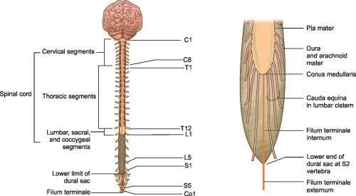
The spinal cord is a cylindrical tube of nervous tissue that extends from the base of the brain (specifically, from the medulla oblongata at the foramen magnum) down to approximately the level of the first or second lumbar vertebra (L1-L2) in adults. In infants, it may extend down to L2-L3 due to differences in vertebral growth rates.
2. Segmentation
The spinal cord is divided into 31 segments, each corresponding to a pair of spinal nerves that emerge from it. These segments are categorized into:
- Cervical Segments (C1-C8): There are eight cervical segments, with C1-C7 exiting above their respective vertebrae and C8 exiting between C7 and T1.
- Thoracic Segments (T1-T12): Twelve thoracic segments follow, with each nerve emerging below its corresponding vertebra.
- Lumbar Segments (L1-L5): Five lumbar segments provide innervation to lower back and leg muscles.
- Sacral Segments (S1-S5): Five sacral segments contribute to pelvic and lower limb function.
- Coccygeal Segment (Co): One coccygeal segment is primarily vestigial.
3. Conus Medullaris
At its terminal end, the spinal cord tapers into a cone-shaped structure known as the conus medullaris. This marks where the spinal cord transitions into individual nerve roots that continue downward.
4. Cauda Equina
Below the conus medullaris lies a collection of nerve roots called the cauda equina (“horse’s tail”). This structure consists of lumbar and sacral nerve roots that travel downward before exiting through their respective intervertebral foramina.
5. Protective Layers
The spinal cord is encased in protective layers known as meninges, which include:
- Dura Mater: The outermost tough layer.
- Arachnoid Mater: The middle web-like layer.
- Pia Mater: The innermost delicate layer that adheres closely to the surface of the spinal cord.
Between these layers lies cerebrospinal fluid (CSF), which provides cushioning and protection against mechanical injury.
6. Spinal Nerves
Each segment of the spinal cord gives rise to a pair of spinal nerves composed of sensory (dorsal) and motor (ventral) roots:
- Dorsal Roots: Carry sensory information from peripheral receptors to the spinal cord; their cell bodies reside in dorsal root ganglia.
- Ventral Roots: Emerge from anterior horn cells within the spinal cord and carry motor commands to skeletal muscles.
7. Gray Matter and White Matter
The internal structure of the spinal cord consists of gray matter arranged in an “H” or butterfly shape surrounded by white matter:
- Gray Matter: Contains neuronal cell bodies, dendrites, and synapses; it is organized into horns—dorsal horns for sensory processing and ventral horns for motor output.
- White Matter: Composed mainly of myelinated axons forming ascending sensory pathways and descending motor pathways.
These features collectively enable the spinal cord to perform its essential functions in reflex actions, sensation transmission, and motor control throughout the body.
Comparison of Spinal Segments to Vertebral Levels
The spinal cord is organized into segments that correspond to specific pairs of spinal nerves, and these segments do not always align directly with the vertebral levels. Below is a detailed comparison of the different spinal segments and their respective vertebral levels:
- Cervical Segments (C1-C8):
- The cervical spine consists of 7 cervical vertebrae (C1-C7) but has 8 cervical spinal nerves (C1-C8).
- The C1 nerve exits above the first cervical vertebra (C1), while C2 through C7 exit above their corresponding vertebrae.
- The C8 nerve exits below the C7 vertebra, specifically between the C7 and T1 vertebrae.
- Thoracic Segments (T1-T12):
- There are 12 thoracic vertebrae (T1-T12) and 12 thoracic spinal nerves.
- Each thoracic nerve exits at the level of its corresponding vertebra. For example, T1 exits between T1 and T2, T2 exits between T2 and T3, continuing this pattern down to T12.
- Lumbar Segments (L1-L5):
- The lumbar region consists of 5 lumbar vertebrae (L1-L5) and has 5 lumbar spinal nerves.
- Similar to the thoracic region, each lumbar nerve exits at its corresponding level: L1 exits between L1 and L2, L2 between L2 and L3, up to L5 which exits between L5 and S1.
- Sacral Segments (S1-S5):
- There are 5 sacral vertebrae fused together to form the sacrum, with 5 sacral spinal nerves.
- Each sacral nerve also exits at its corresponding level: S1 exits between S1 and S2, S2 between S2 and S3, continuing this pattern through S5.
- Coccygeal Segment:
- There is one coccygeal nerve that corresponds to the coccyx but does not have a direct bony counterpart as it is a single structure.
In summary, while cervical segments have an atypical arrangement where some nerves exit above their respective vertebrae due to anatomical differences in development, thoracic, lumbar, and sacral segments generally align with their respective vertebral levels. This discrepancy is particularly notable in the cervical region where there are more nerves than there are vertebrae.
Important Gross Features of the Spinal Cord, Nerve Roots, and Spinal Ganglia
1. Spinal Cord Structure
The spinal cord is a cylindrical structure composed of nervous tissue that serves as a critical communication pathway between the brain and the body. It extends from the foramen magnum at the base of the skull to approximately the level of the first or second lumbar vertebrae in adults. The spinal cord is about 40 to 50 cm long and has a diameter ranging from 1 cm to 1.5 cm. It is organized into four main regions: cervical (C), thoracic (T), lumbar (L), and sacral (S). Each region contains several segments, with a total of 31 pairs of spinal nerves emerging from it.
2. Enlargements
There are two notable enlargements in the spinal cord:
- Cervical Enlargement: This region extends from C3 to T1 and corresponds to the nerves that innervate the upper limbs.
- Lumbar Enlargement: This region extends from L1 to S2 and corresponds to nerves that innervate the lower limbs.
3. Nerve Roots
Each spinal nerve emerges from the spinal cord through two roots:
- Anterior (Ventral) Roots: These roots contain efferent motor fibers that carry signals away from the central nervous system (CNS) to skeletal muscles and autonomic targets. The cell bodies for these motor neurons reside in the anterior horn of gray matter within the spinal cord.
- Posterior (Dorsal) Roots: These roots contain afferent sensory fibers that transmit sensory information from peripheral receptors back to the CNS. The cell bodies for these sensory neurons are located in the dorsal root ganglion, which is situated outside of the spinal cord.
4. Spinal Ganglia
Spinal ganglia, also known as dorsal root ganglia, are clusters of neuronal cell bodies located along each posterior root of a spinal nerve. They play a crucial role in processing sensory information before it reaches the CNS. Each ganglion contains sensory neurons that relay information regarding touch, pain, temperature, and proprioception from various parts of the body.
5. Meninges
The spinal cord is protected by three layers of meninges:
- Dura Mater: The tough outer layer.
- Arachnoid Mater: The middle layer that provides cushioning.
- Pia Mater: The delicate inner layer that closely adheres to the surface of the spinal cord.
6. Cauda Equina
In addition to these structures, below L2, there exists a collection of nerve roots known as the cauda equina (“horse’s tail”). These roots extend downward within the vertebral canal before exiting at their respective intervertebral foramina.
In summary, understanding these gross features—such as structural organization, nerve root composition, and protective coverings—is essential for comprehending how signals travel between different parts of the body and how injuries or diseases affecting these areas can impact overall function.
Internal Features of the Spinal Cord: Gray Matter and White Matter in Different Regions
The spinal cord is a vital structure within the central nervous system (CNS) that serves as a conduit for nerve signals between the brain and the body. It is organized into distinct regions, each characterized by specific arrangements of gray matter and white matter.
1. General Structure of the Spinal Cord
The spinal cord extends from the foramen magnum at the base of the skull to approximately the level of the first or second lumbar vertebrae. It is divided into four main regions: cervical, thoracic, lumbar, and sacral. Each region has unique features regarding its gray and white matter composition.
2. Gray Matter
Gray matter in the spinal cord primarily consists of neuronal cell bodies, dendrites, and unmyelinated axons. It forms an H-shaped structure in cross-section, with two dorsal (posterior) horns and two ventral (anterior) horns:
- Cervical Region: The gray matter is relatively large due to a higher number of motor neurons that innervate upper limb muscles. The anterior horns are prominent, reflecting extensive motor control.
- Thoracic Region: The gray matter is smaller compared to cervical levels. The lateral horns are present here, which contain sympathetic neurons involved in autonomic functions.
- Lumbar Region: Similar to the cervical region, there is a significant amount of gray matter due to innervation of lower limb muscles. The anterior horns are well-developed.
- Sacral Region: The gray matter is also prominent here but smaller than in lumbar levels. It contains neurons that control pelvic organs and lower limbs.
3. White Matter
White matter consists mainly of myelinated axons organized into tracts that facilitate communication between different parts of the CNS:
- Cervical Region: White matter is abundant here due to numerous ascending sensory tracts (such as posterior columns) and descending motor tracts (like corticospinal tracts). This region has a larger volume of white matter compared to other regions because it carries signals from both upper and lower body segments.
- Thoracic Region: There is less white matter than in cervical levels since fewer ascending fibers are present from lower body segments. However, it still contains important tracts for autonomic functions.
- Lumbar Region: The amount of white matter decreases further as you move downwards; however, it still contains essential pathways for motor control and sensory information from lower limbs.
- Sacral Region: This region has the least amount of white matter due to its position at the end of the spinal cord where fewer ascending or descending pathways are present.
4. Summary
In summary, each segment of the spinal cord exhibits distinct characteristics in terms of gray and white matter organization:
- In general, gray matter houses neuronal cell bodies crucial for processing sensory input and generating motor output.
- White matter facilitates communication through myelinated axons organized into specific tracts that carry information up and down the spinal cord.
Understanding these internal features helps elucidate how injuries or diseases affecting different regions can lead to specific functional deficits.
Location, Origin, Course, and Termination of Ascending and Descending Tracts of the Spinal Cord
Ascending Tracts
The ascending tracts of the spinal cord are responsible for transmitting sensory information from the body to the brain. They are primarily located in the white matter of the spinal cord and can be categorized based on their specific functions.
- Dorsal Column-Medial Lemniscus Pathway
- Location: Dorsal funiculus (dorsal columns) of the spinal cord.
- Origin: The first-order neurons originate from sensory receptors in the skin, muscles, and joints.
- Course: These fibers ascend through the dorsal columns as fasciculus gracilis (medially) and fasciculus cuneatus (laterally). They synapse in the medulla oblongata at the nucleus gracilis and nucleus cuneatus.
- Termination: Second-order neurons decussate (cross over) in the medulla and ascend to the thalamus via the medial lemniscus. Third-order neurons then project from the thalamus to the primary somatosensory cortex.
- Spinothalamic Tract
- Location: Lateral and anterior funiculi of the spinal cord.
- Origin: First-order neurons originate from nociceptors and thermoreceptors in peripheral tissues.
- Course: These fibers enter the spinal cord through dorsal roots, synapse with second-order neurons in the dorsal horn, and then decussate immediately within one or two segments before ascending through the lateral spinothalamic tract.
- Termination: Second-order neurons ascend to terminate in the thalamus, where third-order neurons relay information to specific areas of the cerebral cortex.
- Spinocerebellar Tracts
- Location: Lateral funiculus of the spinal cord.
- Origin: First-order neurons arise from muscle spindle receptors and Golgi tendon organs.
- Course: There are two main spinocerebellar tracts: dorsal spinocerebellar tract (which ascends ipsilaterally) and ventral spinocerebellar tract (which crosses over before entering cerebellum).
- Termination: Both tracts terminate in different regions of the cerebellum, providing proprioceptive information necessary for coordination.
Descending Tracts
The descending tracts are responsible for transmitting motor commands from the brain to various parts of the body. They also reside primarily within white matter but can be divided into several key pathways.
- Corticospinal Tract
- Location: Lateral funiculus (lateral corticospinal tract) and anterior funiculus (anterior corticospinal tract).
- Origin: The upper motor neurons originate in the primary motor cortex of the brain.
- Course: The fibers descend through internal capsule, brainstem, and decussate at either pyramidal decussation (for lateral corticospinal tract) or remain ipsilateral until they reach lower levels (for anterior corticospinal tract).
- Termination: The lateral corticospinal tract terminates on lower motor neurons in anterior horn cells across multiple spinal segments; anterior corticospinal fibers terminate bilaterally on lower motor neurons.
- Extrapyramidal Tracts These include several important pathways that modulate voluntary movement:a. Rubrocortical Tract
- Originates from red nucleus; involved in motor control.
- Descends through midbrain into lateral funiculus.
b. Reticulospinal Tract
- Originates from reticular formation; involved in reflexive movements.
- Descends bilaterally throughout all levels of spinal cord.
- Vestibulospinal Tract
- Location: Anterior funiculus.
- Origin: Arises from vestibular nuclei located in pons/medulla.
- Course: Descends ipsilaterally along entire length of spinal cord.
- Termination: Terminates on lower motor neurons controlling posture and balance.
- Tectospinal Tract
- Originates from superior colliculus; involved in reflexive head movements toward visual stimuli.
- Crosses over immediately after origin before descending into cervical regions.
In summary, both ascending and descending tracts play crucial roles in sensory perception and motor control by connecting various parts of our body with higher centers within our central nervous system.


