Nerve Supply of the Gastrointestinal Tract (GIT)
The gastrointestinal tract (GIT) is innervated by a complex network of nerves that coordinate its functions, including motility, secretion, and blood flow. The nerve supply can be divided into two main components: the intrinsic nervous system (enteric nervous system) and the extrinsic nervous system (autonomic nervous system).
1. Enteric Nervous System (ENS)
The enteric nervous system is often referred to as the “second brain” of the body due to its ability to operate independently of the central nervous system. It consists of two main plexuses:
- Myenteric Plexus (Auerbach’s Plexus): Located between the longitudinal and circular muscle layers throughout most of the GIT, this plexus primarily controls gastrointestinal motility. It regulates peristalsis and segmentation movements.
- Submucosal Plexus (Meissner’s Plexus): Found in the submucosa layer, this plexus mainly regulates enzyme secretion, blood flow, and absorption processes within the gut.
Both plexuses contain sensory neurons that respond to changes in the chemical composition of the gut contents and mechanical stretch from food intake.
2. Autonomic Nervous System (ANS)
The autonomic nervous system provides extrinsic control over the GIT through sympathetic and parasympathetic pathways:
- Parasympathetic Nervous System: The vagus nerve (cranial nerve X) is primarily responsible for parasympathetic innervation from the esophagus down to the transverse colon. This stimulation promotes digestive activities such as increased peristalsis, glandular secretions, and relaxation of sphincters.
- Sympathetic Nervous System: Sympathetic fibers originate from thoracic and lumbar spinal cord segments (T5-L2). They travel via splanchnic nerves to synapse in prevertebral ganglia such as the celiac, superior mesenteric, and inferior mesenteric ganglia. Sympathetic stimulation generally inhibits digestive activities by decreasing motility and secretions while constricting blood vessels supplying the GIT.
3. Specific Innervation by Regions:
- Mouth: The trigeminal nerve (cranial nerve V) provides sensory innervation to most structures in the mouth. The facial nerve (cranial nerve VII) supplies taste sensations from anterior two-thirds of the tongue.
- Esophagus: The esophagus receives both sympathetic fibers from cervical ganglia and parasympathetic fibers from the vagus nerve.
- Stomach: The stomach is innervated by branches of the vagus nerve for parasympathetic control and sympathetic fibers from thoracic splanchnic nerves.
- Small Intestine: Similar to the stomach, it receives parasympathetic input from the vagus nerve and sympathetic input from thoracic splanchnics.
- Large Intestine: The proximal part up to two-thirds of transverse colon receives parasympathetic innervation via vagal fibers; beyond this point, sacral outflow via pelvic splanchnics takes over. Sympathetic innervation comes from lumbar splanchnics for all parts of the large intestine.
- Rectum and Anus: The rectum is innervated by pelvic splanchnics for parasympathetic control while sympathetic fibers come from lumbar splanchnics. Somatic control for voluntary anal sphincter function is provided by branches of sacral spinal nerves S2-S4 through pudendal nerves.
This intricate interplay between intrinsic and extrinsic neural inputs ensures that various regions of the GIT can effectively coordinate their functions during digestion.
Innervations of Associated Digestive Organs
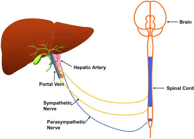
1. Liver Innervation
The liver is primarily innervated by the autonomic nervous system, which includes both sympathetic and parasympathetic fibers. The sympathetic innervation comes from the thoracic splanchnic nerves, specifically the greater splanchnic nerve (T5-T9), which synapses in the celiac ganglion. This sympathetic input generally inhibits liver functions such as bile secretion and glycogen synthesis. On the other hand, the parasympathetic innervation is provided by the vagus nerve (cranial nerve X). This stimulation promotes liver functions, including bile production and metabolic processes.
2. Gallbladder Innervation
The gallbladder receives its innervation mainly from the autonomic nervous system as well. The sympathetic fibers arise from the celiac plexus and are responsible for inhibiting gallbladder contraction. Conversely, parasympathetic fibers from the vagus nerve stimulate gallbladder contraction and bile release into the duodenum during digestion. Additionally, sensory fibers from the phrenic nerve can also provide some level of sensation to the gallbladder.
3. Pancreas Innervation
The pancreas is similarly innervated by both sympathetic and parasympathetic fibers. Sympathetic innervation comes from the thoracic splanchnic nerves (greater splanchnic nerve) that synapse in celiac ganglia. This sympathetic input tends to inhibit pancreatic secretions. In contrast, parasympathetic innervation is provided by the vagus nerve, which stimulates pancreatic enzyme secretion and bicarbonate production necessary for digestion in the small intestine.
4. Spleen Innervation
The spleen’s innervation is predominantly sympathetic, originating from T5-T9 spinal segments via splenic nerves that synapse in celiac ganglia. These sympathetic fibers help regulate blood flow through splenic arteries and can influence immune responses by modulating splenic activity. Unlike other digestive organs, there is no significant parasympathetic innervation to the spleen; thus, its functions are primarily controlled through sympathetic pathways.
In summary, all these associated digestive organs receive a combination of sympathetic and parasympathetic innervations that regulate their respective functions during digestion and metabolic processes.
Lymphatic Drainage and Regional Lymph Nodes of the Gastrointestinal Tract and Associated Abdominal Organs
The lymphatic system plays a crucial role in the immune response and fluid balance within the body. It consists of a network of lymph vessels, lymph nodes, and organs that help transport lymph, a fluid containing infection-fighting white blood cells, throughout the body. The gastrointestinal tract (GIT) and associated abdominal organs have specific patterns of lymphatic drainage that are essential for understanding their immunological functions and potential disease processes.
1. Overview of Lymphatic Drainage in the GIT
The lymphatic drainage of the GIT is primarily organized into two main trunks: the thoracic duct and the right lymphatic duct. The thoracic duct is responsible for draining lymph from most of the body, including all regions of the GIT except for parts of the right upper quadrant.
2. Major Regions of Lymphatic Drainage
- Esophagus:
- The esophagus drains its lymph primarily through cervical, mediastinal, and abdominal nodes.
- Cervical nodes include deep cervical nodes.
- Mediastinal nodes include tracheobronchial nodes.
- Abdominal drainage involves gastric nodes.
- Stomach:
- The stomach’s lymphatic drainage is divided into several regions:
- Cardiac region: Drains to left gastric nodes.
- Fundus: Drains to splenic nodes.
- Body: Drains to pyloric nodes.
- Pylorus: Drains to pancreaticoduodenal nodes.
- All these converge into celiac nodes.
- The stomach’s lymphatic drainage is divided into several regions:
- Small Intestine:
- The small intestine (duodenum, jejunum, ileum) has a complex drainage system:
- Duodenum drains into pancreaticoduodenal and superior mesenteric nodes.
- Jejunum drains into jejunal nodes which then drain into superior mesenteric nodes.
- Ileum drains into ileal nodes which also lead to superior mesenteric nodes.
- The small intestine (duodenum, jejunum, ileum) has a complex drainage system:
- Large Intestine:
- The large intestine’s drainage includes:
- Cecum and appendix drain into ileocecal nodes.
- Ascending colon drains into right colic nodes.
- Transverse colon drains into middle colic nodes.
- Descending colon drains into left colic nodes.
- Sigmoid colon drains into sigmoid nodes leading to inferior mesenteric nodes.
- The large intestine’s drainage includes:
- Rectum:
- The rectum has both superficial and deep lymphatics:
- Superficial rectal lymphatics drain to inguinal lymph nodes via perineal vessels.
- Deep rectal lymphatics drain to inferior mesenteric or internal iliac lymph nodes.
- The rectum has both superficial and deep lymphatics:
3. Associated Abdominal Organs
- Liver:
- Lymph from the liver primarily drains through hepatic lymphatics to celiac and hepatic hilar (porta hepatis) lymph nodes.
- Pancreas:
- Pancreatic drainage occurs through pancreaticoduodenal and splenic groups leading to celiac or superior mesenteric trunks.
- Spleen:
- The spleen’s drainage goes directly to splenic lymphatics which then connect with celiac trunk pathways.
- Kidneys:
- Renal lymphatics drain primarily through lateral aortic (lumbar) or renal hilar groups leading towards lumbar trunks.
4. Major Lymph Trunks
The major trunks involved in draining these regions include:
- Celiac trunk: Drains stomach, liver, pancreas, spleen, duodenum, and part of small intestine.
- Superior Mesenteric trunk: Drains most of small intestine and proximal large intestine (cecum through transverse colon).
- Inferior Mesenteric trunk: Drains distal large intestine (descending colon through rectum).
These trunks ultimately converge at larger ducts such as the thoracic duct which empties into the venous circulation at the junction between the left subclavian vein and internal jugular vein.
In summary, understanding the intricate network of regional lymphatic drainage in relation to various components of the GIT is vital for clinical assessments related to infections, cancers, or other pathological conditions affecting these organs.


