Clinical Manifestations of Chronic Lymphocytic Leukemia (CLL)
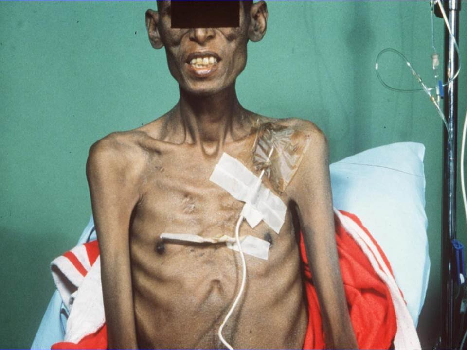
Chronic lymphocytic leukemia (CLL) often presents with a variety of clinical manifestations, which can vary significantly among individuals. Many patients may remain asymptomatic for extended periods, and the disease is frequently discovered incidentally during routine blood tests. When symptoms do occur, they can include:
- Lymphadenopathy: Swelling of lymph nodes is common, particularly in the neck, armpits, and groin.
- Splenomegaly: Enlargement of the spleen can occur, leading to discomfort or a feeling of fullness.
- Fatigue: Patients often report persistent fatigue that does not improve with rest.
- Weight Loss: Unintentional weight loss may be observed.
- Night Sweats: Excessive sweating during the night can be a symptom.
- Fever: Recurrent fevers without an apparent cause may occur.
- Infections: Increased susceptibility to infections due to compromised immune function.
These manifestations are primarily due to the accumulation of malignant B-lymphocytes that interfere with normal hematopoiesis and immune function.
Laboratory Findings in CLL
The diagnosis of CLL is typically confirmed through various laboratory tests:
- Complete Blood Count (CBC): This test often reveals lymphocytosis, characterized by elevated levels of lymphocytes (greater than 5,000 lymphocytes/mm³). Additionally, there may be low levels of red blood cells (anemia) and platelets (thrombocytopenia).
- Peripheral Blood Smear: A microscopic examination may show abnormal lymphocytes known as smudge cells, which are fragile and break apart during preparation.
- Flow Cytometry: This test identifies specific cell surface markers on leukemic cells. In CLL, flow cytometry typically shows a homogeneous population of B-lymphocytes expressing certain markers such as CD5, CD23, and weak expression of surface immunoglobulin.
- Beta-2-Microglobulin Levels: Elevated levels are associated with more advanced disease and poorer prognosis.
- Bone Marrow Biopsy: While not always necessary for diagnosis, it can provide additional information regarding the extent of infiltration by leukemic cells.
- Immunoglobulin Levels: Testing for immunoglobulin levels helps assess the patient’s ability to fight infections; low levels indicate compromised immunity.
Complications Associated with CLL
Patients with CLL face several potential complications stemming from both the disease itself and its treatment:
- Infections: Due to impaired immune function from both the disease process and potential treatments like chemotherapy or immunotherapy, patients are at increased risk for bacterial, viral, and fungal infections.
- Autoimmune Disorders: Some patients develop autoimmune hemolytic anemia or thrombocytopenia as a result of their immune system mistakenly attacking healthy blood cells.
- Transformation to More Aggressive Disease: A small percentage of patients may experience transformation from CLL to a more aggressive form known as Richter’s transformation or prolymphocytic leukemia (PLL), which has a poorer prognosis.
- Organ Dysfunction: As CLL progresses, it can lead to organ dysfunction due to infiltration by leukemic cells or complications from treatment-related toxicity affecting organs such as the liver or kidneys.
- Secondary Malignancies: There is an increased risk for developing other types of cancers in patients with CLL compared to the general population.
In summary, chronic lymphocytic leukemia presents with diverse clinical manifestations primarily related to lymphocyte proliferation and immune dysfunction; laboratory findings confirm diagnosis through characteristic blood counts and cellular markers; complications arise mainly from immunosuppression and disease progression.
Morphologic Characteristics of Chronic Lymphocytic Leukemia Cells
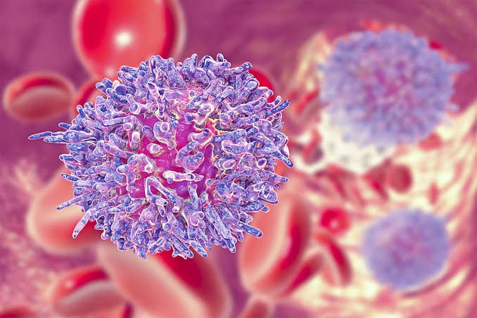
Chronic lymphocytic leukemia (CLL) is characterized by the accumulation of mature B lymphocytes in the blood, bone marrow, and lymphoid tissues. The morphology of CLL cells is typically distinctive, with most cases exhibiting a characteristic appearance. The circulating cells are usually small to medium-sized lymphocytes with a high nucleus-to-cytoplasm ratio. The nuclei are often round or slightly irregular, containing clumped chromatin that appears coarse and dense. In approximately 15% of patients, atypical morphology may be observed, which can manifest as:
- Increased Prolymphocytes: A significant presence (>10%) of prolymphocytes in the blood can indicate a variant known as CLL/PL.
- Lymphoplasmacytic and Cleaved Cells: An increased number (>15%) of these atypical cells can also be seen in some cases.
These morphological features correlate with clinical characteristics such as disease progression and survival rates.
Immunophenotypic Characteristics of Chronic Lymphocytic Leukemia Cells
The immunophenotype of CLL cells is defined by their expression of specific surface markers that help distinguish them from other B-cell malignancies. The typical immunophenotypic profile includes:
- CD5+: CLL cells express CD5, a marker not commonly found on normal B cells.
- CD23+: This marker is also expressed on CLL cells and helps differentiate them from other leukemias.
- FMC7-: Most CLL cases show negative expression for FMC7, which aids in diagnosis.
- Weak Surface Immunoglobulin (sIg): There is generally weak expression of sIg on CLL cells.
- Weak or Absent CD22 and CD79b Expression: These markers are associated with the B-cell receptor (BCR), and their low levels may indicate abnormal signal transduction similar to anergic B lymphocytes.
Because no single marker is specific for CLL, a composite phenotype using multiple markers is employed to improve diagnostic accuracy. Additionally, certain markers like FMC7 or CD38 have prognostic implications; their expression levels can indicate disease outcomes.
Overall, both morphological and immunophenotypic analyses are essential for diagnosing CLL and understanding its clinical behavior.
Clinical Manifestations and Laboratory Findings
1. Hairy Cell Leukemia
Clinical Manifestations: Hairy cell leukemia (HCL) is a rare type of chronic lymphoid leukemia characterized by the proliferation of abnormal B-lymphocytes. Patients often present with:
- Splenomegaly: This is one of the most common findings, and patients may experience abdominal discomfort or fullness.
- Cytopenias: Due to bone marrow infiltration, patients frequently exhibit anemia, thrombocytopenia (low platelet count), and neutropenia (low white blood cell count), leading to symptoms such as fatigue, increased bleeding tendency, and recurrent infections.
- Fatigue and Weakness: Generalized weakness is common due to anemia.
Laboratory Findings:
- Peripheral Blood Smear: The hallmark finding is the presence of “hairy” cells, which are abnormal lymphocytes with cytoplasmic projections.
- Bone Marrow Biopsy: Shows infiltrates of hairy cells, often with a characteristic “fried egg” appearance.
- Immunophenotyping: Hairy cells typically express CD19, CD20 (dim), CD22, and CD11c; they are usually negative for FMC-7.
2. Large Granular Lymphocyte Disorders
Clinical Manifestations: Large granular lymphocyte (LGL) disorders can be classified into two main types: LGL leukemia and LGL lymphoma. Common clinical features include:
- Cytopenias: Similar to HCL, patients may present with anemia and neutropenia.
- Recurrent Infections: Due to impaired immune function from low neutrophil counts.
- Splenomegaly or Lymphadenopathy: Some patients may have enlarged spleens or lymph nodes.
Laboratory Findings:
- Peripheral Blood Smear: Presence of large granular lymphocytes that are typically T-cells or NK-cells.
- Flow Cytometry: Identifies the clonal population of LGLs; they often express CD3 (T-cell markers) or CD56 (NK-cell markers).
- Bone Marrow Biopsy: May show increased numbers of large granular lymphocytes.
3. Mycosis Fungoides
Clinical Manifestations: Mycosis fungoides is a form of cutaneous T-cell lymphoma that primarily affects the skin. Its manifestations include:
- Skin Rash: Initially presents as patches or plaques that can be itchy; these lesions can evolve into tumors over time.
- Erythroderma: In advanced stages, widespread redness and scaling can occur.
- Lymphadenopathy: Patients may develop swollen lymph nodes as the disease progresses.
Laboratory Findings:
- Skin Biopsy: Histological examination reveals atypical T-cells in the epidermis (epidermotropism).
- Immunophenotyping: Tumor cells typically express CD4 and lack CD7 in many cases.
- T-cell Receptor Gene Rearrangement Studies: These tests help confirm clonality in suspected cases.
4. Adult T-cell Leukemia/Lymphoma
Clinical Manifestations: Adult T-cell leukemia/lymphoma (ATLL) is associated with human T-cell lymphotropic virus type I (HTLV-I). Clinical features include:
- Lymphadenopathy and Splenomegaly: Often prominent at diagnosis.
- Skin Lesions: Can appear as plaques or nodules in some subtypes.
- Hypercalcemia and Bone Lesions: Patients may experience elevated calcium levels due to osteolytic lesions.
Laboratory Findings:
- Peripheral Blood Smear: May show atypical lymphocytes with multilobed nuclei (“flower” cells).
- Flow Cytometry Analysis: Typically shows a predominance of CD4+ T-cells expressing activation markers like CD25.
- Serology for HTLV-I Antibodies: Positive serological tests confirm infection with HTLV-I.
Clinical Manifestations of Plasma Cell Tumors
Plasma cell tumors, which include multiple myeloma and plasmacytomas, can present with a variety of clinical manifestations. The symptoms often arise from the accumulation of abnormal plasma cells in the bone marrow and their effects on normal blood cell production and organ function.
- Bone Pain: One of the most common symptoms is bone pain, particularly in the back or ribs. This pain arises due to lesions formed by myeloma cells that weaken the bone structure.
- Fractures: Patients may experience fractures more easily due to weakened bones, a condition known as osteopenia or osteoporosis caused by myeloma.
- Anemia: As myeloma cells proliferate, they can inhibit the production of healthy red blood cells, leading to anemia. Symptoms include fatigue, weakness, and pallor.
- Infections: The immune system may be compromised due to reduced white blood cell production, making patients more susceptible to infections.
- Hypercalcemia: Myeloma can lead to elevated calcium levels in the blood (hypercalcemia), which can cause nausea, vomiting, confusion, and increased thirst.
- Kidney Dysfunction: The buildup of M protein (an abnormal antibody) can damage kidney tissues leading to renal impairment or failure.
- Neurological Symptoms: In some cases, peripheral neuropathy may occur due to amyloidosis or direct nerve compression from plasmacytomas.
- Amyloidosis: This condition occurs when abnormal proteins accumulate in organs and tissues, potentially leading to organ dysfunction such as heart failure or kidney failure.
Laboratory Findings
Diagnosis of plasma cell tumors typically involves several laboratory tests that help identify abnormalities associated with these conditions:
- Complete Blood Count (CBC): This test assesses levels of red blood cells, white blood cells, and platelets. Anemia is commonly detected through low red blood cell counts.
- Serum Protein Electrophoresis (SPEP): This test identifies the presence of M proteins in the serum and helps confirm a diagnosis of multiple myeloma by showing monoclonal gammopathy.
- Serum Immunofixation: This further characterizes the type of M protein present and confirms its monoclonality.
- Beta-2 Microglobulin Levels: Elevated levels are indicative of disease activity and prognosis; higher levels suggest more advanced disease.
- Electrolyte Levels: Monitoring electrolytes is crucial since plasma cell disorders can affect kidney function and lead to imbalances like hypercalcemia.
- Urine Tests for Bence Jones Proteins: These are free light chains that may be present in urine when there is significant myeloma activity.
Complications
The complications arising from plasma cell tumors can significantly impact patient health:
- Renal Failure: Due to high levels of M protein causing damage to renal tubules or secondary effects from hypercalcemia.
- Bone Complications: Osteolytic lesions can lead not only to pain but also increase fracture risk and spinal cord compression if vertebrae collapse.
- Infection Risk: Increased susceptibility due to impaired immune function leads to recurrent infections which can complicate treatment regimens.
- Amyloidosis Complications: Organ dysfunction resulting from amyloid deposits can severely affect quality of life and overall prognosis depending on which organs are involved (e.g., heart failure).
- Hyperviscosity Syndrome: High levels of M protein can thicken the blood leading to complications such as headaches, blurred vision, or even stroke-like symptoms due to reduced circulation efficiency.
In summary, plasma cell tumors manifest through various clinical signs primarily related to bone health, hematological status, infection risk, organ function impairment due to amyloidosis or hypercalcemia complications; laboratory findings support diagnosis through specific tests identifying abnormal proteins and cellular deficiencies; while complications pose significant risks that require careful management during treatment.
Overview of Bence Jones Proteins, Monoclonal Spike, M Proteins, Heavy Chain Disease and Waldenström’s Macroglobulinemia
1. Bence Jones Proteins
Bence Jones proteins are free light chains of immunoglobulins that are produced by abnormal plasma cells, typically found in conditions such as multiple myeloma and Waldenström macroglobulinemia (WM). These proteins can be detected in urine through a test known as urine immunofixation electrophoresis. The presence of Bence Jones proteins is significant because it indicates the overproduction of monoclonal antibodies, which can lead to various complications, including kidney damage and increased risk of infections. In the context of WM, Bence Jones proteins may also be present due to the underlying lymphoplasmacytic infiltration.
2. Monoclonal Spike
A monoclonal spike refers to an abnormal increase in a specific type of immunoglobulin (antibody) detected in serum protein electrophoresis. This spike indicates the presence of a monoclonal gammopathy, where a single clone of plasma cells produces an excess amount of one type of immunoglobulin. Monoclonal spikes are commonly associated with conditions like multiple myeloma, MGUS (monoclonal gammopathy of undetermined significance), and Waldenström macroglobulinemia. In WM, the monoclonal spike is typically associated with elevated levels of IgM.
3. M Proteins
M proteins are another term for monoclonal proteins or paraproteins produced by neoplastic plasma cells. These proteins can be identified in blood tests and are indicative of various hematological malignancies, including multiple myeloma and Waldenström macroglobulinemia. In WM specifically, M proteins are predominantly IgM types, which contribute to symptoms such as hyperviscosity syndrome due to their high molecular weight. Monitoring M protein levels is crucial for assessing disease progression and response to treatment.
4. Heavy Chain Disease
Heavy chain disease is a rare type of lymphoproliferative disorder characterized by the production of abnormal heavy chains without accompanying light chains. It is classified into different types based on the heavy chain involved (e.g., alpha, gamma). Heavy chain diseases can lead to various clinical manifestations depending on the type and extent of disease involvement. While not directly related to Waldenström macroglobulinemia, it shares some similarities with other B-cell disorders and may present with overlapping symptoms such as anemia or hyperviscosity.
5. Waldenström’s Macroglobulinemia
Waldenström’s macroglobulinemia (WM) is a low-grade lymphoproliferative disorder characterized by the proliferation of lymphoplasmacytic cells in the bone marrow and high levels of IgM monoclonal protein in the blood. Symptoms often include fatigue due to anemia, bleeding tendencies from hyperviscosity syndrome, peripheral neuropathy, and occasionally splenomegaly or lymphadenopathy. Diagnosis involves confirming the presence of IgM monoclonal protein along with characteristic bone marrow infiltration patterns. Treatment options include alkylating agents like chlorambucil, purine nucleoside analogs, rituximab therapy, interferon, corticosteroids, and emerging therapies like stem cell transplantation for younger patients.


