Functions of the Autonomic Nervous System (ANS)
The autonomic nervous system (ANS) is a critical component of the peripheral nervous system that regulates involuntary physiological functions, including heart rate, blood pressure, respiration, digestion, and sexual arousal. It operates largely below the level of consciousness and is divided into two main divisions: the sympathetic division and the parasympathetic division. Each division has distinct functions and utilizes different neurotransmitters to communicate with effector organs.
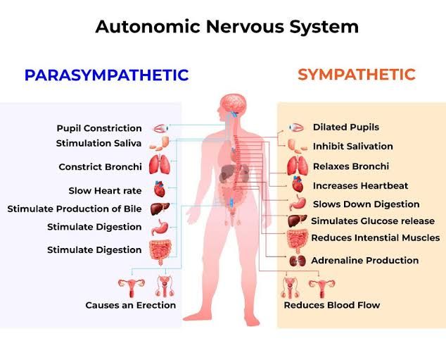
- Sympathetic Division: Often referred to as the “fight or flight” system, the sympathetic division prepares the body for stressful or emergency situations. When activated, it increases heart rate, dilates airways to improve oxygen intake, inhibits digestive processes, and mobilizes energy stores. This response is mediated primarily by the release of norepinephrine (noradrenaline) from postganglionic neurons and epinephrine (adrenaline) from the adrenal medulla into the bloodstream.
- Parasympathetic Division: Known as the “rest and digest” system, this division conserves energy and promotes maintenance activities when the body is at rest. It decreases heart rate, stimulates digestive processes, and enhances glandular activity. The primary neurotransmitter involved in parasympathetic responses is acetylcholine (ACh), which is released from postganglionic neurons to act on muscarinic receptors in target tissues.
Response of Effector Organs
- Heart: The sympathetic division increases heart rate and force of contraction through β1-adrenergic receptors activated by norepinephrine. In contrast, the parasympathetic division decreases heart rate via muscarinic receptors activated by acetylcholine.
- Lungs: Sympathetic activation causes bronchodilation through β2-adrenergic receptors while parasympathetic activation leads to bronchoconstriction via muscarinic receptors.
- Digestive System: The sympathetic response inhibits peristalsis and secretion in digestive organs while diverting blood flow away from these systems. Conversely, parasympathetic stimulation enhances digestive activity by increasing peristalsis and stimulating glandular secretions.
- Blood Vessels: Sympathetic stimulation generally causes vasoconstriction through α1-adrenergic receptors; however, certain blood vessels can experience vasodilation due to β2-adrenergic receptor activation during stress responses. The parasympathetic system has minimal direct effects on blood vessels but can influence them indirectly through other mechanisms.
Neurotransmitters Involved
The key neurotransmitters involved in ANS signaling include:
- Norepinephrine (NE): Released by most postganglionic sympathetic fibers; acts on adrenergic receptors.
- Epinephrine (E): Released into circulation from adrenal medulla during sympathetic activation; also acts on adrenergic receptors.
- Acetylcholine (ACh): Released by all preganglionic fibers (both divisions) and postganglionic fibers of the parasympathetic division; acts on nicotinic receptors at ganglia and muscarinic receptors at effector organs.
In summary, the autonomic nervous system plays a vital role in maintaining homeostasis through its two divisions—sympathetic and parasympathetic—each utilizing specific neurotransmitters to elicit appropriate physiological responses in various effector organs.
ANS as a Reflex-Based Control System
The ANS can be understood as a reflex-based control system due to its reliance on reflex arcs to initiate and regulate responses to internal and external stimuli.
General Features of Autonomic Neuronal Reflexes
- Components of Reflex Arcs:
- A typical autonomic reflex arc consists of several key components: a sensory receptor, an afferent pathway (sensory neuron), an integration center (often within the spinal cord or brainstem), an efferent pathway (motor neuron), and an effector organ (such as smooth muscle, cardiac muscle, or glands).
- For example, when blood pressure drops, baroreceptors in the carotid arteries detect this change and send signals via afferent neurons to the central nervous system. The CNS processes this information and activates efferent pathways that stimulate heart rate increase and vasoconstriction to restore blood pressure.
- Involuntary Responses:
- Unlike somatic reflexes that involve voluntary control over skeletal muscles, autonomic reflexes are involuntary. This means they occur automatically without conscious thought. For instance, when food enters the stomach, stretch receptors activate a reflex that stimulates digestive secretions and peristalsis.
- Dual Innervation:
- Many organs receive innervation from both branches of the ANS: the sympathetic and parasympathetic systems. This dual innervation allows for fine-tuned regulation of organ function. For example, during stress or danger, sympathetic activation increases heart rate and dilates airways; conversely, parasympathetic activation promotes rest-and-digest functions like slowing heart rate and stimulating digestion.
- Feedback Mechanisms:
- Autonomic reflexes often incorporate feedback mechanisms that help maintain homeostasis. For instance, thermoregulation involves sensors detecting body temperature changes; if the body overheats, autonomic responses such as sweating are triggered to cool it down.
- Integration with Higher Brain Centers:
- While many autonomic reflexes are mediated at the spinal cord or brainstem level, higher brain centers such as the hypothalamus play a significant role in modulating these responses based on emotional states or environmental conditions. This integration allows for complex behaviors like stress responses that involve both autonomic regulation and conscious awareness.
- Adaptability:
- The ANS exhibits plasticity; it can adapt its responses based on experience or changes in internal state over time. For example, chronic stress can lead to long-term alterations in autonomic function that may affect cardiovascular health.
In summary, the ANS functions as a sophisticated reflex-based control system characterized by involuntary responses mediated through complex neural circuits involving sensory input and motor output pathways. Its ability to maintain homeostasis through feedback mechanisms while integrating higher brain functions underscores its essential role in human physiology.
Autonomic Reflexes Integrated at the Level of Spinal Cord and Brain Stem
Autonomic reflexes are integrated at both the spinal cord and brain stem levels, allowing for rapid responses to internal stimuli without conscious thought.
1. Structure of Autonomic Reflex Arcs
Autonomic reflex arcs typically consist of a two-neuron pathway: the preganglionic neuron and the postganglionic neuron. The preganglionic neuron originates in either the lateral horn of the spinal cord or specific nuclei within the brain stem. Its axon extends to an autonomic ganglion where it synapses with the postganglionic neuron. The postganglionic neuron then projects to target tissues such as smooth muscle, cardiac muscle, or glands.
2. Integration at the Spinal Cord Level
At the spinal cord level, autonomic reflexes can be categorized into short reflexes and long reflexes:
- Short Reflexes: These occur entirely within the spinal cord without involving higher brain centers. For example, a visceral reflex like micturition (urination) can be initiated by stretch receptors in the bladder wall that send signals directly to spinal interneurons. This leads to coordinated contraction of bladder smooth muscle and relaxation of sphincters without conscious input.
- Long Reflexes: These involve pathways that extend beyond the spinal cord to include brain regions for more complex processing. For instance, baroreceptor reflexes that help regulate blood pressure involve sensory neurons from baroreceptors in carotid arteries sending signals to the medulla oblongata in the brain stem before eliciting responses through autonomic pathways.
3. Integration at the Brain Stem Level
The brain stem houses critical centers for autonomic control:
- Medulla Oblongata: This region contains vital centers for cardiovascular regulation (e.g., heart rate and blood vessel diameter) and respiratory control. It integrates sensory information from peripheral receptors (like baroreceptors) and modulates sympathetic or parasympathetic output accordingly.
- Pons: The pons also contributes to autonomic functions by regulating aspects of respiration alongside other cranial nerve nuclei involved in facial expressions and swallowing.
- Hypothalamus: Although not part of the brain stem per se, it plays a pivotal role in integrating autonomic functions with endocrine responses and emotional states. It influences autonomic activity through connections with both spinal cord circuits and brain stem nuclei.
4. Feedback Mechanisms
Autonomic reflexes often utilize feedback mechanisms to maintain homeostasis. For example, changes in blood pressure detected by baroreceptors lead to adjustments in heart rate via sympathetic or parasympathetic pathways mediated by both spinal cord circuits and brain stem centers.
5. Clinical Relevance
Understanding these integrative processes is essential for diagnosing conditions related to dysautonomia or other disorders affecting autonomic regulation. Interventions targeting these reflex pathways can help manage conditions such as hypertension or heart failure.
In summary, autonomic reflexes are integrated at both spinal cord and brain stem levels through distinct neural pathways that allow for rapid adjustments in bodily functions based on internal stimuli.
Central Regulation of Autonomic Output
Central regulation of autonomic output involves several key brain structures that integrate sensory information and coordinate appropriate autonomic responses.
Nucleus of the Solitary Tract (NST)
The nucleus of the solitary tract (NST) is a critical structure located in the medulla oblongata. It serves as a primary relay center for visceral sensory information from the body. The NST receives input from various cranial nerves that convey signals related to cardiovascular function, respiratory status, and gastrointestinal activity.
- Integration of Sensory Input: The NST integrates this sensory information and plays a pivotal role in reflexive autonomic responses. For instance, it can modulate heart rate through parasympathetic pathways by influencing the vagus nerve.
- Cardiovascular Control: The NST is particularly important in maintaining cardiovascular homeostasis. It processes baroreceptor signals that detect changes in blood pressure and adjusts sympathetic and parasympathetic outputs accordingly.
- Connection to Other Brain Regions: The NST has extensive connections with other brain regions involved in autonomic regulation, including the hypothalamus and limbic system, facilitating coordinated responses to internal and external stimuli.
Limbic System
The limbic system is a complex set of structures located deep within the brain that is primarily associated with emotion, behavior, motivation, long-term memory, and olfaction. Its role in autonomic function is significant due to its influence on emotional states which can affect physiological responses.
- Emotional Influence on Autonomic Responses: The limbic system can modulate autonomic output based on emotional states; for example, stress or fear can trigger sympathetic activation leading to increased heart rate and blood pressure.
- Connections with Hypothalamus: Structures within the limbic system such as the amygdala and hippocampus communicate with the hypothalamus to influence autonomic functions based on emotional context.
- Behavioral Responses: The limbic system also contributes to behavioral responses that have autonomic implications; for instance, feelings of hunger or satiety can influence digestive processes through autonomic pathways.
Hypothalamus
The hypothalamus is a small but crucial region located below the thalamus that plays a central role in homeostasis by regulating various bodily functions through its control over the ANS.
- Homeostatic Regulation: The hypothalamus integrates signals related to temperature regulation, thirst, hunger, sleep-wake cycles, and stress response. It acts as a command center for maintaining homeostasis by adjusting autonomic outputs accordingly.
- Endocrine Control: In addition to its direct effects on the ANS, the hypothalamus regulates hormonal outputs via connections with the pituitary gland which further influences bodily functions such as metabolism and stress response.
- Autonomic Pathways: The hypothalamus sends descending projections to various brainstem nuclei (including NST) that control sympathetic and parasympathetic outflows to different organs throughout the body.
In summary, central regulation of autonomic output involves an intricate interplay between several key brain structures—primarily the nucleus of the solitary tract for processing visceral sensory information; the limbic system for integrating emotional states; and the hypothalamus for maintaining homeostasis through both neural and hormonal pathways.
Major Functions of the Hypothalamus
The hypothalamus is a small but crucial part of the brain located below the thalamus and above the brainstem. It plays a vital role in maintaining homeostasis by regulating various physiological processes. Here are the major functions of the hypothalamus:
1. Body Rhythm Regulation
The hypothalamus is instrumental in regulating circadian rhythms, which are physical, mental, and behavioral changes that follow a daily cycle. These rhythms respond primarily to light and darkness in the environment. The suprachiasmatic nucleus (SCN), a group of neurons within the hypothalamus, acts as the body’s master clock. It receives direct input from retinal cells that detect light, allowing it to synchronize bodily functions with day-night cycles.
Circadian rhythms influence sleep-wake cycles, hormone release, eating habits, and other bodily functions. Disruptions to these rhythms can lead to sleep disorders, metabolic issues, and other health problems.
2. Temperature Regulation
The hypothalamus plays a critical role in thermoregulation—the process of maintaining an optimal body temperature. It contains specialized neurons that act as thermoreceptors, detecting changes in blood temperature and external environmental conditions.
When body temperature rises above normal (around 37°C or 98.6°F), the hypothalamus triggers mechanisms such as sweating and increased blood flow to the skin to dissipate heat. Conversely, if body temperature drops too low, it initiates responses like shivering and constricting blood vessels to conserve heat. This feedback loop helps maintain homeostasis despite external temperature fluctuations.
3. Appetite Control
The hypothalamus regulates appetite through various neuropeptides and hormones that signal hunger or satiety. Two key areas within the hypothalamus involved in appetite regulation are:
- Lateral Hypothalamus (LH): This area stimulates hunger when activated by neuropeptides like orexin.
- Ventromedial Hypothalamus (VMH): This area signals satiety when activated by hormones such as leptin.
These regions work together with peripheral signals from hormones like ghrelin (which stimulates hunger) and insulin (which promotes satiety) to regulate food intake effectively.
4. Water Intake Regulation
The hypothalamus also plays a vital role in osmoregulation—maintaining fluid balance within the body. Osmoreceptors located in the hypothalamus detect changes in blood osmolarity (the concentration of solutes). When osmolarity increases (indicating dehydration), these receptors stimulate thirst and promote water-seeking behavior.
Additionally, the hypothalamus controls the release of antidiuretic hormone (ADH) from the posterior pituitary gland. ADH helps regulate water retention by increasing water reabsorption in the kidneys, thus reducing urine output and conserving body fluids during times of dehydration.
In summary, the major functions of the hypothalamus include regulating body rhythms through circadian clocks, maintaining temperature homeostasis via thermoregulation mechanisms, controlling appetite through complex hormonal signaling pathways, and managing water intake through osmoregulation processes.


