Gracile and Cuneate Tracts: Pathways for Conscious Proprioception, Touch, Pressure, and Vibration
Introduction to Gracile and Cuneate Tracts
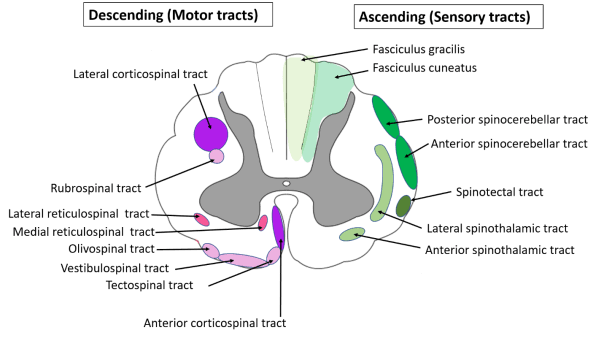
The gracile and cuneate tracts are essential pathways in the central nervous system that carry sensory information related to conscious proprioception, touch, pressure, and vibration from the limbs and trunk to the brain. These tracts are part of the dorsal column-medial lemniscus pathway, which is crucial for transmitting fine touch and proprioceptive information.
Gracile Tract
The gracile tract primarily transmits sensory information from the lower body (below T6 level). It originates from the primary afferent neurons located in the dorsal root ganglia. The pathway can be described in several steps:
- Afferent Fiber Entry: Sensory fibers enter the spinal cord through the dorsal roots.
- Dorsal Column Ascension: These fibers ascend ipsilaterally (on the same side) in the dorsal column of the spinal cord as part of the gracile fasciculus.
- Gracile Nucleus: Upon reaching the medulla oblongata, these fibers synapse in the gracile nucleus.
- Decussation: The second-order neurons then cross over (decussate) at the medulla’s junction to form the medial lemniscus.
- Thalamic Relay: The medial lemniscus ascends to synapse in the ventral posterolateral nucleus (VPL) of the thalamus.
- Cortical Projection: Finally, third-order neurons project from VPL to specific areas of the primary somatosensory cortex (S1), where conscious perception occurs.
The gracile tract is responsible for conveying sensations such as fine touch, pressure, vibration sense, and proprioceptive feedback from lower extremities and lower trunk regions.
Cuneate Tract
In contrast, the cuneate tract carries sensory information from the upper body (above T6 level). Its pathway follows a similar structure:
- Afferent Fiber Entry: Sensory fibers from upper body regions also enter through dorsal roots.
- Dorsal Column Ascension: These fibers ascend ipsilaterally but travel within a different fasciculus known as the cuneate fasciculus.
- Cuneate Nucleus: In the medulla oblongata, these fibers synapse in the cuneate nucleus.
- Decussation: Like those of the gracile tract, second-order neurons decussate at this level to form part of the medial lemniscus.
- Thalamic Relay: The medial lemniscus continues its ascent to synapse in VPL of thalamus.
- Cortical Projection: Third-order neurons project from VPL to S1.
The cuneate tract similarly conveys sensations such as fine touch, pressure, vibration sense, and proprioceptive feedback but specifically from upper extremities and upper trunk regions.
Functional Importance
Both tracts play a critical role in our ability to perceive tactile stimuli accurately and maintain balance through proprioception. Damage or lesions along these pathways can lead to deficits such as loss of fine touch discrimination or impaired proprioception on one side of the body.
In summary, both gracile and cuneate tracts are vital components of our sensory system that ensure we can consciously perceive various forms of tactile stimuli across different parts of our body.
Dorsal and Ventral Spinocerebellar Tracts and Pathways for Unconscious Proprioception from the Limbs and Trunk
The spinocerebellar tracts are essential pathways in the nervous system that convey unconscious proprioceptive information from the body to the cerebellum, which plays a critical role in coordinating movement and maintaining balance. There are two primary spinocerebellar tracts: the dorsal spinocerebellar tract and the ventral spinocerebellar tract. Each of these tracts has distinct pathways and functions.
Dorsal Spinocerebellar Tract
The dorsal spinocerebellar tract (DSCT) is responsible for transmitting proprioceptive information from the lower limbs and trunk to the cerebellum. It carries signals from muscle spindles and Golgi tendon organs, which are sensory receptors located in muscles and tendons, respectively.
- Origin: The DSCT originates from first-order neurons whose cell bodies reside in the dorsal root ganglia. These neurons receive input from peripheral proprioceptors.
- Pathway: The central processes of these first-order neurons enter the spinal cord through the dorsal roots and synapse with second-order neurons located in Clarke’s nucleus (located between C8 to L2/L3).
- Ascension: The axons of these second-order neurons then ascend ipsilaterally (on the same side) through the lateral funiculus of the spinal cord.
- Brainstem Passage: Upon reaching the medulla oblongata, these fibers pass through the inferior cerebellar peduncle to enter the cerebellum.
- Termination: The DSCT terminates in specific regions of the cerebellum where it contributes to processing unconscious proprioceptive information related to limb position and movement.
This tract does not decussate (cross over) at any point along its pathway, meaning that it transmits information from one side of the body directly to the same side of the cerebellum.
Ventral Spinocerebellar Tract
The ventral spinocerebellar tract (VSCT) also conveys proprioceptive information but differs significantly in its pathway compared to the DSCT.
- Origin: Like DSCT, VSCT begins with first-order neurons whose cell bodies are located in dorsal root ganglia, receiving input from Golgi tendon organs as well as other proprioceptors.
- Pathway: After entering through dorsal roots, these first-order neurons synapse with second-order neurons primarily located in laminae VII of the spinal cord.
- Decussation: A key feature of VSCT is that it crosses over (decussates) at this level within the anterior white commissure before ascending through the spinal cord.
- Ascension: The axons then ascend contralaterally (to opposite sides) through both lateral funiculi before reaching higher centers.
- Brainstem Passage: As they approach their termination point, they enter via superior cerebellar peduncle into the cerebellum.
- Double Cross Mechanism: Interestingly, after entering into cerebellum, there is a “double cross” where fibers may cross back again within certain regions of cerebellum before terminating there.
The VSCT provides important feedback about motor commands generated by central nervous system structures regarding limb movements, thus playing a role not only in proprioception but also in motor coordination.
In summary, both tracts work together to ensure that unconscious proprioceptive information is accurately relayed to the cerebellum for effective motor control:
- The dorsal spinocerebellar tract carries information ipsilaterally without crossing over.
- The ventral spinocerebellar tract, on the other hand, involves a double crossing mechanism allowing for complex integration of motor commands with sensory feedback.
Lateral Spinothalamic Tract and Pathways for Pain and Temperature from the Limbs and Trunk
Overview of the Lateral Spinothalamic Tract
The lateral spinothalamic tract is a critical component of the anterolateral system, which is responsible for transmitting sensory information related to pain and temperature from the body (limbs and trunk) to the brain. This tract specifically carries nociceptive (pain) and thermoreceptive (temperature) signals, allowing the central nervous system to process these sensations.
Anatomy of the Lateral Spinothalamic Tract
- First-Order Neurons: The pathway begins with first-order neurons that are located in the dorsal root ganglia. These neurons are specialized nociceptive receptors that respond to painful stimuli (mechanical, thermal, or chemical). They have peripheral processes that extend to the skin and deeper tissues, where they detect noxious stimuli.
- Second-Order Neurons: Once activated, first-order neurons transmit their signals into the spinal cord through their central processes. They synapse with second-order neurons in the posterior grey horn of the spinal cord. The cell bodies of these second-order neurons reside in lamina I, II (substantia gelatinosa), or V of the spinal cord.
- Decussation: After synapsing with first-order neurons, second-order neurons immediately decussate (cross over) at their respective segmental level in the spinal cord via the ventral commissure. This crossing over is crucial as it allows pain and temperature sensations from one side of the body to be processed by the opposite side of the brain.
- Ascending Pathway: Following decussation, these second-order neurons ascend through the lateral spinothalamic tract located anteriolaterally within the spinal cord. As they ascend, they travel through various segments of the spinal cord towards higher centers in the brain.
- Third-Order Neurons: The lateral spinothalamic tract ultimately projects to third-order neurons located in the ventral posterolateral nucleus (VPL) of the thalamus after passing through several relay stations along its path.
- Cortical Projection: Finally, third-order neurons send their axons from the thalamus to specific areas of the primary somatosensory cortex (Brodmann areas 3, 1, and 2), where pain and temperature sensations are consciously perceived and interpreted.
Functionality
The lateral spinothalamic tract is primarily responsible for transmitting two key types of sensory information:
- Pain Sensation: This includes sharp pain or acute discomfort that can arise from various stimuli such as cuts or burns.
- Temperature Sensation: This involves both hot and cold sensations detected by specialized thermoreceptors.
Both types of information are essential for protective reflexes and overall bodily awareness regarding harmful stimuli.
Clinical Relevance
Damage or lesions affecting any part of this pathway can lead to significant clinical symptoms:
- Contralateral Loss of Pain and Temperature Sensation: If there is damage to one side of this tract after it has crossed over in the spinal cord, individuals may experience a loss of pain and temperature sensation on the opposite side of their body below that level.
- Conditions Affecting Pathway Integrity: Conditions such as syringomyelia or multiple sclerosis can compromise this pathway leading to altered sensory perception.
In summary, understanding how pain and temperature pathways operate through structures like the lateral spinothalamic tract is vital for diagnosing neurological conditions that affect sensory processing.
Ventral Spinothalamic Tract and Pathways for Simple Touch from the Limbs and Trunk
Overview of the Ventral Spinothalamic Tract
The ventral spinothalamic tract is a component of the anterolateral system, which is primarily responsible for transmitting sensory information related to crude touch and pressure. It works in conjunction with the lateral spinothalamic tract, which carries pain and temperature sensations. The ventral spinothalamic tract specifically conveys information about simple touch from the limbs and trunk to higher brain centers.
Anatomy and Pathway of the Ventral Spinothalamic Tract
- First-Order Neurons: The pathway begins with first-order neurons that are located in the dorsal root ganglia. These neurons receive sensory input from mechanoreceptors in the skin, which respond to light touch and pressure. The cell bodies of these neurons reside outside the spinal cord, while their peripheral processes extend into the skin.
- Synapse in the Spinal Cord: Upon entering the spinal cord through the dorsal roots, first-order neurons synapse onto second-order neurons located in the posterior grey horn of the spinal cord. This synaptic connection occurs at or near the level where they enter.
- Decussation: After synapsing, second-order neurons immediately decussate (cross over) to the opposite side of the spinal cord at their segmental level. This crossing occurs within a few segments of entry into the spinal cord.
- Ascent to Thalamus: Following decussation, these second-order neurons ascend through the ventral spinothalamic tract on their way to higher brain regions. They travel upward through various levels of the spinal cord and brainstem until they reach the thalamus.
- Third-Order Neurons: In the thalamus, specifically within the ventral posterior nucleus (VPN), second-order neurons synapse with third-order neurons. These third-order neurons then project to specific areas of the primary somatosensory cortex (Brodmann areas 3, 1, and 2).
- Somatotopic Organization: The organization of this pathway is somatotopically arranged, meaning that different parts of the body are represented in specific regions of both thalamus and cortex. For example, areas corresponding to more sensitive parts like fingers or lips occupy larger cortical representations compared to less sensitive areas like legs.
Functionality Related to Simple Touch
The ventral spinothalamic tract plays a crucial role in our ability to perceive simple touch sensations from various parts of our body including limbs and trunk:
- Crude Touch Sensation: The tract is responsible for transmitting crude touch sensations—these are non-discriminative tactile stimuli that do not provide detailed information about texture or shape but indicate that contact has occurred.
- Pressure Sensation: It also conveys pressure sensations that can be felt when an object presses against or deforms skin tissue.
This pathway allows individuals to detect basic tactile stimuli essential for interactions with their environment without requiring fine discrimination capabilities provided by other pathways such as those involved in proprioception or fine touch (which are transmitted via different pathways like dorsal column-medial lemniscal system).
In summary, the ventral spinothalamic tract is integral for transmitting simple touch sensations from limbs and trunk by relaying signals from peripheral mechanoreceptors through a series of first-, second-, and third-order neurons ultimately reaching specific areas in the primary somatosensory cortex where these sensations are processed and perceived.


