Pathways of Protein Turnover
Protein turnover is a critical biological process that involves the continuous synthesis and degradation of proteins within cells. This balance is essential for maintaining cellular homeostasis, responding to environmental changes, and regulating various physiological functions. The pathways involved in protein turnover can be broadly categorized into two main processes: protein synthesis and protein degradation.
1. Protein Synthesis Pathways
Protein synthesis is primarily regulated by several signaling pathways that promote the translation of mRNA into proteins. Key pathways include:
- Insulin/IGF-1 Signaling Pathway: This pathway plays a significant role in promoting growth and metabolism. Insulin and IGF-1 activate downstream signaling cascades that enhance protein synthesis by stimulating ribosomal biogenesis and increasing the translation efficiency of mRNAs.
- TOR (Target Of Rapamycin) Pathway: TOR is a central regulator of cell growth and metabolism in response to nutrient availability. When nutrients are abundant, TOR signaling promotes protein synthesis by activating key translational machinery components, such as S6 kinase and eIF4E-binding proteins.
- RTK/Ras/MAPK Pathway: Receptor Tyrosine Kinases (RTKs) activate Ras, which in turn activates the MAPK cascade. This pathway influences gene expression related to cell growth and differentiation, thereby enhancing protein synthesis.
- TGF-beta Signaling Pathway: Transforming Growth Factor-beta (TGF-beta) can also influence protein synthesis through its effects on cell proliferation and differentiation.
2. Protein Degradation Pathways
The degradation of proteins is equally important for maintaining cellular health by removing damaged or misfolded proteins. The primary mechanisms for protein degradation include:
- Ubiquitin-Proteasome System (UPS): This system tags unwanted proteins with ubiquitin molecules, marking them for degradation by the proteasome. The UPS is crucial for regulating various cellular processes, including the cell cycle, DNA repair, and responses to stress.
- Lysosomal Autophagy: Autophagy is a catabolic process where cells degrade their own components through lysosomes. It plays a vital role in recycling cellular materials during starvation or stress conditions. There are different types of autophagy, including macroautophagy, microautophagy, and chaperone-mediated autophagy.
3. Interplay Between Synthesis and Degradation
An important aspect of protein turnover is the regulatory relationship between protein synthesis and degradation pathways. Under certain conditions, increased protein synthesis can lead to an accumulation of damaged proteins if not balanced by adequate degradation mechanisms. For instance:
- Nutrient Availability: In nutrient-rich environments, enhanced protein synthesis may occur; however, if this increase outpaces degradation processes like autophagy or the UPS, it can result in cellular stress due to the accumulation of nonfunctional proteins.
- Genetic Interventions: Studies have shown that reducing overall protein synthesis through genetic modifications or dietary restrictions can extend lifespan in various organisms by decreasing the load on degradation systems while enhancing their efficiency.
In summary, effective regulation of both protein synthesis and degradation pathways is essential for maintaining cellular function and longevity. Disruptions in these processes can contribute to aging and age-related diseases due to the accumulation of damaged proteins.
Four Levels of Protein Structure
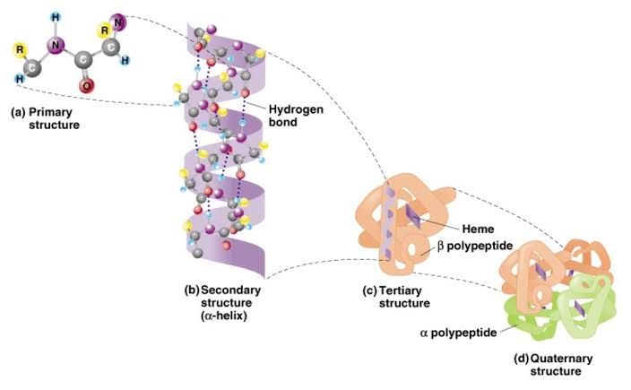
Proteins are complex molecules that play critical roles in biological systems. Their structure is organized into four distinct levels: primary, secondary, tertiary, and quaternary. Each level of structure is essential for the protein’s function and is determined by the sequence of amino acids.
1. Primary Structure
The primary structure of a protein refers to its unique sequence of amino acids. Amino acids are linked together by peptide bonds to form a polypeptide chain. The specific order of these amino acids is dictated by the genetic code within an organism’s DNA. This sequence determines how the protein will fold and ultimately its function. Any change in this sequence, even a single amino acid substitution, can lead to significant changes in the protein’s properties and functions.
2. Secondary Structure
The secondary structure involves local folding patterns within the polypeptide chain, primarily stabilized by hydrogen bonds between the backbone atoms in the polypeptide chain. The most common types of secondary structures are alpha helices and beta sheets:
- Alpha Helices: These are coiled structures where each turn of the helix is stabilized by hydrogen bonds between every fourth amino acid.
- Beta Sheets: These consist of strands lying alongside each other, connected through hydrogen bonds forming a sheet-like structure.
These secondary structures contribute to the overall stability and shape of the protein but do not represent its complete three-dimensional configuration.
3. Tertiary Structure
The tertiary structure refers to the overall three-dimensional shape formed by the entire polypeptide chain as it folds upon itself. This folding is driven by various interactions among side chains (R groups) of amino acids, including:
- Hydrophobic interactions: Nonpolar side chains tend to cluster away from water.
- Ionic bonds: Attractions between positively and negatively charged side chains.
- Hydrogen bonds: Interactions between polar side chains.
- Disulfide bridges: Covalent bonds that can form between cysteine residues.
The tertiary structure is crucial because it determines how a protein interacts with other molecules and performs its biological functions.
4. Quaternary Structure
Some proteins consist of more than one polypeptide chain, which come together to form a functional unit known as a multimeric protein. The quaternary structure describes how these individual polypeptides (subunits) interact and assemble into a larger complex. The interactions that stabilize quaternary structures are similar to those found in tertiary structures (hydrophobic interactions, ionic bonds, hydrogen bonds). Hemoglobin is a classic example; it consists of four subunits that work together to transport oxygen in the blood.
How Amino Acid Sequence Leads to 3D Configuration
The final three-dimensional configuration of a protein is largely determined by its amino acid sequence due to several factors:
- Chemical Properties: Each amino acid has distinct chemical properties based on its side chain (R group), influencing how it interacts with neighboring residues and with water.
- Folding Pathways: As a polypeptide chain emerges from ribosomes during translation, it begins folding into secondary structures almost immediately due to local interactions among nearby amino acids.
- Energetics: Proteins tend to fold into conformations that minimize their free energy state; this means they adopt shapes that are thermodynamically favorable under physiological conditions.
- Chaperones: Molecular chaperones assist in proper folding and prevent misfolding or aggregation during synthesis or stress conditions.
In summary, while the primary structure provides the blueprint for folding, it is through intricate interactions at all levels—from primary through quaternary—that proteins achieve their functional forms.
Protein Folding and Misfolding
Introduction to Protein Folding
Proteins are essential macromolecules that perform a vast array of functions in biological systems. For proteins to function correctly, they must fold into specific three-dimensional structures. This process is known as protein folding, which is guided by the sequence of amino acids that make up the protein. The correct folding of proteins is crucial because their functionality is directly linked to their structure.
The folding process can be understood through the concept of an “energetic funnel,” where proteins navigate through a vast conformational space to reach a stable, low-energy state. Most proteins can achieve their native conformation spontaneously due to the inherent properties of their amino acid sequences, which dictate how they interact with each other and their environment.
Causes of Protein Misfolding
Despite the robust mechanisms that guide proper protein folding, errors can occur, leading to misfolded proteins. Several factors contribute to this phenomenon:
- Genetic Mutations: Changes in the DNA sequence can lead to alterations in the amino acid sequence of proteins, resulting in instability or incorrect folding.
- Environmental Factors: Conditions such as temperature fluctuations, pH changes, and oxidative stress can disrupt normal folding processes.
- Cellular Crowding: The intracellular environment is highly crowded with various macromolecules, which can hinder proper protein folding and increase the likelihood of misfolding.
- Aging: As organisms age, the efficiency of cellular machinery responsible for protein folding (such as chaperones) may decline, increasing the risk of misfolding.
Clinical Implications of Protein Misfolding
Protein misfolding has significant clinical implications and is associated with various degenerative diseases known as protein-misfolding disorders. Some notable examples include:
- Alzheimer’s Disease: Characterized by the accumulation of amyloid-beta plaques and tau tangles in the brain, both resulting from misfolded proteins that disrupt neuronal function.
- Parkinson’s Disease: Involves the aggregation of alpha-synuclein into Lewy bodies within neurons, leading to neurodegeneration and motor dysfunction.
- Huntington’s Disease: Caused by mutations in the huntingtin gene that result in abnormal protein aggregates disrupting cellular processes.
- Type 2 Diabetes: Misfolded insulin or other related proteins can lead to amyloid formation in pancreatic cells, impairing insulin secretion and glucose metabolism.
- Cystic Fibrosis: A genetic disorder caused by mutations in the CFTR gene leading to improper folding and degradation of cystic fibrosis transmembrane conductance regulator protein.
The accumulation of misfolded proteins not only leads to cell death but also triggers inflammatory responses that exacerbate tissue damage. Understanding these mechanisms has opened avenues for therapeutic interventions aimed at enhancing proper protein folding or clearing misfolded proteins from cells.
Conclusion
In summary, protein folding is a critical biological process influenced by genetic information and environmental conditions. Misfolding can lead to severe clinical consequences manifesting as various degenerative diseases. Ongoing research aims to unravel these complex pathways further and develop effective treatments targeting these fundamental issues at a molecular level.
Analysis of Zwitterions and Their Clinical Applications
Definition of Zwitterions
A zwitterion is a molecule that contains both positive and negative electrical charges, resulting in an overall neutral charge. The term originates from the German word “zwitter,” meaning hybrid or hermaphrodite. This unique structure allows zwitterions to exist in various forms depending on the pH of their environment. For example, amino acids, which are the building blocks of proteins, commonly exist as zwitterions in their solid state due to the presence of both an amino group (-NH3+) that carries a positive charge and a carboxyl group (COO-) that carries a negative charge.
Characteristics of Zwitterions
Zwitterions possess several key characteristics:
- Dual Charge: They contain both positively and negatively charged regions within the same molecule.
- Stability: The charges are held together by covalent bonds, leading to stable structures.
- Isoelectric Point (pI): Each zwitterion has an isoelectric point, which is the pH at which it has no net charge. The pI can be calculated using the equilibrium constants of the acidic and basic groups present in the molecule.
The behavior of zwitterions is highly dependent on their surrounding environment’s pH. At low pH levels, they tend to gain protons and become cations, while at high pH levels, they can lose protons and become anions.
Clinical Applications of Zwitterions
Zwitterions have significant clinical applications across various fields:
- Drug Delivery Systems: Due to their amphoteric nature (ability to act as both acids and bases), zwitterionic compounds can enhance drug solubility and stability in biological environments. This property is particularly useful for formulating medications that require precise delivery mechanisms.
- Biomedical Implants: Zwitterionic materials are used in medical implants because they can reduce protein adsorption and biofilm formation on surfaces. This antifouling property helps prevent complications associated with infections when implants are introduced into the body.
- Separation Techniques: In molecular biology, zwitterionic compounds play a crucial role in techniques such as SDS-PAGE (sodium dodecyl sulfate-polyacrylamide gel electrophoresis). This method separates proteins based on their size and charge, facilitating research in protein analysis and purification.
- Sensors: Zwitterionic polymers are utilized in biosensors for detecting biomolecules due to their biocompatibility and ability to maintain stable interactions with biological fluids.
- Marine Applications: In marine industries, zwitterionic coatings are applied to boats and piers to prevent biofouling by aquatic organisms, thereby enhancing longevity and performance.
In summary, zwitterions exhibit unique properties that make them valuable in various clinical applications ranging from drug delivery systems to biomedical devices and separation techniques.


