Enzymatic Defects in the Heme Biosynthesis Pathway Leading to Porphyrias
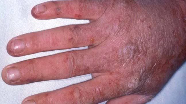
Porphyrias are a group of metabolic disorders caused by defects in the enzymes involved in the heme biosynthesis pathway. Each type of porphyria is associated with a specific enzymatic defect, leading to the accumulation of porphyrin precursors and resulting in various clinical manifestations. Below is a detailed explanation of the enzymatic defects that lead to different forms of porphyria:
- Congenital Erythropoietic Porphyria (CEP):
- Enzyme Defect: Uroporphyrinogen III synthase (UROS).
- Mechanism: This enzyme catalyzes the conversion of hydroxymethylbilane to uroporphyrinogen III. A deficiency leads to the accumulation of uroporphyrin I and other precursors, causing severe photosensitivity and hemolytic anemia.
- Erythropoietic Protoporphyria (EPP):
- Enzyme Defect: Ferrochelatase (FECH).
- Mechanism: Ferrochelatase catalyzes the insertion of ferrous iron into protoporphyrin IX to form heme. A deficiency results in the accumulation of protoporphyrin IX, which causes skin sensitivity to sunlight.
- Acute Intermittent Porphyria (AIP):
- Enzyme Defect: Porphobilinogen deaminase (PBGD), also known as hydroxymethylbilane synthase.
- Mechanism: This enzyme converts porphobilinogen into hydroxymethylbilane. A deficiency leads to an accumulation of porphobilinogen and δ-aminolevulinic acid (ALA), resulting in acute neurovisceral attacks characterized by abdominal pain, neurological symptoms, and psychiatric disturbances.
- Hereditary Coproporphyria (HCP):
- Enzyme Defect: Coproporphyrinogen oxidase (CPOX).
- Mechanism: CPOX catalyzes the conversion of coproporphyrinogen III to protoporphyrinogen IX. A deficiency leads to an accumulation of coproporphyrinogen III and can cause both neurovisceral symptoms and cutaneous manifestations.
- Variegate Porphyria (VP):
- Enzyme Defect: Protoporphyrinogen oxidase (PPOX).
- Mechanism: This enzyme converts protoporphyrinogen IX into protoporphyrin IX. A deficiency results in the accumulation of protoporphyrinogen IX, leading to both acute neurovisceral symptoms and cutaneous photosensitivity.
- Porphyria Cutanea Tarda (PCT):
- Enzyme Defect: Uroporphyrinogen decarboxylase (UROD).
- Mechanism: UROD catalyzes the decarboxylation of uroporphyrinogen III to coproporphyrinogen III. In PCT, reduced activity can be due to genetic factors or environmental influences such as alcohol consumption or exposure to certain chemicals, leading to an accumulation of uroporphyrins primarily in urine.
- ALA Dehydratase Deficiency Porphyria (ADP):
- Enzyme Defect: ALA dehydratase.
- Mechanism: This enzyme catalyzes the condensation of two molecules of δ-aminolevulinic acid into porphobilinogen. Its deficiency leads to increased levels of ALA and is associated with neurological symptoms but not typically with photosensitivity.
These enzymatic defects result in varying clinical presentations depending on which intermediates accumulate and where they are primarily deposited within the body.
Introduction to Jaundice
Jaundice is characterized by the yellow discoloration of the skin and sclera (the white part of the eyes) due to elevated levels of bilirubin in the blood, a condition known as hyperbilirubinemia. This yellowing typically becomes noticeable when serum bilirubin levels exceed approximately 50 µmol/L. The underlying cause of jaundice can be attributed to various disruptions in the normal metabolism and excretion of bilirubin.
Bilirubin Metabolic Pathway
Bilirubin is a product formed from the breakdown of heme, which is found in hemoglobin within red blood cells. The metabolic pathway for bilirubin involves several key steps:
- Heme Catabolism: When red blood cells reach the end of their lifespan (approximately 120 days), they are phagocytosed by macrophages, primarily in the spleen and liver. Heme is released from hemoglobin and converted into biliverdin through the action of heme oxygenase.
- Conversion to Bilirubin: Biliverdin is then reduced to unconjugated bilirubin (also known as indirect bilirubin) by biliverdin reductase. Unconjugated bilirubin is lipid-soluble and cannot be excreted directly into urine.
- Transport to Liver: Unconjugated bilirubin binds to albumin in the bloodstream for transport to the liver.
- Conjugation in Liver: In hepatocytes (liver cells), unconjugated bilirubin undergoes conjugation with glucuronic acid via the enzyme UDP-glucuronosyltransferase, resulting in conjugated bilirubin (direct bilirubin), which is water-soluble.
- Excretion into Bile: Conjugated bilirubin is secreted into bile and stored in the gallbladder or directly released into the intestine, where it contributes to stool color through its metabolic breakdown products, urobilinogen and stercobilin.
- Reabsorption and Excretion: Approximately 10% of urobilinogen can be reabsorbed back into circulation and eventually excreted by the kidneys.
Defects in Bilirubin Metabolism
Defects in any part of this metabolic pathway can lead to different types of jaundice:
- Pre-Hepatic Jaundice: This occurs due to excessive breakdown of red blood cells (hemolysis), overwhelming the liver’s capacity to conjugate bilirubin. As a result, there is an increase in unconjugated (indirect) bilirubin levels while conjugated levels remain normal.
- Causes: Hemolytic anemia, sickle cell disease, thalassemia, etc.
- Hepatocellular Jaundice: This type arises from liver cell dysfunction where hepatocytes lose their ability to conjugate bilirubin effectively. Both unconjugated and conjugated forms may be present due to liver damage or disease processes affecting hepatic function.
- Causes: Viral hepatitis, alcoholic liver disease, cirrhosis, etc.
- Post-Hepatic Jaundice: Also known as obstructive jaundice, this occurs when there is an obstruction in bile flow after it has been conjugated by the liver. This leads to an accumulation of conjugated (direct) bilirubin in the bloodstream since it cannot be excreted into bile or intestines.
- Causes: Gallstones, tumors compressing bile ducts, strictures, etc.
In addition to these types of jaundice caused by defects along this pathway, hereditary conditions such as Gilbert’s syndrome or Crigler-Najjar syndrome can also lead to abnormal elevations in serum bilirubin levels due to genetic defects affecting enzymes involved in its metabolism.
Understanding these pathways and defects provides critical insights into diagnosing and managing jaundice effectively based on its underlying causes.
Understanding Bilirubin Glucuronyl Transferase Enzyme and Jaundice in Newborns
Bilirubin is a yellow compound that occurs in the body as a result of the breakdown of heme, which is found in hemoglobin. In newborns, jaundice is characterized by the yellowing of the skin and sclera (the white part of the eyes) due to elevated levels of bilirubin in the blood. This condition is common in infants and can be classified into two main types: physiological (non-pathologic) jaundice and pathological jaundice.
Role of Bilirubin Glucuronyl Transferase
The enzyme bilirubin glucuronyl transferase (UDPGT) plays a critical role in the metabolism of bilirubin. It catalyzes the conjugation of unconjugated bilirubin with glucuronic acid, transforming it into water-soluble conjugated bilirubin. This process occurs primarily in the liver and is essential for the excretion of bilirubin through bile into the intestines.
In newborns, particularly during the first few days after birth, UDPGT activity is low. This limited enzymatic activity can lead to an accumulation of unconjugated bilirubin because the liver’s capacity to conjugate and excrete bilirubin is not fully developed at this stage. As a result, many newborns experience physiological hyperbilirubinemia, where bilirubin levels rise but typically resolve without intervention.
Pathophysiology of Neonatal Jaundice
Neonatal jaundice arises from two primary factors:
- Low Hepatic Excretory Capacity: Newborns have lower concentrations of ligandin, a binding protein that helps transport bilirubin within liver cells. Additionally, UDPGT activity is initially low, which means that even normal amounts of bilirubin produced from hemolysis (breakdown of red blood cells) can overwhelm the liver’s ability to conjugate it.
- Increased Bilirubin Production: Newborns have a higher rate of red blood cell destruction compared to adults due to their shorter lifespan and higher turnover rates. Conditions such as bruising from birth trauma or cephalohematoma can further increase this destruction, leading to elevated levels of unconjugated bilirubin.
Consequences of Elevated Unconjugated Bilirubin
When serum levels of unconjugated bilirubin exceed albumin’s binding capacity, free bilirubin can cross lipid membranes, including the blood-brain barrier. This neurotoxic effect can lead to kernicterus—a severe form of brain damage characterized by permanent neurological impairment or even death if not addressed promptly.
The threshold for concern regarding hyperbilirubinemia varies; for term infants, levels above 20 mg/dl are often considered dangerous. In preterm infants or those with other risk factors, lower thresholds may apply.
Physiological vs Pathological Hyperbilirubinemia
- Physiological Hyperbilirubinemia: Typically appears around two days after birth, peaks at about 3-4 days with levels around 10-12 mg/dl, and resolves within one week.
- Pathological Hyperbilirubinemia: Indicated by jaundice appearing within the first 24 hours after birth or total serum bilirubin exceeding 12 mg/dl in term infants. It may persist beyond ten days or arise from conditions such as hemolytic disease or metabolic disorders affecting UDPGT function.
Conclusion
Understanding the role of UDPGT in bilirubin metabolism is crucial for recognizing and managing jaundice in newborns effectively. The balance between production and conjugation/excretion determines whether an infant will experience physiological or pathological jaundice.


