Understanding Anesthesia Depth
Anesthesia depth refers to the degree of central nervous system (CNS) depression induced by anesthetic agents. This depth is influenced by the potency of the anesthetic agent and its concentration during administration. The concept encompasses both hypnosis (loss of consciousness) and analgesia (pain relief), which are critical for ensuring a patient remains unaware and unresponsive during surgical procedures.
Components of Anesthesia Depth
The effective management of anesthesia depth requires two primary components:
- Hypnosis: This is achieved through agents such as propofol or inhalational anesthetics, which induce unconsciousness.
- Analgesia: This is provided by opioids or nitrous oxide, which alleviate pain sensations.
Both components must be balanced to suppress hemodynamic responses to noxious stimuli effectively, ensuring that the patient does not experience awareness or pain during surgery.
Measurement of Anesthesia Depth
Measuring anesthesia depth is complex and cannot rely on a single stimulus-response measurement. Instead, it requires a comprehensive assessment involving various stimuli and responses at well-defined drug concentrations. Common indicators include:
- Verbal responses
- Movement
- Physiological responses such as tachycardia
Clinicians must continuously adjust the administered dose based on these observations to achieve the desired level of anesthesia.
Clinical Implications of Inadequate Anesthesia Depth
Inadequate anesthesia can lead to intraoperative awareness, where patients may become conscious during surgery and recall events afterward. The incidence of this phenomenon ranges from 0.2% to 2%, but it may be underreported due to masking effects from muscle relaxants and other medications used during procedures. Patients experiencing awareness may exhibit physiological signs such as hypertension, tachycardia, sweating, or lacrimation.
Mechanism of Action
General anesthetics work by modulating synaptic transmission in the CNS. They affect specific brain regions responsible for maintaining consciousness and arousal, leading to unconsciousness through disruption of cortical integration. These agents cross the blood-brain barrier and interact with neurotransmitter receptors, particularly enhancing inhibitory signals while reducing excitatory ones.
Electroencephalogram (EEG) activity serves as an important indicator of anesthesia depth; as anesthetic doses increase, EEG patterns shift from high-frequency wakefulness to slow-wave sleep and eventually burst suppression patterns indicative of deep anesthesia.
Factors Influencing Anesthesia Depth
Several factors can influence how an individual responds to anesthetics:
- Physiological Variations: Age, gender, race, hypothermia, acid-base imbalances, hypoglycemia, and cerebral ischemia can all affect drug metabolism and response.
- Drug Interactions: The use of multiple drugs in a balanced technique can obscure classical stages of anesthesia.
- Patient-Specific Factors: Individual variations in drug requirements can lead to either overdosage or underdosage.
Effective monitoring techniques are essential for adjusting anesthetic delivery based on these variables to maintain appropriate anesthesia depth throughout surgical procedures.
Guidelines to the Practice of Anesthesia and Patient Monitoring
The practice of anesthesia is governed by a set of guidelines that ensure patient safety and quality care during surgical procedures. These guidelines are designed to address various aspects of anesthesia, including monitoring, medication safety, and the responsibilities of healthcare personnel.
1. Continuous Presence of Qualified Personnel
One of the fundamental principles in anesthesia practice is that qualified anesthesia personnel must be present throughout the administration of all types of anesthetics—general, regional, and monitored anesthesia care. This continuous presence is crucial because patient status can change rapidly during anesthesia. The anesthesiologist must be able to respond immediately to any changes in the patient’s condition.
2. Monitoring Objectives
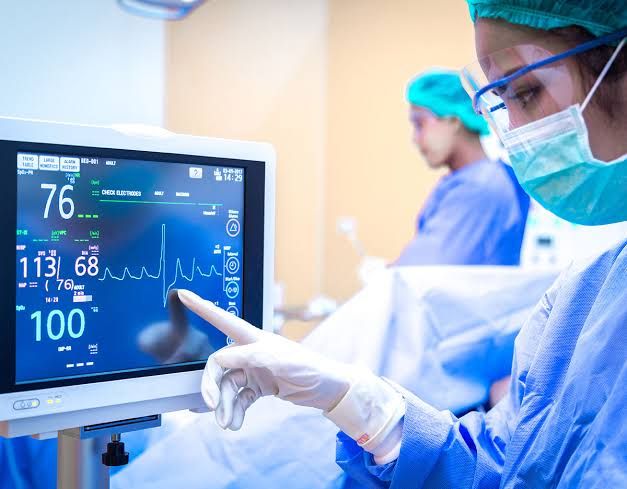
Monitoring during anesthesia focuses on four key areas: oxygenation, ventilation, circulation, and temperature. Each area has specific objectives and methods for assessment:
- Oxygenation: The primary goal is to ensure adequate oxygen concentration in both inspired gas and blood. This involves using an oxygen analyzer with alarms for low oxygen concentration during general anesthesia and employing pulse oximetry for blood oxygenation assessment.
- Ventilation: Adequate ventilation must be continually evaluated through qualitative clinical signs (e.g., chest excursion) and quantitative measures such as expired carbon dioxide monitoring. Capnography or capnometry should be used when appropriate to assess end-tidal CO2 levels.
- Circulation: It is essential to monitor the adequacy of circulatory function throughout the anesthetic process. This includes assessing heart rate, blood pressure, and other vital signs continuously.
- Temperature: Maintaining normothermia is also important during surgery; therefore, temperature monitoring should be part of standard practice.
3. Emergency Situations
In emergency situations where immediate action is required, certain monitoring protocols may need to be adjusted based on clinical judgment. If an anesthesiologist must temporarily leave the room due to an emergency, they must ensure that another qualified individual assumes responsibility for patient monitoring.
4. Guidelines Evolution
The guidelines are subject to revision based on advancements in technology and evolving practices within the field of anesthesiology. The Canadian Anesthesiologists’ Society (CAS) publishes updated versions annually to reflect these changes while emphasizing that adherence does not guarantee specific patient outcomes.
5. Medication Safety
Recent updates have included additional recommendations related to medication safety and error prevention within healthcare facilities, highlighting the importance of proper protocols in administering anesthetic agents.
In summary, adherence to established guidelines for anesthesia practice ensures that patients receive safe and effective care through continuous monitoring by qualified personnel across all phases of their treatment.
Monitoring in Anesthesia: Oxygenation, Ventilation, Circulation, and Temperature
1. Monitoring Oxygenation
Oxygenation is crucial during anesthesia to ensure that the patient receives adequate oxygen concentration in both the inspired gas and the blood. The following methods are employed:
- Inspired Gas Monitoring: An oxygen analyzer is used to measure the concentration of oxygen in the patient’s breathing system. This device should have a low oxygen concentration limit alarm to alert the anesthesia team if levels fall below safe thresholds.
- Blood Oxygenation Monitoring: Pulse oximetry is utilized as a quantitative method for assessing blood oxygen levels. The pulse oximeter provides continuous readings of arterial oxygen saturation (SpO2) and should be equipped with an audible alarm for low threshold levels. Adequate lighting and exposure of the patient are necessary for accurate assessment of skin color, which can indicate oxygenation status.
2. Monitoring Ventilation
Ventilation monitoring ensures that the patient is adequately ventilated throughout the anesthetic procedure. Key methods include:
- Clinical Signs Evaluation: Continuous evaluation of qualitative clinical signs such as chest excursion, observation of the reservoir breathing bag, and auscultation of breath sounds are essential indicators of ventilation adequacy.
- Expired Carbon Dioxide Monitoring: Continuous monitoring for expired carbon dioxide (CO2) is performed using capnography or capnometry. This involves analyzing exhaled gases to confirm proper ventilation and detect any abnormalities in respiratory function.
- Endotracheal Tube Verification: When an endotracheal tube or laryngeal mask airway is used, its correct placement must be verified through clinical assessment and identification of CO2 in expired gas.
- Mechanical Ventilation Monitoring: If mechanical ventilation is employed, devices capable of detecting disconnections within the breathing system must be continuously used, providing audible alarms when thresholds are exceeded.
3. Monitoring Circulation
Circulatory monitoring focuses on ensuring adequate blood flow and heart function during anesthesia:
- Heart Rate and Rhythm Monitoring: Electrocardiogram (ECG) monitoring provides continuous assessment of heart rate and rhythm, allowing for early detection of arrhythmias or other cardiac issues.
- Blood Pressure Measurement: Non-invasive or invasive blood pressure monitors are used to continuously assess blood pressure levels throughout the procedure. This helps identify hypotension or hypertension that may require intervention.
- Peripheral Perfusion Assessment: Observing peripheral circulation through capillary refill time and skin temperature can provide additional information about circulatory status.
4. Monitoring Temperature
Temperature monitoring is essential to prevent hypothermia or hyperthermia during anesthesia:
- Continuous Temperature Measurement: A temperature probe is typically placed in a suitable location (e.g., esophageal, rectal) to provide continuous readings throughout the procedure. Maintaining normothermia is critical as it affects metabolic rates and recovery times post-anesthesia.
In summary, effective monitoring during anesthesia involves a combination of technologies and clinical assessments focused on oxygenation, ventilation, circulation, and temperature to ensure patient safety throughout surgical procedures.
Monitoring Techniques in Anesthesia
Monitoring during anesthesia is critical for ensuring patient safety and effective management of physiological parameters. Various monitoring techniques are employed to assess the cardiopulmonary status of patients undergoing surgical procedures. Here, we will discuss five key monitoring methods: ECG (Electrocardiography), Pulse Oximetry, Blood Pressure Monitoring, Central Venous Pressure (CVP) Monitoring, and Capnography (EtCO2).
1. ECG (Electrocardiography)
ECG is a fundamental tool used to monitor the electrical activity of the heart. It provides real-time information about heart rate and rhythm, which is crucial during anesthesia as changes can indicate potential complications such as arrhythmias or ischemia. The heart rate should be continuously monitored, with specific thresholds established for different species; for instance, a heart rate below 55 beats per minute in dogs or 80 beats per minute in cats may indicate bradycardia unless premedicated with certain drugs like medetomidine.
2. Pulse Oximetry
Pulse oximetry measures the oxygen saturation level in the blood non-invasively. This method uses light absorption characteristics of oxygenated and deoxygenated hemoglobin to provide an estimate of arterial oxygen saturation (SpO2). Normal values typically range from 95% to 100%. Values below this range may indicate hypoxemia, necessitating immediate intervention to ensure adequate oxygen delivery to tissues.
3. Blood Pressure Monitoring
Blood pressure monitoring is essential for assessing cardiovascular stability during anesthesia. It can be measured non-invasively using a sphygmomanometer or invasively through an arterial catheter for continuous monitoring. The systolic, diastolic, and mean arterial pressures provide insights into perfusion status and help detect hypotension or hypertension that could compromise organ function.
4. Central Venous Pressure (CVP) Monitoring
CVP monitoring involves measuring the pressure in the central venous system, which reflects right atrial pressure and indirectly assesses fluid status and cardiac function. This invasive technique requires placement of a catheter into a central vein (e.g., jugular or subclavian vein). Normal CVP values typically range from 0 to 8 mmHg; deviations from this range can indicate hypovolemia or fluid overload.
5. Capnography (EtCO2)
Capnography measures the concentration of carbon dioxide (CO2) in exhaled air, providing valuable information about ventilation status and metabolic activity. The end-tidal CO2 (EtCO2) value reflects the partial pressure of CO2 at the end of expiration and is typically around 38 mmHg or 5%. Abnormal EtCO2 levels can indicate respiratory issues; increased levels may suggest hypoventilation while decreased levels may indicate hyperventilation or other respiratory complications.
In conclusion, effective monitoring using these techniques allows anesthetists to make informed decisions regarding patient management throughout the perioperative period. Continuous assessment helps identify abnormalities early on, enabling timely interventions that enhance patient safety.
Identifying Cyanosis
Cyanosis is a clinical sign characterized by a bluish or purplish discoloration of the skin and mucous membranes, indicating inadequate oxygenation of the blood. In the context of anesthesia, identifying cyanosis is crucial for monitoring patient safety and ensuring adequate oxygen delivery during surgical procedures. Here’s how to identify cyanosis step by step:
1. Understanding Cyanosis Types:
- Central Cyanosis: This form is typically observed in the lips, tongue, and sublingual tissues. It indicates systemic hypoxemia and is more concerning than peripheral cyanosis.
- Peripheral Cyanosis: This occurs in the extremities (hands and feet) and may be due to peripheral vasoconstriction rather than systemic hypoxemia.
2. Visual Inspection:
- During anesthesia, continuous visual inspection of the patient’s face, particularly around the lips and mucous membranes, is essential. Look for any bluish discoloration that may indicate central cyanosis.
- Peripheral cyanosis can be assessed by examining the fingers and toes. Note that peripheral cyanosis can occur without central cyanosis.
3. Monitoring Oxygen Saturation:
- Use pulse oximetry to measure arterial oxygen saturation (SaO2). A SaO2 level below 90% often correlates with the presence of cyanosis.
- Be aware that patients with anemia may not exhibit visible cyanosis despite having low oxygen levels because they require a higher concentration of deoxygenated hemoglobin to show clinical signs.
4. Assessing Skin Color:
- Consider natural skin pigmentation when assessing for cyanosis; darker skin tones may mask subtle changes in color.
- Evaluate under consistent lighting conditions to avoid misinterpretation caused by shadows or poor lighting.
5. Recognizing Associated Symptoms:
- Monitor for other signs of hypoxemia such as tachycardia, tachypnea, altered mental status, or respiratory distress. These symptoms can accompany or precede visible signs of cyanosis.
6. Utilizing Capillary Refill Time:
- Assess capillary refill time as an additional indicator of perfusion status; prolonged refill time may suggest inadequate circulation leading to peripheral cyanosis.
7. Clinical Context:
- Always consider the clinical context; for instance, if a patient has known respiratory or cardiac issues, they may be at higher risk for developing hypoxemia and subsequent cyanosis during anesthesia.
In summary, identifying cyanosis during anesthesia involves careful visual inspection for discoloration in key areas (lips and extremities), monitoring oxygen saturation levels using pulse oximetry, considering skin pigmentation variations, recognizing associated symptoms of hypoxemia, evaluating capillary refill time, and understanding the patient’s clinical background.
Oxygen-Hemoglobin Dissociation Curve
The oxygen-hemoglobin dissociation curve (ODC) is a critical concept in understanding how hemoglobin (Hb) transports oxygen (O2) throughout the body. This curve illustrates the relationship between the partial pressure of oxygen (PO2) and the saturation of hemoglobin with oxygen (SO2). The ODC is typically represented as a sigmoid (S-shaped) curve, which reflects the cooperative binding nature of hemoglobin.
Background on Hemoglobin and Oxygen Transport
Hemoglobin is a protein found in red blood cells that can bind up to four molecules of oxygen. Each hemoglobin molecule consists of four globin chains, each containing a heme group capable of binding one oxygen molecule. When no oxygen is bound, hemoglobin has an unbound conformation. The binding of the first oxygen molecule induces a conformational change that increases the affinity for subsequent oxygen molecules. This phenomenon is known as cooperative binding.
As blood circulates through the lungs, where PO2 is high, hemoglobin binds to oxygen readily. Conversely, when blood reaches tissues with lower PO2, hemoglobin releases its bound oxygen. The efficiency of this process is influenced by several factors including pH, temperature, carbon dioxide levels, and 2,3-diphosphoglycerate (2,3-DPG).
Shape and Characteristics of the Curve
The ODC has a characteristic sigmoidal shape due to the cooperative nature of oxygen binding:
- Flat Upper Portion: At high PO2 levels (above approximately 60 mmHg), further increases in PO2 result in minimal changes in SO2 because hemoglobin becomes nearly saturated with oxygen.
- Steep Lower Portion: In contrast, at lower PO2 levels (around 20-40 mmHg), small decreases in PO2 lead to significant releases of O2 from hemoglobin. This allows peripheral tissues to extract more oxygen efficiently even with slight drops in available PO2.
Factors Affecting the Dissociation Curve
Several physiological factors can shift the ODC either to the left or right:
- Temperature: An increase in temperature shifts the curve to the right, indicating decreased affinity for oxygen and promoting unloading at tissues.
- Carbon Dioxide Levels: The Bohr effect describes how increased CO2 concentration and lower pH (higher H+ ion concentration) shift the curve to the right, enhancing O2 release in metabolically active tissues.
- Hydrogen Ion Concentration: Similar to CO2 effects, increased acidity promotes O2 unloading by shifting the curve rightward.
- 2,3-Diphosphoglycerate (2,3-DPG): Produced during glycolysis under conditions like hypoxia or anemia, elevated levels of 2,3-DPG also shift the curve to the right by decreasing Hb’s affinity for O2.
- Carbon Monoxide: CO binds strongly to hemoglobin and shifts the curve leftward by increasing Hb’s affinity for O2 but reducing overall O2 transport capacity.
Clinical Relevance
Understanding shifts in the ODC is crucial for clinical applications such as assessing respiratory function and managing conditions like chronic obstructive pulmonary disease (COPD), anemia, and other disorders affecting gas exchange.
In summary, the oxyhemoglobin dissociation curve illustrates how hemoglobin’s affinity for oxygen varies with changes in partial pressure of oxygen and other physiological factors, facilitating efficient delivery of oxygen from lungs to tissues while adapting to metabolic demands.
Normal Values of Monitored Parameters for a Healthy Adult Undergoing General Anesthesia
When a healthy adult is undergoing general anesthesia, several physiological parameters are continuously monitored to ensure the patient’s safety and well-being. The normal values for these monitored parameters can vary slightly depending on the specific monitoring equipment used and the individual patient characteristics. However, the following ranges are generally accepted as normal values:
1. Oxygenation
- Arterial Oxygen Saturation (SpO2): 95% to 100%
- Inspired Oxygen Concentration (FiO2): Typically set between 21% (room air) to 100%, depending on the clinical scenario.
2. Ventilation
- End-Tidal Carbon Dioxide (EtCO2): 35 to 45 mmHg (or approximately 4.7 to 6.0 kPa). This value indicates adequate ventilation.
- Respiratory Rate: Generally between 12 to 20 breaths per minute for adults, but this may be adjusted based on anesthetic depth and patient condition.
3. Circulation
- Heart Rate: Normal resting heart rate for adults is typically between 60 to 100 beats per minute.
- Blood Pressure: Normal systolic blood pressure ranges from 90 to 120 mmHg, while diastolic pressure ranges from 60 to 80 mmHg.
- Cardiac Output: Normal values range from about 4 to 8 liters per minute, depending on body size and physical condition.
4. Temperature
- Core Body Temperature: Normal range is approximately 36°C to 37.5°C (96.8°F to 99.5°F). Hypothermia can occur during anesthesia due to environmental factors and should be monitored closely.
These parameters are essential for assessing the patient’s physiological status during anesthesia and guiding any necessary interventions by the anesthesia care team.
The monitoring standards emphasize that qualified anesthesia personnel must be present throughout the procedure, continuously evaluating these parameters to ensure patient safety.
In summary, maintaining these normal values is crucial during general anesthesia as they provide vital information regarding the patient’s respiratory, circulatory, and overall physiological status.


