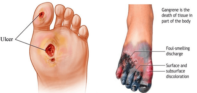
Causes of Chronic Leg Ulcers and Differences in Appearance
1. Venous Leg Ulcers (VLUs)
Venous leg ulcers are the most common type, accounting for 60-80% of all leg ulcers. They typically arise due to chronic venous insufficiency, where veins struggle to return blood to the heart, leading to increased pressure in the veins. This condition can be exacerbated by factors such as obesity, immobility, and a history of deep vein thrombosis (DVT).
Appearance:
- Located between the knee and ankle.
- The wound is often shallow with irregular edges.
- Surrounding skin may appear discolored (brownish pigmentation) and swollen.
- There may be signs of varicose veins nearby.
- Exudate can be yellowish or serous.
2. Arterial Ulcers
Arterial ulcers occur due to inadequate blood supply from arterial disease, often linked to conditions like peripheral artery disease (PAD). Risk factors include smoking, diabetes, high cholesterol, and hypertension.
Appearance:
- Typically found on the toes, feet, or lower legs.
- The wound is usually well-defined with a “punched-out” appearance.
- Edges are smooth and may have a necrotic base.
- Surrounding skin may be pale or cool to touch due to poor circulation.
- Minimal exudate is present.
3. Diabetic Ulcers
Diabetic ulcers result from neuropathy and poor circulation associated with diabetes mellitus. These ulcers commonly develop on pressure points of the foot.
Appearance:
- Often located on the plantar surface of the foot or around bony prominences.
- The ulcer may have a calloused rim with a deep base.
- Surrounding skin might show signs of infection or inflammation.
- Exudate can vary but is often purulent if infected.
4. Pressure Ulcers (Bedsores)
Pressure ulcers develop when sustained pressure impairs blood flow to an area of skin, commonly affecting individuals who are bedridden or immobile.
Appearance:
- Usually found over bony areas like heels, sacrum, or elbows.
- Stages range from non-blanchable redness (stage 1) to full-thickness skin loss (stage 4).
- Wounds can appear as open sores with varying degrees of tissue loss and necrosis depending on the stage.
5. Mixed Ulcers
Some patients may experience mixed ulcers that have characteristics of both venous and arterial causes due to underlying circulatory issues.
Appearance:
- May exhibit features from both venous and arterial ulcers depending on which pathology predominates at any given time.
In summary, chronic leg ulcers can arise from various causes including venous insufficiency, arterial disease, diabetes complications, pressure-related injuries, or a combination thereof. Their appearances differ significantly based on their etiology—ranging from irregularly shaped wounds with surrounding discoloration in venous ulcers to well-defined “punched-out” lesions in arterial cases.
Comparison of Venous and Arterial Leg Ulcers
Venous and arterial ulcers are both types of chronic wounds that typically occur on the lower extremities, particularly the legs and feet. However, they arise from different underlying pathophysiological processes and exhibit distinct clinical presentations.
Causes
- Arterial Ulcers: These ulcers develop due to insufficient blood flow caused by arterial disease. Conditions such as peripheral artery disease (PAD), atherosclerosis, high blood pressure, diabetes, and smoking contribute to reduced oxygen-rich blood reaching the tissues.
- Venous Ulcers: In contrast, venous ulcers result from chronic venous insufficiency, where the veins fail to return blood effectively to the heart. This can lead to pooling of blood in the legs, causing increased pressure in the veins (venous hypertension) and subsequent ulcer formation.
Location
- Arterial Ulcers: Typically found on the outer side of the ankle, toes, heels, or other areas of the feet. They may also appear on areas with less fat cushion.
- Venous Ulcers: Commonly located below the knee, particularly around the inner ankle area. They can also occur on other parts of the lower leg.
Appearance
- Arterial Ulcers: Characterized by small, deep “punched-out” wounds with well-defined borders. The surrounding skin may appear pale or grayish due to poor circulation and can feel cool to touch.
- Venous Ulcers: Present as larger, shallow wounds with irregular edges. The surrounding skin often shows signs of inflammation and discoloration (brown or black staining) due to hemosiderin deposition from leaked red blood cells.
Symptoms
- Arterial Ulcers: Often very painful; patients may experience severe pain in the affected area that worsens with activity or elevation.
- Venous Ulcers: Pain levels can vary; they may cause a dull ache throughout the leg but are generally less painful than arterial ulcers unless infected.
Surrounding Skin Changes
- Arterial Ulcers: The skin around these ulcers is usually hairless and may feel tight or itchy. There might be signs of atrophy or thinning skin.
- Venous Ulcers: The surrounding skin often shows signs of swelling (edema), inflammation, itching, scabbing or flaking skin. It may also appear thickened or hardened.
Healing Potential
- Arterial Ulcers: Healing is often slow and requires restoration of adequate blood flow through medical or surgical interventions.
- Venous Ulcers: While they can take months to heal and sometimes do not heal completely, treatment focuses on improving venous return through compression therapy and addressing underlying venous insufficiency.
In summary, while both types of ulcers are chronic wounds found primarily in the lower extremities, their causes, locations, appearances, symptoms, surrounding skin changes, and healing potentials differ significantly.
Pathogenesis of Ischaemic, Venous, and Diabetic Ulcers
Ischaemic Ulcers
Ischaemic ulcers are primarily caused by inadequate blood supply to the tissues, often due to peripheral artery disease (PAD). The pathogenesis involves several key factors:
- Reduced Blood Flow: In PAD, atherosclerosis leads to narrowing or blockage of arteries, reducing blood flow to the extremities. This lack of perfusion results in tissue ischemia.
- Tissue Hypoxia: The reduced blood flow causes insufficient oxygen delivery to the tissues, leading to hypoxia. Cells begin to die due to lack of oxygen and nutrients.
- Necrosis and Ulcer Formation: As ischemia persists, necrosis occurs in the affected tissues. The skin becomes fragile and breaks down under pressure or trauma, leading to ulcer formation.
- Infection Risk: Ischaemic ulcers are prone to infection due to compromised blood supply that limits immune response and healing capabilities.
- Clinical Presentation: These ulcers typically appear on pressure points such as the heels or toes and are characterized by a pale base with well-defined edges.
Venous Ulcers
Venous ulcers arise from chronic venous insufficiency (CVI), where veins cannot effectively return blood from the lower extremities back to the heart. The pathogenesis includes:
- Venous Hypertension: In CVI, valves within the veins become incompetent, causing blood pooling in the lower legs and increased venous pressure.
- Capillary Leakage: Elevated venous pressure leads to increased hydrostatic pressure in capillaries, resulting in leakage of fluid and proteins into surrounding tissues.
- Tissue Edema: The accumulation of interstitial fluid causes edema, which compromises local tissue health and contributes to skin changes such as discoloration and thickening.
- Skin Breakdown: Prolonged edema can lead to skin breakdown and ulceration as the skin becomes fragile and unable to withstand normal stresses.
- Clinical Presentation: Venous ulcers typically occur around the medial malleolus (ankle area) and have irregular borders with a moist base covered by yellow slough or granulation tissue.
Diabetic Ulcers
Diabetic ulcers are common complications in individuals with diabetes mellitus due to multiple interrelated factors:
- Neuropathy: Diabetic neuropathy impairs sensation in the feet, leading patients to be unaware of injuries or pressure points that can cause ulceration.
- Peripheral Artery Disease (PAD): Many diabetic patients also suffer from PAD, which reduces blood flow and exacerbates tissue ischemia similar to ischaemic ulcers.
- Foot Deformities: Diabetes can lead to foot deformities (e.g., Charcot foot) that alter weight distribution during walking, increasing pressure on specific areas of the foot.
- Infection Susceptibility: High glucose levels impair immune function, making diabetic patients more susceptible to infections once an ulcer develops.
- Clinical Presentation: Diabetic ulcers often present as deep lesions on weight-bearing surfaces of the foot with a characteristic “punched-out” appearance; they may also have callus formation around them due to abnormal pressures.
In summary, ischaemic ulcers result from inadequate blood supply leading to tissue death; venous ulcers stem from chronic venous insufficiency causing fluid leakage and edema; while diabetic ulcers arise from a combination of neuropathy, vascular disease, foot deformities, and infection susceptibility associated with diabetes mellitus.
Investigations and Treatment Options for Chronic Leg Ulcers
Chronic leg ulcers are a common clinical problem that can significantly impact a patient’s quality of life. The management of these ulcers involves a comprehensive approach that includes identifying and treating underlying causes, appropriate dressing and bandaging techniques, and potential surgical reconstruction if necessary.
a. Management of Underlying Cause
The first step in managing chronic leg ulcers is to identify the underlying cause. Common causes include venous insufficiency, arterial disease, diabetes mellitus, and pressure ulcers.
- Assessment: A thorough assessment should be conducted, including a detailed patient history and physical examination. This may involve:
- Ankle-brachial index (ABI) testing to assess arterial circulation.
- Doppler ultrasound to evaluate venous reflux.
- Blood tests to check for diabetes or other systemic conditions.
- Skin biopsy if there is suspicion of malignancy or atypical ulceration.
- Treatment of Underlying Conditions:
- Venous Insufficiency: Compression therapy is critical in managing venous ulcers. This may involve the use of graduated compression stockings or bandages to improve venous return.
- Arterial Disease: For patients with arterial ulcers, revascularization procedures such as angioplasty or bypass surgery may be indicated if there is significant limb ischemia.
- Diabetes Management: Tight glycemic control is essential for diabetic patients to promote wound healing and prevent further complications.
- Infection Control: If an infection is present, appropriate antibiotic therapy should be initiated based on culture results.
- Lifestyle Modifications: Patients should be advised on lifestyle changes such as smoking cessation, weight management, and regular exercise to improve overall vascular health.
b. Dressings and Bandaging
The choice of dressings and bandaging plays a crucial role in the management of chronic leg ulcers:
- Types of Dressings:
- Hydrocolloid Dressings: These are beneficial for maintaining a moist wound environment and are often used for granulating wounds.
- Foam Dressings: They provide cushioning and absorb exudate while maintaining moisture.
- Alginate Dressings: Suitable for highly exudative wounds; they help absorb fluid while promoting healing.
- Antimicrobial Dressings: These can be used when there is evidence of infection or high risk for infection.
- Bandaging Techniques:
- Proper application techniques are vital to ensure effective compression without compromising blood flow.
- Multi-layer compression bandaging systems are often recommended for venous ulcers to provide sustained pressure over time.
- Frequency of Dressing Changes: The frequency will depend on the type of dressing used and the level of exudate from the ulcer but typically ranges from every 1-7 days.
- Monitoring Progress: Regular assessment of the ulcer’s size, depth, and signs of infection should guide ongoing treatment decisions.
c. Reconstruction
In cases where conservative measures fail or when there is significant tissue loss, surgical reconstruction may be considered:
- Debridement: Surgical debridement may be necessary to remove necrotic tissue that impedes healing.
- Skin Grafting:
- Split-thickness skin grafts can be applied over granulating wounds once they have been adequately prepared.
- Full-thickness grafts may also be used depending on the size and location of the ulcer.
- Flap Surgery:
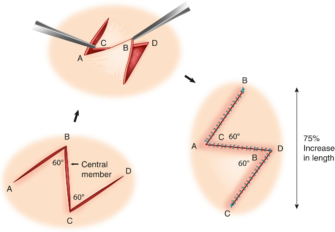
- Local flap procedures can provide coverage for larger defects by transferring adjacent tissue with its blood supply intact.
- Free flap techniques may also be employed in more complex cases where local options are insufficient.
- Postoperative Care: After surgical intervention, careful monitoring is required to ensure proper healing and prevent complications such as infection or graft failure.
In conclusion, managing chronic leg ulcers requires a multifaceted approach focusing on underlying causes, appropriate dressing techniques, and surgical options when necessary to promote healing effectively.
Overview of Gangrene Associated with Chronic Ischaemia
Gangrene is a serious condition characterized by the death of body tissue due to a lack of blood flow (ischemia) or severe bacterial infection. When gangrene is associated with chronic ischemia, it typically arises from prolonged inadequate blood supply to a specific area, leading to tissue necrosis. This condition often affects the extremities, such as the toes and fingers, and can result in significant morbidity if not addressed promptly.
Causes of Chronic Ischaemia Leading to Gangrene
Chronic ischemia occurs when there is a gradual reduction in blood flow to tissues over time. This can be caused by various factors:
- Atherosclerosis: The most common cause of chronic ischemia is atherosclerosis, where fatty deposits build up in the arteries, narrowing them and restricting blood flow.
- Diabetes Mellitus: Diabetes can lead to vascular complications that impair circulation and increase the risk of gangrene.
- Peripheral Artery Disease (PAD): This condition involves narrowed arteries reducing blood flow to the limbs, which can lead to chronic ischemia.
- Other Vascular Diseases: Conditions like vasculitis or thromboangiitis obliterans (Buerger’s disease) can also contribute to reduced blood supply.
Types of Gangrene Related to Chronic Ischaemia
- Dry Gangrene: This type develops slowly and is characterized by dry, shriveled skin that may appear brownish to purplish blue or black. It typically occurs in individuals with diabetes or peripheral artery disease due to insufficient blood supply over time.
- Wet Gangrene: If an infection occurs alongside chronic ischemia, wet gangrene may develop. This type is marked by swelling, blistering, and a foul-smelling discharge from the affected area. Wet gangrene requires immediate medical attention as it can spread rapidly and become life-threatening.
- Gas Gangrene: Although less common in cases of chronic ischemia alone, gas gangrene can occur if anaerobic bacteria infect necrotic tissue. It primarily affects deep muscle tissues and produces gas within tissues.
Symptoms of Gangrene Associated with Chronic Ischaemia
The symptoms may vary depending on the type of gangrene but generally include:
- Changes in skin color (pale gray to blue, purple, black)
- Swelling
- Blisters
- Severe pain followed by numbness
- Foul-smelling discharge from sores
- Coolness or coldness of the affected area
In cases where internal organs are involved or if septic shock develops due to systemic infection from gangrenous tissue, additional symptoms such as fever, low blood pressure, rapid heart rate, lightheadedness, shortness of breath, and confusion may occur.
Diagnosis and Treatment
Diagnosis typically involves clinical evaluation based on symptoms and physical examination findings. Imaging studies may be used to assess blood flow and identify areas of necrosis.
Treatment for gangrene associated with chronic ischemia focuses on restoring blood flow and managing infection:
- Surgical Intervention: Debridement (removal of dead tissue) is often necessary; in severe cases, amputation may be required.
- Antibiotics: Broad-spectrum antibiotics are administered if an infection is present.
- Oxygen Therapy: Hyperbaric oxygen therapy may be utilized for certain types of gangrene.
- Management of Underlying Conditions: Addressing risk factors such as diabetes management or lifestyle changes related to cardiovascular health is crucial for preventing recurrence.
Early identification and treatment are vital for improving outcomes in patients suffering from gangrene associated with chronic ischemia.


