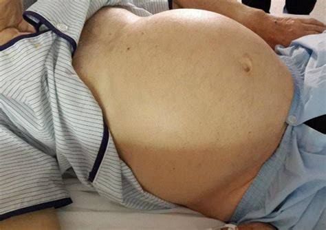
Pathophysiological Causes of Abdominal Swelling
Abdominal swelling, often referred to as abdominal distension or bloating, can arise from a variety of pathophysiological mechanisms. Understanding these causes is crucial for effective diagnosis and treatment. The primary causes can be categorized into functional gastrointestinal disorders (FGIDs) and organic diseases.
- Functional Gastrointestinal Disorders (FGIDs):
- Irritable Bowel Syndrome (IBS): A common FGID characterized by abdominal pain and altered bowel habits, IBS often presents with bloating due to gut hypersensitivity and abnormal motility. Patients may experience visceral hypersensitivity, leading to an exaggerated perception of normal intestinal gas.
- Functional Dyspepsia (FD): Similar to IBS, FD involves discomfort in the upper abdomen without any identifiable organic cause. Symptoms may include bloating due to delayed gastric emptying or increased sensitivity to gastric distension.
- Functional Bloating: This condition can occur independently of other gastrointestinal disorders. It is characterized by the sensation of bloating without significant abdominal distension or identifiable pathology.
- Organic Diseases:
- Gastrointestinal Obstruction: Mechanical obstruction in the intestines can lead to significant abdominal swelling due to the accumulation of gas and fluids proximal to the blockage.
- Ascites: The accumulation of fluid in the peritoneal cavity, often due to liver cirrhosis, heart failure, or malignancy, results in noticeable abdominal distension.
- Inflammatory Conditions: Conditions such as pancreatitis or inflammatory bowel disease (IBD) can cause swelling due to inflammation and fluid accumulation in the abdominal cavity.
- Tumors: Abdominal masses from tumors can lead to physical obstruction or displacement of intestinal structures, contributing to swelling.
- Other Factors:
- Dietary Influences: Consumption of certain foods that are high in fermentable carbohydrates (FODMAPs) can lead to increased gas production and subsequent bloating.
- Altered Gut Microbiota: Dysbiosis or an imbalance in gut microbiota may contribute to excessive gas production and bloating symptoms.
Relevant Investigations
To accurately diagnose the underlying cause of abdominal swelling, several investigations may be employed:
- Clinical Evaluation:
- Detailed patient history focusing on symptom patterns, dietary habits, and associated symptoms is essential for initial assessment.
- Physical Examination:
- A thorough physical examination helps identify signs such as tenderness, masses, or fluid wave indicating ascites.
- Laboratory Tests:
- Blood tests may include complete blood count (CBC), liver function tests (LFTs), and markers for inflammation (e.g., C-reactive protein).
- Imaging Studies:
- Ultrasound: Useful for detecting ascites or organ enlargement.
- CT Scan: Provides detailed images that help identify obstructions, tumors, or inflammatory conditions within the abdomen.
- Endoscopy Procedures:
- Upper endoscopy (esophagogastroduodenoscopy) or colonoscopy may be indicated if there is suspicion of structural abnormalities or malignancy.
- Specialized Tests:
- Breath tests for carbohydrate malabsorption (e.g., hydrogen breath test for lactose intolerance).
- Motility studies might be considered for patients with suspected dysmotility syndromes.
In summary, understanding both functional and organic causes of abdominal swelling is critical for effective management. Investigations should be tailored based on clinical findings and patient history.
Aetiology of Intestinal Obstruction
Intestinal obstruction is a blockage that prevents the normal passage of contents through the intestines. The aetiology can be classified into several categories based on the underlying causes:
- Mechanical Causes: These involve physical blockages in the intestinal lumen.
- Adhesions: Scar tissue from previous abdominal or pelvic surgeries is the most common cause in adults, forming fibrous bands that can constrict or obstruct the intestine.
- Hernias: Portions of the intestine can protrude through weak spots in the abdominal wall, leading to incarceration or strangulation.
- Tumors: Both benign and malignant tumors, particularly colon cancer, can obstruct bowel passages.
- Intussusception: This occurs when one segment of the intestine telescopes into another, commonly seen in children.
- Volvulus: Twisting of the intestine can lead to obstruction and compromised blood supply.
- Functional Causes (Pseudo-obstruction): These do not involve a physical blockage but rather a failure of normal peristalsis.
- Conditions such as paralytic ileus, which may result from surgery, infections, or certain medications (e.g., opioids), disrupt normal intestinal motility.
- Inflammatory Conditions: Diseases like Crohn’s disease and diverticulitis can cause strictures due to inflammation and scarring.
- Other Causes: Impacted feces or foreign bodies can also lead to obstruction.
Presentation of Intestinal Obstruction
The clinical presentation of intestinal obstruction varies depending on its location (small vs. large intestine) and severity:
- Symptoms:
- Abdominal Pain: Crampy pain that may come and go; small bowel obstructions often present with intermittent pain while large bowel obstructions may have more constant discomfort.
- Nausea and Vomiting: Often occurs as contents back up behind the obstruction.
- Constipation or Inability to Pass Gas: A hallmark sign; complete obstructions typically prevent any stool passage.
- Abdominal Distension: Swelling due to trapped gas and fluid.
- Signs on Examination:
- Abdominal tenderness upon palpation.
- Bowel sounds may be hyperactive initially but can diminish with prolonged obstruction.
- Complications if Untreated:
- Tissue death (necrosis) due to compromised blood flow.
- Perforation leading to peritonitis, a life-threatening infection.
Management of Intestinal Obstruction
Management strategies depend on the cause, severity, and presence of complications:
- Initial Assessment and Stabilization:
- Immediate medical evaluation is crucial for suspected cases.
- Vital signs monitoring and intravenous fluids are essential for hydration and electrolyte balance.
- Diagnostic Imaging:
- X-rays or CT scans are used to confirm diagnosis and determine the location and cause of obstruction.
- Non-Surgical Management:
- For partial obstructions without signs of ischemia or perforation, conservative management may include bowel rest (nothing by mouth), nasogastric tube placement for decompression, and close monitoring.
- Surgical Intervention:
- Indicated for complete obstructions, those with signs of ischemia, perforation, or failure to improve with conservative measures.
- Surgical options may include resection of obstructed segments, removal of adhesions, or hernia repair depending on the underlying cause.
- Postoperative Care:
- Monitoring for complications such as infection or recurrence is vital after surgical intervention.
In summary, intestinal obstruction requires prompt recognition due to its potential complications. Treatment strategies vary widely based on etiology but often necessitate both medical management and surgical intervention when indicated.
Differential Diagnosis, Investigation, and Management of Patients Presenting with Left or Right Iliac Fossa Masses
1. Differential Diagnosis
When evaluating a patient with an iliac fossa mass, it is crucial to consider various potential causes based on the location of the mass (left or right). The differential diagnosis can be categorized into several groups:
- Gastrointestinal Causes:
- Appendiceal Mass: Commonly presents in the right iliac fossa due to appendicitis.
- Diverticular Disease: Can cause localized masses in the left iliac fossa due to diverticulitis.
- Colorectal Cancer: Tumors can present as palpable masses in either iliac fossa depending on their location.
- Ileal Obstruction: May lead to distension and palpable masses.
- Genitourinary Causes:
- Vascular Causes:
- Aneurysms: Abdominal aortic aneurysms may present as pulsatile masses, typically more common on the left side.
- Musculoskeletal Causes:
- Hernias: Inguinal hernias can present as bulges in the iliac region.
- Infectious Causes:
- Abscesses: Such as psoas abscesses or intra-abdominal abscesses from diverticulitis or appendicitis.
2. Investigation
The investigation of an iliac fossa mass involves a combination of clinical assessment and imaging studies:
- Clinical Assessment:
- Detailed history taking including onset, duration, associated symptoms (pain, fever, changes in bowel habits).
- Physical examination focusing on abdominal palpation and signs of peritonitis.
- Imaging Studies:
- Ultrasound (US): First-line imaging for assessing soft tissue masses, particularly useful for ovarian pathology and fluid collections.
- Computed Tomography (CT) Scan: Provides detailed information about abdominal organs and is essential for diagnosing appendicitis, diverticulitis, tumors, and vascular abnormalities. CT scans are particularly useful for differentiating between solid and cystic masses.
- Magnetic Resonance Imaging (MRI): May be used for further characterization of soft tissue masses when ultrasound results are inconclusive or when there is suspicion of malignancy.
- Laboratory Tests:
- Blood tests including complete blood count (CBC), inflammatory markers (CRP), liver function tests, renal function tests, and tumor markers if malignancy is suspected (e.g., CA-125 for ovarian cancer).
3. Management
Management strategies depend on the underlying cause identified through investigations:
- Surgical Intervention:
- For acute conditions such as appendicitis or diverticulitis with complications (abscess formation), surgical intervention may be necessary (appendectomy or resection).
- Ovarian masses that are symptomatic or suspicious for malignancy may require surgical evaluation.
- Medical Management:
- Antibiotics are indicated for infectious causes such as diverticulitis or abscesses.
- Pain management should also be addressed according to the patient’s needs.
- Oncological Management:
- If malignancy is diagnosed (e.g., colorectal cancer), referral to oncology for further management including chemotherapy or radiotherapy may be warranted.
In summary, patients presenting with an iliac fossa mass require a thorough evaluation involving differential diagnosis based on clinical presentation, appropriate imaging studies for accurate diagnosis, and tailored management strategies based on the underlying condition identified.
Pathophysiological Causes of Swelling in the Epigastrium
Swelling in the epigastric region can arise from various pathophysiological processes, including those related to the liver and other abdominal organs. Understanding these causes involves examining the anatomical structures in this area and how different conditions can lead to swelling.
1. Liver-Related Causes
The liver is a significant organ located in the upper right quadrant of the abdomen, but its enlargement or dysfunction can cause swelling that may be perceived in the epigastric area. The following liver-related conditions can contribute to this swelling:
- Hepatomegaly: This condition refers to an enlarged liver, which can occur due to various factors such as viral hepatitis (A, B, C), alcoholic liver disease, nonalcoholic fatty liver disease (NAFLD), cirrhosis, and hepatic tumors. Hepatomegaly can lead to a palpable mass in the epigastrium.
- Cirrhosis: Advanced scarring of the liver tissue due to chronic liver diseases leads to cirrhosis. This condition often results in portal hypertension (increased blood pressure in the portal venous system), which can cause ascites (fluid accumulation) that may manifest as abdominal distension.
- Liver Tumors: Both benign tumors (like hemangiomas and adenomas) and malignant tumors (such as hepatocellular carcinoma) can cause localized swelling due to their mass effect on surrounding structures.
2. Gastrointestinal Causes
Several gastrointestinal disorders can also lead to swelling or distension in the epigastric region:
- Gastric Distension: Conditions such as gastric outlet obstruction or pyloric stenosis can lead to excessive gas or fluid accumulation within the stomach, causing visible swelling.
- Pancreatitis: Inflammation of the pancreas may result in pain and swelling in the upper abdomen due to edema and fluid accumulation around the pancreas.
- Bowel Obstruction: Obstructions within the small intestine or colon can lead to significant distension due to trapped gas and fluid, resulting in abdominal swelling that may extend into the epigastric area.
3. Cardiovascular Causes
Conditions affecting blood flow and pressure within abdominal vessels can also contribute:
- Congestive Heart Failure: Right-sided heart failure may lead to systemic venous congestion, resulting in fluid retention and ascites that could cause abdominal distension.
- Budd-Chiari Syndrome: This rare condition involves occlusion of hepatic veins leading to increased pressure within these vessels, potentially causing hepatomegaly and abdominal swelling.
4. Other Causes
Other potential causes of epigastric swelling include:
- Infections: Abdominal infections such as peritonitis or abscess formation can result in localized inflammation and fluid accumulation.
- Trauma: Abdominal trauma may lead to hematoma formation or organ rupture, contributing to swelling.
- Tumors Outside of Liver/GI Tract: Tumors originating from adjacent organs (e.g., spleen or kidneys) may exert pressure on surrounding tissues leading to perceived swelling in the epigastrium.
In summary, a variety of conditions ranging from liver diseases like hepatomegaly and cirrhosis, gastrointestinal disorders such as pancreatitis and bowel obstruction, cardiovascular issues like congestive heart failure, infections, trauma, and tumors outside these systems contribute to swelling in the epigastrium. Each condition has distinct pathophysiological mechanisms that ultimately result in fluid accumulation or organ enlargement leading to noticeable abdominal distension.
Imaging in the Investigation of Acute Abdominal Pain
Acute abdominal pain is a common clinical presentation that can arise from various underlying conditions, necessitating a systematic approach to imaging for accurate diagnosis. The choice of imaging modality depends on the clinical context, patient history, and physical examination findings. Below is a detailed explanation of the appropriate imaging techniques used in the investigation of acute abdominal pain.
1. Plain Radiography
Plain radiography includes both erect chest X-rays and abdominal X-rays, which are often used as initial imaging modalities.
- Erect Chest X-ray: This view is primarily utilized to identify free air under the diaphragm, which may indicate perforation of a hollow viscera (e.g., perforated peptic ulcer). The presence of free air is best visualized in an upright position due to its tendency to rise to the highest point in the abdominal cavity. Additionally, this X-ray can help assess for other thoracic issues that may mimic or contribute to abdominal pain, such as pneumonia or pleural effusion.
- Abdominal X-ray: This modality can reveal bowel obstruction through signs such as dilated loops of bowel and air-fluid levels. It may also show calcifications (e.g., gallstones or kidney stones) and certain masses. However, its sensitivity and specificity are limited compared to other imaging techniques, making it less reliable for definitive diagnosis.
2. Abdominal Ultrasound Scan
Ultrasound is a non-invasive imaging technique that uses sound waves to produce images of internal structures. It is particularly useful in specific scenarios:
- Gallbladder Disease: Ultrasound is the first-line imaging modality for suspected cholecystitis or choledocholithiasis (bile duct stones). It can visualize gallstones, thickening of the gallbladder wall, and pericholecystic fluid.
- Appendicitis: In children and pregnant women, ultrasound can be helpful in diagnosing appendicitis without exposing them to radiation.
- Other Conditions: Ultrasound can also evaluate other causes of acute abdominal pain such as ovarian cysts or ectopic pregnancy in females.
The advantages of ultrasound include its availability, lack of ionizing radiation, and ability to provide real-time images. However, it has limitations related to operator dependency and difficulty visualizing certain areas due to gas-filled intestines or obesity.
3. CT Scanning
Computed Tomography (CT) scanning has become a cornerstone in the evaluation of acute abdominal pain due to its high sensitivity and specificity.
- Indications: CT scans are particularly useful when there is a high suspicion of serious conditions such as appendicitis, diverticulitis, pancreatitis, bowel obstruction, or malignancy.
- Technique: A CT scan can be performed with or without contrast material. Intravenous contrast enhances vascular structures and helps delineate organs more clearly; oral contrast may be used for gastrointestinal tract evaluation but is less common in emergency settings due to time constraints.
- Findings: CT scans provide detailed cross-sectional images that can reveal inflammation, abscesses, perforations, tumors, and other pathologies effectively.
Despite its advantages, CT scanning involves exposure to ionizing radiation; thus it should be used judiciously based on clinical indications.
4. Contrast Studies
Contrast studies involve using contrast agents during radiographic examinations to enhance visualization of specific anatomical structures:
- Barium Studies: Barium swallow or enema studies are traditionally used for evaluating esophageal or colonic pathology but are less commonly employed in acute settings due to their time-consuming nature and potential complications if perforation is present.
- Intravenous Contrast CT: As mentioned earlier with CT scanning, intravenous contrast enhances diagnostic accuracy by providing better visualization of vascular structures and organ perfusion.
In summary, each imaging modality plays a vital role in assessing acute abdominal pain:
- Plain Radiography provides initial insights but has limited diagnostic capability.
- Abdominal Ultrasound Scan offers a safe option for specific conditions like gallbladder disease.
- CT Scanning, especially with contrast enhancement, serves as a comprehensive tool for diagnosing various acute abdominal conditions.
- Contrast Studies, while useful in select cases, are generally not first-line options in acute presentations but provide valuable information when indicated.
In practice, the choice among these modalities will depend on clinical judgment based on patient presentation and history while considering factors such as age, sex (especially reproductive status), prior medical history, and potential need for surgical intervention.
Differential Diagnoses for Bowel Obstruction
Bowel obstruction can arise from various underlying conditions, and it is crucial to consider a comprehensive differential diagnosis when evaluating a patient. Below are the main categories of differential diagnoses for bowel obstruction, along with explanations for each.
1. Mechanical Obstruction
Mechanical obstructions occur when there is a physical blockage in the intestines. Common causes include:
- Adhesions: Scar tissue from previous surgeries can bind loops of intestine together, leading to obstruction.
- Hernias: A hernia occurs when an internal organ pushes through a weak spot in the abdominal wall, potentially obstructing the bowel.
- Tumors: Benign or malignant tumors can grow within or outside the bowel, compressing it and causing obstruction.
- Intussusception: This condition occurs when one segment of the intestine telescopes into another, leading to blockage and potential ischemia.
- Volvulus: Twisting of the intestine can obstruct blood flow and lead to necrosis if not treated promptly.
2. Functional Obstruction (Ileus)
Functional obstructions do not involve a physical blockage but rather a failure of normal peristalsis:
- Postoperative Ileus: After abdominal surgery, temporary paralysis of the bowel may occur, preventing normal movement of contents.
- Medications: Certain medications, particularly opioids and anticholinergics, can slow down intestinal motility.
- Electrolyte Imbalances: Conditions such as hypokalemia can impair muscle contractions in the gastrointestinal tract.
3. Inflammatory Conditions
Inflammation in or around the bowel can lead to obstruction:
- Diverticulitis: Inflammation of diverticula (small pouches) in the colon can cause swelling and narrowing of the intestinal lumen.
- Inflammatory Bowel Disease (IBD): Conditions like Crohn’s disease can lead to strictures and narrowing due to chronic inflammation.
4. Vascular Causes
Vascular issues may also result in bowel obstruction:
- Mesenteric Ischemia: Reduced blood flow to the intestines due to arterial occlusion can lead to ischemic changes and subsequent obstruction.
5. Other Causes
Other less common causes include:
- Foreign Bodies: Ingested objects may obstruct the bowel if they cannot pass through.
- Gallstone Ileus: A gallstone may erode into the intestine and cause obstruction.
In summary, understanding these differential diagnoses is essential for clinicians when assessing patients with suspected bowel obstruction. Proper identification allows for timely intervention and management tailored to the specific underlying cause.
Complications of Bowel Obstruction
Bowel obstruction can lead to several serious complications if not treated promptly. The primary complications include ischaemia, perforation, and biochemical derangement. Each of these complications arises from the blockage that prevents normal intestinal function and blood flow.
(a) Ischaemia
Ischaemia refers to a reduction in blood supply to tissues, which can occur in the intestines due to bowel obstruction. When an obstruction occurs, the affected segment of the intestine may become distended with gas and fluid, leading to increased pressure within the bowel. This pressure can compromise the blood vessels supplying that segment of the intestine. If blood flow is significantly reduced or cut off entirely, it can result in tissue damage due to lack of oxygen and nutrients.
The consequences of ischaemia can be severe; if not resolved quickly, it can progress to necrosis (tissue death) of the affected bowel segment. This condition is critical because necrotic tissue can lead to further complications such as infection and sepsis.
(b) Perforation
Perforation is another grave complication that may arise from bowel obstruction. As pressure builds up within the obstructed segment of the intestine, the wall may weaken and eventually rupture (perforate). A perforation allows intestinal contents, including bacteria and toxins, to spill into the abdominal cavity. This situation leads to peritonitis, an inflammation of the peritoneum (the lining of the abdominal cavity), which is a life-threatening condition requiring immediate surgical intervention.
Symptoms of perforation may include sudden severe abdominal pain, fever, rapid heart rate, and signs of shock. The risk factors for perforation increase with prolonged obstruction and delayed treatment.
(c) Biochemical Derangement
Biochemical derangement refers to disturbances in metabolic processes caused by bowel obstruction. When food and fluids cannot pass through the intestines due to an obstruction, various metabolic imbalances can occur:
- Electrolyte Imbalance: The inability to absorb nutrients and fluids leads to dehydration and electrolyte imbalances (such as low potassium or sodium levels), which are critical for normal cellular function.
- Acid-Base Disturbances: Obstruction can also lead to metabolic acidosis or alkalosis depending on fluid loss or retention patterns in different parts of the gastrointestinal tract.
- Toxin Accumulation: As waste products accumulate behind an obstruction, there is a risk that toxins will enter systemic circulation, potentially leading to multi-organ dysfunction.
These biochemical changes can have widespread effects on bodily functions and may contribute further to morbidity if not addressed swiftly.
In summary, ischaemia, perforation, and biochemical derangement are significant complications associated with bowel obstruction that require urgent medical attention to prevent severe outcomes.


