Cofactors Required for Pyruvate Dehydrogenase Complex
The pyruvate dehydrogenase complex (PDC) is a crucial multienzyme complex that catalyzes the conversion of pyruvate into acetyl-CoA, linking glycolysis to the citric acid cycle. This process is essential for cellular respiration and energy production. The PDC requires several cofactors to function effectively, each playing a specific role in the enzymatic reactions involved. The five key cofactors required for the pyruvate dehydrogenase complex are:
- Thiamine Pyrophosphate (TPP)
TPP is derived from vitamin B1 (thiamine) and acts as a coenzyme for the E1 component of the PDC, known as pyruvate dehydrogenase. It facilitates the decarboxylation of pyruvate by forming a covalent bond with the carbonyl group of pyruvate, leading to the release of carbon dioxide and the formation of an acyl-TPP intermediate. - Lipoic Acid
Lipoic acid serves as a cofactor for the E2 component, known as dihydrolipoamide acetyltransferase. It exists in two forms: oxidized lipoic acid and reduced dihydrolipoamide. In its oxidized form, lipoic acid accepts the acyl group from TPP, forming an acyl-lipoamide intermediate. Subsequently, it undergoes reduction to dihydrolipoamide during this transfer process. - Coenzyme A (CoA)
CoA is essential for transferring acyl groups within metabolic pathways. In the context of PDC, CoA reacts with the acyl-lipoamide intermediate formed in the previous step to produce acetyl-CoA and regenerate lipoic acid in its oxidized form. This reaction is catalyzed by E2. - Flavin Adenine Dinucleotide (FAD)
FAD functions as a cofactor for the E3 component of PDC, known as dihydrolipoamide dehydrogenase. It plays a critical role in reoxidizing dihydrolipoamide back to lipoic acid after it has transferred its acyl group to CoA. FAD accepts electrons during this process and is subsequently reduced to FADH2. - Nicotinamide Adenine Dinucleotide (NAD)
NAD acts as an electron acceptor in reactions catalyzed by E3 after FAD has been reduced to FADH2. NAD+ accepts electrons from FADH2, regenerating FAD and producing NADH in the process. This reaction is vital for maintaining redox balance within cells and contributes to ATP production through oxidative phosphorylation.
In summary, these five cofactors—Thiamine Pyrophosphate (TPP), Lipoic Acid, Coenzyme A (CoA), Flavin Adenine Dinucleotide (FAD), and Nicotinamide Adenine Dinucleotide (NAD)—are integral to the functioning of the pyruvate dehydrogenase complex, facilitating critical biochemical transformations necessary for energy metabolism.
Regulation of Pyruvate Dehydrogenase Enzyme
The regulation of the pyruvate dehydrogenase complex (PDC) is crucial for controlling metabolic pathways in mammalian cells, particularly in the context of carbohydrate utilization and energy production. The PDC catalyzes the conversion of pyruvate into acetyl-CoA, linking glycolysis to the citric acid cycle. This regulation involves multiple mechanisms, including allosteric control, covalent modification, and hormonal signaling.
1. Allosteric Regulation
The activity of the PDC is influenced by several metabolites that act as allosteric regulators. Key substrates and products play significant roles:
- Inhibitors: High levels of ATP, NADH, and acetyl-CoA indicate a sufficient energy supply and substrate availability, leading to inhibition of PDC activity. These molecules activate pyruvate dehydrogenase kinase (PDK), which phosphorylates and inactivates the E1 component of the PDC.
- Activators: Conversely, ADP and pyruvate serve as activators. Increased levels of ADP signal low energy status, promoting PDC activation through inhibition of PDK. This allows for enhanced conversion of pyruvate to acetyl-CoA when energy demand is high.
2. Covalent Modification
Covalent modification through phosphorylation plays a pivotal role in regulating PDC activity:
- Phosphorylation: The enzyme is regulated by PDK, which phosphorylates specific serine residues on the E1 component (pyruvate dehydrogenase). Phosphorylation leads to a decrease in enzyme activity.
- Dephosphorylation: Pyruvate dehydrogenase phosphatase (PDP) counteracts this effect by removing phosphate groups from E1, thereby reactivating the complex. The balance between these two enzymes determines the active state of the PDC.
3. Hormonal Regulation
Hormones also significantly influence PDC activity through signaling pathways:
- Insulin: Insulin stimulates PDP activity without altering calcium levels directly. This action promotes dephosphorylation and activation of the PDC primarily in adipose tissue and liver cells capable of lipogenesis.
- Calcium Ions: Hormones that increase cytoplasmic calcium levels can activate PDP as well. Calcium acts as a secondary messenger that enhances PDP activity, thus facilitating increased flux through the TCA cycle during periods of heightened cellular demand for ATP.
4. Metabolic State Influence
The regulation of PDC is also affected by the metabolic state of tissues:
- During starvation or low glucose availability, there is an increase in PDK expression due to transcriptional changes, leading to reduced PDC activity and a shift towards fat metabolism instead of glucose catabolism.
- In contrast, during feeding or high carbohydrate intake, decreased levels of fatty acids and increased insulin lead to enhanced activation of PDC.
In summary, the regulation of pyruvate dehydrogenase involves intricate mechanisms including allosteric modulation by metabolites, covalent modifications via phosphorylation/dephosphorylation processes mediated by specific kinases and phosphatases, hormonal influences particularly from insulin and calcium signaling pathways, along with adaptations based on metabolic states.
Causes of Congenital and Acquired Lactic Acidosis
Congenital Lactic Acidosis: Congenital lactic acidosis is primarily caused by genetic defects that affect the body’s ability to metabolize lactate. The following are key causes:
- Mitochondrial Disorders: These are genetic conditions that impair mitochondrial function, leading to reduced energy production and increased lactate levels.
- Glycogen Storage Diseases: Conditions such as Pompe disease or McArdle disease can disrupt normal glucose metabolism, resulting in lactic acid accumulation.
- Pyruvate Dehydrogenase Deficiency: This enzyme deficiency prevents the conversion of pyruvate into acetyl-CoA, causing pyruvate to be converted into lactate instead.
- Biotin Deficiency: A lack of biotin can lead to impaired metabolism of certain amino acids and carbohydrates, contributing to lactic acidosis.
- Inborn Errors of Metabolism: Various metabolic disorders present at birth can lead to an inability to properly process lactate.
Acquired Lactic Acidosis: Acquired lactic acidosis occurs due to external factors or conditions that increase lactate production or decrease its clearance. Key causes include:
- Sepsis: Severe infections can lead to tissue hypoxia and increased anaerobic metabolism, resulting in elevated lactate levels.
- Shock: Conditions like septic shock, cardiogenic shock, or hypovolemic shock reduce blood flow and oxygen delivery to tissues, promoting anaerobic metabolism.
- Hypoxia: Any condition that leads to decreased oxygen availability (e.g., respiratory failure) can result in increased lactate production due to anaerobic glycolysis.
- Intense Exercise: Strenuous physical activity can temporarily elevate lactate levels due to increased anaerobic metabolism during exertion.
- Certain Medications and Toxins: Drugs such as metformin (especially in renal impairment) or toxins like cyanide can interfere with normal cellular respiration and lead to lactic acidosis.
In summary, congenital lactic acidosis is often linked to genetic metabolic disorders, while acquired lactic acidosis is typically associated with conditions that impair oxygen delivery or increase metabolic demand.
Citric Acid Cycle and Its Role in Amino Acid Synthesis
The citric acid cycle (CAC), also known as the Krebs cycle or tricarboxylic acid (TCA) cycle, is a fundamental metabolic pathway that plays a crucial role in cellular respiration. It occurs in the mitochondria of eukaryotic cells and is essential for the oxidation of acetyl-CoA derived from carbohydrates, fats, and proteins into carbon dioxide and water, while generating high-energy molecules such as ATP, NADH, and FADH2.
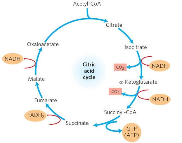
The citric acid cycle consists of a series of enzymatic reactions that convert acetyl-CoA into citrate, which undergoes several transformations to regenerate oxaloacetate. The overall reaction can be summarized as follows:
- Acetyl-CoA + Oxaloacetate → Citrate
- Citrate undergoes isomerization to form isocitrate.
- Isocitrate is oxidized and decarboxylated to α-ketoglutarate.
- α-Ketoglutarate is further oxidized and decarboxylated to succinyl-CoA.
- Succinyl-CoA is converted to succinate, producing GTP or ATP.
- Succinate is oxidized to fumarate.
- Fumarate is hydrated to malate.
- Malate is oxidized back to oxaloacetate.
Each turn of the cycle processes one acetyl-CoA molecule, releasing two molecules of CO2 and regenerating oxaloacetate for another round.
Intermediates of the Citric Acid Cycle
The key intermediates produced during the citric acid cycle include:
- Citrate
- Isocitrate
- α-Ketoglutarate
- Succinyl-CoA
- Succinate
- Fumarate
- Malate
- Oxaloacetate
These intermediates are not only critical for energy production but also serve as precursors for various biosynthetic pathways, including amino acid synthesis.
Role in Amino Acid Synthesis
Several intermediates from the citric acid cycle are directly involved in the synthesis of amino acids:
- α-Ketoglutarate: This intermediate can be transaminated to form glutamate, an important amino acid that serves as a precursor for other amino acids such as glutamine, proline, and arginine.
- Reaction:
- α-Ketoglutarate + NH3 + NADPH → Glutamate + NADP+
- Reaction:
- Oxaloacetate: This compound can be transaminated to produce aspartate, which can then be converted into other amino acids like asparagine through amidation.
- Reaction:
- Oxaloacetate + NH3 + ATP → Asparagine + ADP + Pi
- Reaction:
- Succinyl-CoA: While not directly involved in amino acid synthesis, it plays a role in heme synthesis (which involves glycine) and can influence pathways leading to certain amino acids.
- Citrate: Although primarily associated with energy metabolism, citrate can also lead to the production of fatty acids and cholesterol through acetyl-CoA conversion; however, its direct role in amino acid synthesis is less pronounced compared to α-ketoglutarate and oxaloacetate.
Interconversion with Amino Acids
Conversely, certain amino acids can also contribute back into the citric acid cycle:
- Glutamate: Can be deaminated to form α-ketoglutarate.
- Reaction:
- Glutamate → α-Ketoglutarate + NH3
- Reaction:
- Aspartate: Can be converted back into oxaloacetate through deamination.
- Reaction:
- Aspartate → Oxaloacetate + NH3
- Reaction:
- Other amino acids such as alanine can enter gluconeogenesis pathways that ultimately feed into pyruvate or oxaloacetate.
Conclusion
In summary, the citric acid cycle serves not only as a central hub for energy production but also plays a significant role in synthesizing various amino acids through its intermediates like α-ketoglutarate and oxaloacetate. The interplay between these metabolic pathways highlights the interconnectedness of energy metabolism and biosynthesis within cellular processes.
Citrate Regulation of the TCA Cycle and Fatty Acid Synthesis
Citrate is a key metabolite in cellular metabolism, primarily produced in the mitochondria during the tricarboxylic acid (TCA) cycle (also known as the Krebs cycle). It plays a crucial role in regulating energy production and metabolic pathways, particularly influencing both the TCA cycle and fatty acid synthesis, albeit in opposing manners.
Citrate’s Role in the TCA Cycle
- Production of Citrate: In the TCA cycle, citrate is formed from acetyl-CoA and oxaloacetate through the action of citrate synthase. This reaction marks the entry point of acetyl-CoA into the cycle, which is essential for energy production.
- Regulation of Enzymatic Activity: High levels of citrate indicate an abundance of substrates for energy production. When citrate accumulates, it can inhibit key enzymes in glycolysis, such as phosphofructokinase-1 (PFK-1), thereby reducing glucose breakdown when energy levels are sufficient.
- Feedback Inhibition: Citrate also acts as a feedback inhibitor for certain enzymes within the TCA cycle itself. For example, it inhibits isocitrate dehydrogenase and α-ketoglutarate dehydrogenase when energy levels are high, preventing excessive flux through the cycle and conserving resources.
- Energy Status Indicator: The presence of citrate signals that there is enough energy available; thus, it helps maintain metabolic homeostasis by regulating further entry into the TCA cycle based on cellular energy needs.
Citrate’s Role in Fatty Acid Synthesis
- Transport to Cytosol: When citrate levels are high in mitochondria (often due to excess acetyl-CoA), it can be transported out into the cytosol via specific transporters like the citrate transporter (SLC25A1). This transport occurs when mitochondrial ATP levels are high and indicates an abundance of energy substrates.
- Conversion to Acetyl-CoA: Once in the cytosol, citrate is cleaved by ATP-citrate lyase into acetyl-CoA and oxaloacetate. This conversion provides a source of acetyl-CoA for fatty acid synthesis.
- Activation of Lipogenesis: Acetyl-CoA generated from citrate promotes lipogenesis—the synthesis of fatty acids—by serving as a substrate for fatty acid synthase (FAS). Additionally, elevated levels of malonyl-CoA (produced from acetyl-CoA by acetyl-CoA carboxylase) inhibit carnitine palmitoyltransferase I (CPT1), thus preventing fatty acid oxidation and favoring storage as fat.
- Regulatory Feedback Mechanism: The accumulation of fatty acids leads to increased storage forms like triglycerides while simultaneously inhibiting gluconeogenesis and ketogenesis pathways when energy substrates are plentiful.
Opposing Effects on Metabolism
The contrasting roles that citrate plays in these two pathways highlight its regulatory importance:
- In the TCA cycle, high citrate concentrations signal sufficient energy availability and inhibit further catabolic processes.
- Conversely, when transported to the cytosol, it promotes anabolic processes such as fatty acid synthesis by providing necessary substrates for lipid formation.
This duality allows cells to adapt their metabolic pathways based on nutrient availability and energetic demands effectively.
In summary, while citrate serves as an important intermediate in both processes—regulating catabolism through inhibition within the TCA cycle and promoting anabolism via fatty acid synthesis—it does so in ways that reflect opposite metabolic priorities depending on cellular conditions.
Energy Yield of the TCA Cycle and Its Regulation
The tricarboxylic acid (TCA) cycle, also known as the Krebs cycle or citric acid cycle, is a crucial metabolic pathway that plays a significant role in cellular respiration. It occurs in the mitochondrial matrix of eukaryotic cells and is responsible for the oxidation of acetyl-CoA derived from carbohydrates, fats, and proteins into carbon dioxide and water while generating energy-rich compounds.
Energy Yield of the TCA Cycle
- Input to the Cycle: Each turn of the TCA cycle begins with one molecule of acetyl-CoA, which combines with oxaloacetate to form citrate. The cycle then proceeds through a series of enzymatic reactions.
- Products Generated: For each acetyl-CoA that enters the TCA cycle, the following energy-containing products are generated:
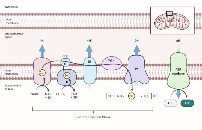
- 3 NADH: Each NADH can yield approximately 2.5 ATP when oxidized in the electron transport chain.
- 1 FADH2: Each FADH2 can yield approximately 1.5 ATP.
- 1 GTP (or ATP): This is directly usable energy.
- Total Energy Yield Calculation:
- From 3 NADH: 3 × 2.5 = 7.5 ATP
- From 1 FADH2: 1 × 1.5 = 1.5 ATP
- From 1 GTP (or ATP): 1 ATP
Therefore, the total energy yield per acetyl-CoA molecule entering the TCA cycle is:Total ATP = 7.5 + 1.5 +1 = 10 ATP
- Overall Yield from Glycolysis: Since each glucose molecule produces two molecules of pyruvate during glycolysis, which are then converted to two molecules of acetyl-CoA, the total yield from one glucose molecule through both glycolysis and the TCA cycle would be:
- From glycolysis: 2 NADH = 2 × 2.5 = 5 ATP
- From two acetyl-CoA in TCA cycle: 10 ATP
Thus, total yield from one glucose molecule: Total from glucose = 5 + (10) = 15 ATP
Regulation of the TCA Cycle
The regulation of the TCA cycle is critical for maintaining metabolic balance and ensuring efficient energy production:
- Allosteric Regulation by Metabolites:
- The activity of enzymes within the TCA cycle is regulated by various metabolites that serve as allosteric effectors.
- Key regulatory points include:
- NADH inhibits several dehydrogenases such as pyruvate dehydrogenase and α-ketoglutarate dehydrogenase.
- ATP acts as an inhibitor for citrate synthase and α-ketoglutarate dehydrogenase under certain conditions.
- Substrate Availability:
- The availability of substrates such as acetyl-CoA and oxaloacetate influences enzyme activity.
- A high concentration of ADP stimulates the conversion of substrates into energy forms by promoting enzyme activity.
- Feedback Inhibition:
- Certain intermediates like succinyl-CoA inhibit their own synthesis pathways to prevent overproduction when their levels are sufficient.
- Hypoxia-Induced Regulation:
- Under low oxygen conditions, cells may exhibit a pseudohypoxic phenotype which stabilizes hypoxia-inducible factors (HIFs), leading to altered metabolism including increased reliance on anaerobic pathways rather than oxidative phosphorylation.
In summary, each turn of the TCA cycle yields approximately 10 ATP per acetyl-CoA, with regulation primarily occurring through allosteric effects by metabolites like NADH and ATP, substrate availability, feedback inhibition mechanisms, and adaptations to oxygen levels.
Shuttle Mechanisms for Transport of NADH into Mitochondria
The transport of NADH into mitochondria is crucial for cellular respiration, particularly in the context of aerobic metabolism. Since NADH cannot directly cross the mitochondrial membrane due to its charged nature, specific shuttle mechanisms are employed to facilitate its transfer. The two primary shuttle systems involved in this process are the Glycerol Phosphate Shuttle and the Malate-Aspartate Shuttle. Below is a detailed examination of these mechanisms.
1. Glycerol Phosphate Shuttle
The Glycerol Phosphate Shuttle primarily operates in muscle and brain tissues. This shuttle allows for the transfer of reducing equivalents from cytosolic NADH into the mitochondria by converting NADH into glycerol-3-phosphate (G3P) and then back to dihydroxyacetone phosphate (DHAP) within the mitochondria.
- Step 1: Conversion of NADH to G3P
- In the cytosol, an enzyme called glycerol-3-phosphate dehydrogenase catalyzes the reduction of DHAP to G3P using electrons from NADH. This reaction produces NAD+ as a byproduct.
DHAP + NADH → G3P + NAD+
- Step 2: Transport into Mitochondria
- G3P can then be transported across the mitochondrial membrane via specific transport proteins.
- Step 3: Conversion back to DHAP
- Once inside the mitochondria, another isoform of glycerol-3-phosphate dehydrogenase oxidizes G3P back to DHAP, transferring electrons to FAD, forming FADH2.
G3P + FAD → DHAP + FADH2
This FADH2 can then enter the electron transport chain (ETC), contributing to ATP production.
2. Malate-Aspartate Shuttle
The Malate-Aspartate Shuttle is more prominent in liver, kidney, and heart tissues and is considered more efficient than the glycerol phosphate shuttle because it transfers reducing equivalents directly as malate.
- Step 1: Conversion of NADH to Malate
- In this system, cytosolic NADH reduces oxaloacetate (OAA) to malate through the action of malate dehydrogenase, generating NAD+.
OAA + NADH → Malate + NAD+
- Step 2: Transport into Mitochondria
- Malate can freely cross the mitochondrial membrane via specific transporters.
- Step 3: Conversion back to OAA
- Inside the mitochondria, malate is reoxidized back to OAA by mitochondrial malate dehydrogenase, producing another molecule of NADH.
Malate + NAD + → OAA + NADH
- Step 4: Transamination Reaction
- OAA can be converted into aspartate through a transamination reaction with glutamate, which regenerates α-ketoglutarate and allows OAA to exit back into the cytosol.
This cycle effectively transfers reducing equivalents from cytosolic NADH into mitochondrial NAD+, allowing for efficient ATP production through oxidative phosphorylation.
Conclusion
Both shuttle systems—Glycerol Phosphate and Malate-Aspartate—play critical roles in transporting reducing equivalents from cytosolic NADH into mitochondria where they can participate in ATP synthesis via oxidative phosphorylation. The choice between these shuttles depends on tissue type and metabolic conditions.


