Definitions and Diagnostic Criteria for Pericarditis
Definition of Pericarditis
Pericarditis is defined as inflammation of the pericardium, the fibrous sac surrounding the heart. This condition can result from various etiologies, including viral infections, bacterial infections, autoimmune diseases, trauma, or post-myocardial infarction. The inflammation can lead to symptoms such as chest pain, fever, and dyspnea.
Diagnostic Criteria for Pericarditis
The diagnosis of pericarditis is primarily clinical but can be supported by various diagnostic tests. The following criteria are commonly used:
- Typical Chest Pain:
- The pain is usually sharp and pleuritic in nature.
- It may worsen with inspiration or coughing and improve when sitting forward.
- Pericardial Friction Rub:
- A characteristic sound heard on auscultation due to the rubbing of the inflamed pericardial layers.
- Electrocardiogram (ECG) Changes:
- Early stages show widespread ST-segment elevation and PR-segment depression.
- These changes are typically diffuse rather than localized to specific leads.
- Imaging Studies:
- Echocardiography can reveal pericardial effusion and assess the size of the pericardium.
- In some cases, a chest X-ray or CT scan may be utilized to evaluate for other causes of chest pain or complications like constrictive pericarditis.
- Laboratory Tests:
- Blood tests may show elevated inflammatory markers (e.g., C-reactive protein, erythrocyte sedimentation rate).
- Specific tests may be conducted based on suspected underlying causes (e.g., viral serologies).
- Exclusion of Other Conditions:
- It is essential to rule out other potential causes of chest pain such as myocardial infarction, pulmonary embolism, or aortic dissection through appropriate clinical evaluation and testing.
Application in Clinical Practice
In clinical practice, recognizing pericarditis involves a thorough history-taking and physical examination followed by targeted investigations:
- History Taking: Clinicians should inquire about the onset, character, duration of chest pain, associated symptoms (fever, malaise), recent infections or illnesses, and any history of autoimmune diseases.
- Physical Examination: Auscultation for a pericardial friction rub is crucial; this finding strongly suggests pericarditis.
- Initial Workup: An ECG should be performed early in the evaluation process to identify characteristic changes associated with pericarditis.
- Echocardiography: If there is suspicion of significant effusion or if symptoms persist despite treatment, an echocardiogram should be ordered to assess cardiac function and fluid accumulation around the heart.
- Management Strategies: Treatment typically involves non-steroidal anti-inflammatory drugs (NSAIDs) for symptom relief. In cases where there is an underlying cause (e.g., infection), specific therapies may be warranted.
- Follow-Up Care: Patients diagnosed with acute pericarditis should be monitored for recurrence or complications such as constrictive pericarditis or cardiac tamponade.
By adhering to these definitions and diagnostic criteria in clinical practice, healthcare providers can effectively identify and manage patients with pericarditis.
Classification of Pericardial Effusion
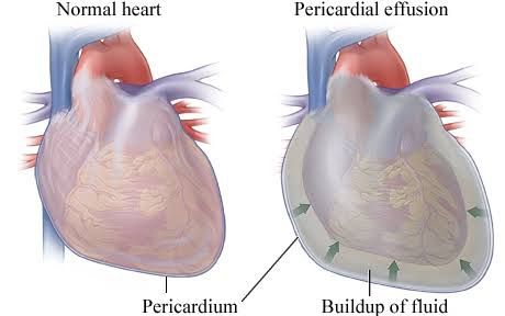
Pericardial effusion refers to the accumulation of fluid in the pericardial space, which can have various causes and implications for cardiac function. The classification of pericardial effusion is essential for diagnosis and management. It can be categorized based on several criteria:
1. Onset
- Acute or Subacute: Acute effusions develop rapidly, often within hours to days, while subacute effusions develop over a period of weeks.
- Chronic: This type lasts longer than three months and may result from ongoing conditions such as malignancy or chronic inflammatory diseases.
2. Distribution
- Circumferential: Fluid surrounds the heart uniformly.
- Loculated: Fluid accumulates in specific pockets or areas, often due to adhesions or previous infections.
3. Hemodynamic Impact
- Cardiac Tamponade: This occurs when the pressure from the fluid accumulation significantly impairs the heart’s ability to fill properly, leading to reduced cardiac output and potential shock.
- No Cardiac Tamponade: In cases where the fluid does not exert enough pressure to affect heart function significantly.
4. Composition
- Serous Fluid: The most common type found in benign conditions.
- Blood: May indicate trauma or malignancy.
- Rarely Air or Gas: Can occur due to bacterial infections leading to an empyema.
5. Size Assessment The size of the effusion can be evaluated using echocardiography, which provides a semiquantitative assessment. The echocardiographic findings help determine the volume of pericardial fluid present and guide clinical decisions regarding intervention.
In summary, understanding these classifications helps healthcare providers assess the severity and potential impact of pericardial effusion on cardiac function, guiding appropriate treatment strategies.
Therapeutic Options for Refractory Recurrent Pericarditis and Pericardial Effusion
Refractory recurrent pericarditis and pericardial effusion present significant challenges in clinical management. The treatment approach must be individualized, taking into account the patient’s history, underlying causes, and response to previous therapies. Below is a detailed overview of both old and new therapeutic options available as of December 2024.
1. Understanding Refractory Recurrent Pericarditis
Recurrent pericarditis is defined as the return of symptoms after an initial episode has resolved. When it becomes refractory, it means that the condition does not respond adequately to standard treatments. The most common symptoms include chest pain, fever, and signs of inflammation.
2. Standard Treatment Options
Historically, the management of recurrent pericarditis has included:
- Nonsteroidal Anti-Inflammatory Drugs (NSAIDs): These are typically the first-line treatment for acute episodes. Common NSAIDs used include ibuprofen and indomethacin.
- Colchicine: This medication has been shown to reduce the recurrence rate of pericarditis when used alongside NSAIDs or as monotherapy.
- Corticosteroids: In cases where NSAIDs and colchicine are ineffective or contraindicated, corticosteroids like prednisone may be prescribed. However, their long-term use can lead to complications.
3. Newer Therapeutic Approaches
Recent advancements have introduced several new therapeutic options for managing refractory recurrent pericarditis:
- Interleukin-1 Inhibitors: Medications such as anakinra (Kineret) and canakinumab (Ilaris) target inflammatory pathways involved in pericarditis. Clinical trials have demonstrated their efficacy in reducing symptoms and preventing recurrences.
- Immunosuppressive Agents: Drugs such as azathioprine or mycophenolate mofetil may be considered in patients who do not respond to conventional therapies. These agents help modulate the immune response but require careful monitoring due to potential side effects.
- Biologics: Emerging biologics targeting specific inflammatory pathways are being studied for their effectiveness in treating refractory cases. For example, agents that inhibit tumor necrosis factor-alpha (TNF-alpha) may offer benefits based on their role in inflammatory processes.
4. Management of Pericardial Effusion
Pericardial effusion can occur alongside recurrent pericarditis and may require different management strategies:
- Observation: Small effusions without significant symptoms often require no immediate intervention but should be monitored with follow-up echocardiograms.
- Pericardiocentesis: This procedure involves using a needle to remove excess fluid from the pericardial space and can provide symptomatic relief, especially if there is cardiac tamponade (pressure on the heart).
- Surgical Options: For recurrent or large effusions that do not respond to medical therapy, surgical interventions such as pericardial window creation or complete pericardiectomy may be necessary.
5. Multidisciplinary Approach
Management of refractory recurrent pericarditis often requires a multidisciplinary team approach involving cardiologists, rheumatologists, and sometimes infectious disease specialists to address underlying causes effectively.
In summary, while traditional treatments like NSAIDs and colchicine remain foundational in managing recurrent pericarditis, newer therapies such as interleukin inhibitors and immunosuppressants provide additional options for those with refractory cases. Continuous research is essential for developing more effective strategies tailored to individual patient needs.
Strengths and Weaknesses of Different Imaging Methods for Pericardial Diseases
When evaluating pericardial diseases, several imaging modalities are available, each with its strengths and weaknesses. The choice of imaging method depends on the specific clinical scenario, the patient’s condition, and the diagnostic target. Below is a detailed analysis of the most commonly used imaging techniques: echocardiography, computed tomography (CT), magnetic resonance imaging (MRI), and chest X-ray.
1. Echocardiography
Strengths:
- Non-invasive: Echocardiography is a bedside procedure that does not involve radiation.
- Real-time imaging: It provides dynamic images of cardiac structures and function.
- Assessment of hemodynamics: Doppler echocardiography can evaluate blood flow across valves and estimate pressures in heart chambers.
- Identification of effusions: It is particularly effective in detecting pericardial effusion and assessing its size and hemodynamic significance.
Weaknesses:
- Operator-dependent: The quality of the images can vary significantly based on the operator’s skill.
- Limited visualization: Certain anatomical areas may be difficult to visualize due to patient factors (e.g., obesity, lung disease).
- Inability to assess complex anatomy: While it can identify effusions, it may struggle with more complex pericardial diseases such as constrictive pericarditis.
2. Computed Tomography (CT)
Strengths:
- High-resolution images: CT provides detailed cross-sectional images of the heart and surrounding structures.
- Assessment of calcification: It is excellent for visualizing calcifications in the pericardium, which can indicate chronic conditions.
- Evaluation of masses: CT can help differentiate between pericardial effusion and masses or tumors adjacent to the heart.
Weaknesses:
- Radiation exposure: CT involves ionizing radiation, which poses risks, especially in younger patients or those requiring multiple scans.
- Limited functional assessment: Unlike echocardiography, CT does not provide real-time functional information about cardiac dynamics.
3. Magnetic Resonance Imaging (MRI)
Strengths:
- Excellent soft tissue contrast: MRI provides superior detail regarding soft tissue structures compared to CT or echocardiography.
- Functional assessment: It allows for evaluation of cardiac function and morphology without radiation exposure.
- Characterization of lesions: MRI can help differentiate between various types of pericardial disease based on tissue characteristics.
Weaknesses:
- Cost and availability: MRI is generally more expensive than other modalities and may not be readily available in all settings.
- Longer acquisition time: The time required for an MRI scan can be a limitation in acute settings.
- Contraindications: Patients with certain implants or devices may not be eligible for MRI.
4. Chest X-ray
Strengths:
- Widely available and quick: Chest X-rays are easily accessible in most healthcare settings and provide rapid results.
- Initial assessment tool: They can help identify gross abnormalities such as significant cardiomegaly or large pleural effusions.
Weaknesses:
- Limited detail: Chest X-rays do not provide sufficient detail for diagnosing specific pericardial diseases.
- Poor sensitivity for small effusions or early disease changes.
Selecting the Best Imaging Method
The selection of the best imaging modality depends on several factors:
- Clinical Presentation:
- For patients presenting with symptoms suggestive of pericarditis or effusion (e.g., chest pain, dyspnea), echocardiography is typically first-line due to its non-invasive nature and ability to assess hemodynamics quickly.
- Diagnostic Target:
- If there is suspicion of constrictive pericarditis or complex anatomy that requires detailed assessment, MRI would be preferred due to its superior soft tissue characterization.
- For cases where calcification needs evaluation or when masses are suspected, CT would be advantageous.
- Patient Factors:
- In patients who cannot undergo MRI due to contraindications (e.g., pacemakers), CT may serve as an alternative despite radiation concerns.
- Availability and Cost Considerations:
- In resource-limited settings where advanced imaging may not be available, echocardiography remains a practical choice for initial evaluation.
In summary, while echocardiography often serves as the first-line imaging modality due to its accessibility and effectiveness in assessing fluid status around the heart, CT or MRI may be necessary depending on specific clinical scenarios requiring further investigation into structural abnormalities or complex pathologies.
Best Imaging Method According to Patient Needs:
For most cases involving suspected pericardial disease where immediate assessment is needed—especially if there are signs of effusion—echocardiography is typically the best initial imaging method, followed by either CT or MRI based on further diagnostic requirements related to patient presentation.


