Understanding Pediatric Chest X-Ray Interpretation
Interpreting pediatric chest X-rays requires a systematic approach due to the unique anatomical and physiological characteristics of children. The process can be broken down into several key steps, which include assessing image quality, identifying normal anatomical structures, and recognizing common abnormalities.
1. Assessing Image Quality
Before interpreting any chest X-ray, it is crucial to evaluate the quality of the image. This involves checking for:
- Rotation: The medial ends of the clavicles should be equidistant from the spinous processes of the vertebrae. If one side appears larger than the other, it indicates rotation.
- Inspiration: A good quality X-ray should show at least 7 anterior ribs and 9 posterior ribs visible above the diaphragm. The diaphragm should ideally be at the level of the 8th to 10th ribs posteriorly or between the 5th and 6th ribs anteriorly.
- Exposure: Adequate exposure allows for visualization of both lung fields and mediastinal structures. Underexposed films appear too bright, while overexposed films appear too dark.
2. Identifying Normal Anatomical Structures
Once image quality is confirmed, identifying normal structures is essential:
- Cardiac Silhouette: In a posteroanterior (PA) view, assess both right and left heart borders. The cardiothoracic ratio (heart width compared to thoracic width) should be less than 0.5 in adults and less than 0.6 in infants.
- Lungs: Evaluate lung fields for symmetry and clarity. Look for vascular markings that indicate normal pulmonary circulation.
- Diaphragm: The position and contour of the diaphragm can provide insights into underlying conditions such as atelectasis or pleural effusion.
3. Recognizing Common Abnormalities
Several common abnormalities can be identified through careful examination:
- Consolidation: Appears as an area of increased opacity in one or more lobes, often indicating pneumonia.
- Atelectasis: Characterized by volume loss in a lung segment or lobe, leading to displacement of fissures and mediastinal shift towards the affected side.
- Pleural Effusion: Appears as blunting of costophrenic angles on upright films; larger effusions may cause mediastinal shift away from the effusion.
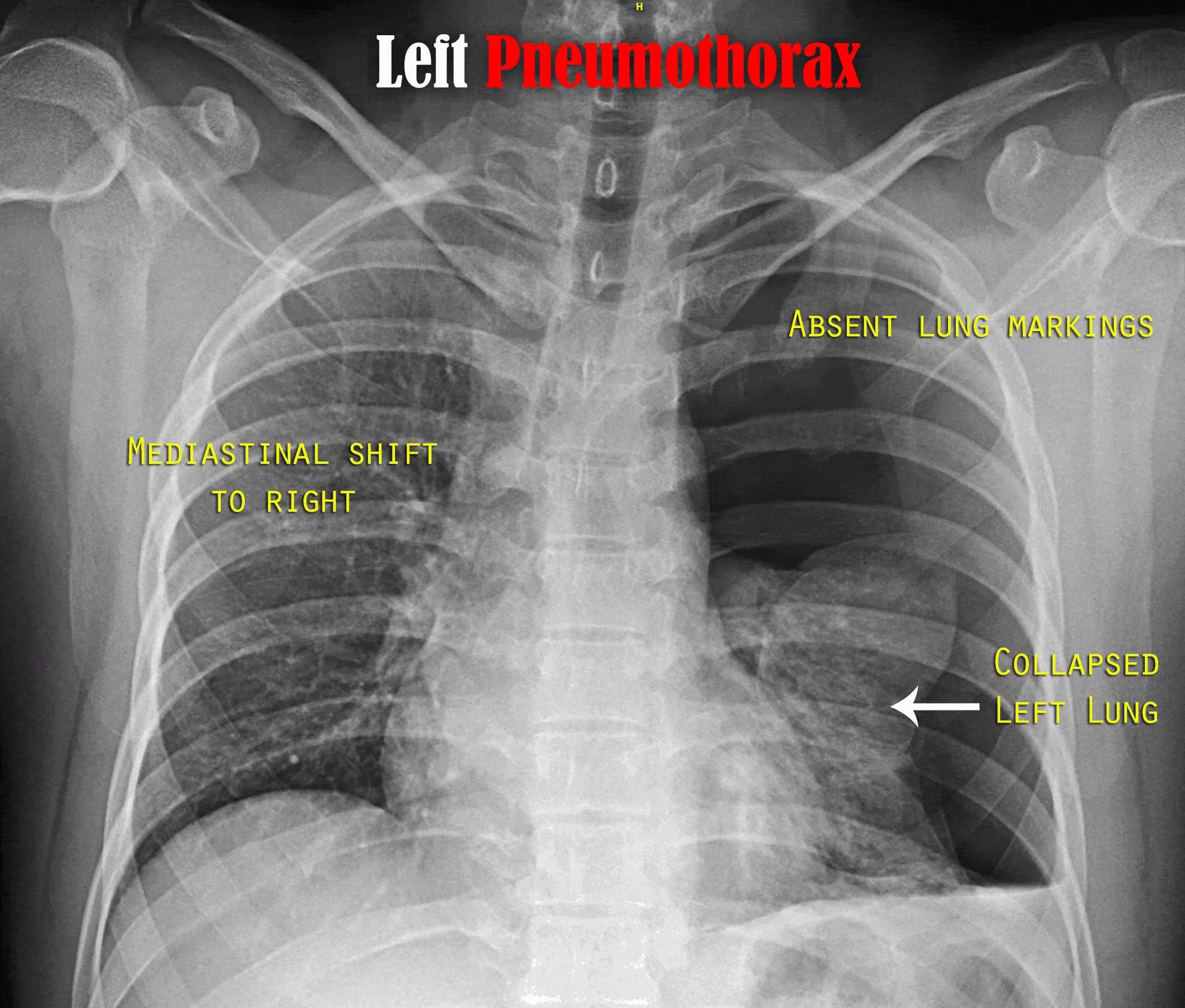
- Pneumothorax: Identified by a visceral pleural line with absence of vascular markings beyond this line; may cause mediastinal shift towards the affected side if large.
- Air Bronchograms: Visible air-filled bronchi surrounded by consolidated lung tissue indicate pneumonia or other forms of consolidation.
4. Special Considerations in Neonates
Neonatal chest X-rays require additional considerations due to their unique physiology:
- Assess lung volumes carefully; under-inflated lungs may suggest respiratory distress.
- Look for air bronchograms which are significant indicators in diagnosing neonatal pneumonia.
- Be aware of complications related to prematurity such as hyaline membrane disease or bronchopulmonary dysplasia.
By following these systematic steps—evaluating image quality, identifying normal anatomy, recognizing common abnormalities, and considering special factors for neonates—clinicians can improve their accuracy in interpreting pediatric chest X-rays effectively.
Identifying Different Types of Tubes and Lines on Chest and Abdomen Radiographs
Introduction to Radiographic Tubes and Lines
Radiographs, commonly known as X-rays, are essential diagnostic tools in medicine that provide images of the internal structures of the body. In particular, chest and abdominal radiographs can reveal various medical devices such as tubes and lines that are used for therapeutic or diagnostic purposes. Understanding how to identify these devices is crucial for healthcare professionals in assessing patient conditions.
Types of Tubes and Lines
- Endotracheal Tube (ET Tube):
- Description: An endotracheal tube is a flexible plastic tube inserted through the mouth or nose into the trachea to maintain an open airway.
- Identification on Radiograph: On a chest X-ray, it appears as a radiopaque line extending from the oral cavity down into the trachea, typically positioned above the carina (the point where the trachea bifurcates into the left and right bronchi). The tip should ideally be 2-5 cm above the carina.
- Central Venous Catheter (CVC):
- Description: A central venous catheter is a long, thin tube inserted into a large vein, often used for administering medication or fluids.
- Identification on Radiograph: On chest X-rays, CVCs appear as straight lines that can be seen traversing from a peripheral insertion site (like the subclavian or jugular vein) towards the superior vena cava. The tip of the catheter should ideally be located at or near the junction of the superior vena cava and right atrium.
- Nasogastric Tube (NG Tube):
- Description: A nasogastric tube is inserted through the nose into the stomach for feeding or draining gastric contents.
- Identification on Radiograph: On abdominal X-rays, an NG tube appears as a radiopaque line that runs from one nostril down through the esophagus into the stomach. The tip should be located in the gastric fundus.
- Chest Tube (Pleural Drainage Tube):
- Description: A chest tube is used to drain air (pneumothorax) or fluid (pleural effusion) from around the lungs.
- Identification on Radiograph: On chest X-rays, it appears as a radiopaque line entering through an intercostal space and extending into either pleural cavity. The position will depend on whether it is draining air or fluid; typically placed in anterior locations for pneumothorax and posterior/inferior locations for fluid drainage.
- Intra-Aortic Balloon Pump (IABP):
- Description: An IABP is used to support cardiac function by inflating and deflating a balloon within the aorta.
- Identification on Radiograph: It appears as a long catheter with an inflatable balloon at its distal end, usually positioned just below the left subclavian artery in chest X-rays.
- Dialysis Catheter:
- Description: These catheters are used for hemodialysis access.
- Identification on Radiograph: They can be identified similarly to CVCs but may have multiple lumens visible on imaging. Their tips are often positioned in central veins like subclavian or femoral veins.
- Feeding Tube (Gastrostomy Tube):
- Description: A gastrostomy tube is placed directly into the stomach through abdominal wall surgery for feeding purposes.
- Identification on Radiograph: On abdominal X-rays, it appears as a radiopaque line leading from an external port directly into the stomach.
- Umbilical Catheter (UAC/UVC):
- Description: Used primarily in neonates for vascular access.
- Identification on Radiograph: UACs appear as lines running up towards thoracic vessels while UVCs run towards inferior vena cava; both should be assessed carefully for proper placement.
- Biliary Drainage Catheter:
- Description: This catheter drains bile when there’s obstruction due to stones or tumors.
- Identification on Radiograph: Appears as a line leading from outside of abdomen into biliary tree structures visible in upper abdomen imaging.
- Transvenous Pacemaker Wire:
- Description: Used to manage heart rhythm disturbances by pacing cardiac activity.
- Identification on Radiograph: Appears as thin wires traversing from insertion site toward heart chambers; careful assessment needed to ensure correct positioning within right atrium/ventricle.
Each type of tube or line has specific characteristics that allow healthcare providers to identify them accurately on radiographs. Proper identification not only aids in confirming correct placement but also helps prevent complications associated with misplaced devices.
Understanding these distinctions enhances clinical decision-making processes regarding patient management based upon imaging findings.
Radiographic Signs of a Pneumothorax
Pneumothorax is characterized by the presence of gas in the pleural space, which can be identified through various radiographic signs. The detection and interpretation of these signs are crucial for diagnosis and management. Below are the key radiographic features associated with pneumothorax:
1. Visible Pleural Line
The most definitive sign of a pneumothorax on a chest X-ray is the visible visceral pleural edge, which appears as a thin, sharp white line. This line indicates the boundary between the lung and the pleural space filled with air.
2. Absence of Vascular Markings
In areas peripheral to the visible pleural line, there should be an absence of vascular markings (the white branching structures that represent blood vessels). This lack of vascularity suggests that there is no lung tissue present in that area, indicating a pneumothorax.
3. Radiolucent Area
The region surrounding the lung where air has accumulated will appear radiolucent (dark) compared to adjacent lung tissue on an X-ray. This contrast helps in identifying the extent of the pneumothorax.
4. Mediastinal Shift
In cases of tension pneumothorax, there may be a noticeable shift of the mediastinum away from the side of the pneumothorax due to increased pressure in one pleural cavity. This is a critical finding as it can indicate life-threatening conditions requiring immediate intervention.
5. Lung Collapse
A significant pneumothorax may lead to complete or partial collapse of the lung on the affected side, which can also be visualized on an X-ray.
While not specific to pneumothorax itself, subcutaneous emphysema may accompany it and can be seen as air tracking into subcutaneous tissues around the thoracic cavity.
7. CT Imaging Findings
On computed tomography (CT), pneumothoraces are typically seen as rims of gas surrounding the edges of the lung and may track along fissures. CT scans are particularly useful for detecting small or occult pneumothoraces that might not be visible on standard chest X-rays.
In summary, recognizing these radiographic signs is essential for diagnosing pneumothorax effectively and determining appropriate treatment strategies.
Radiological Findings of Foreign Body Inhalation in Pediatric Age Groups
Foreign body inhalation is a significant concern in pediatric populations, as children are more likely to place objects in their mouths and airways. Radiological imaging plays a crucial role in diagnosing foreign body aspiration. The following outlines the key radiological findings associated with foreign body inhalation in children.
1. Types of Foreign Bodies
Foreign bodies can vary widely, including food items (e.g., nuts, popcorn), toys, and other small objects. The type of foreign body can influence the radiological findings observed on imaging studies.
2. Initial Imaging: Chest X-ray
The first-line imaging modality for suspected foreign body aspiration is typically a chest X-ray (CXR). Key findings may include:
- Air Trapping: This is often seen as hyperlucency on the affected side due to obstructive atelectasis or air trapping distal to the obstruction.
- Atelectasis: This may present as increased density or opacity in the lung fields, indicating collapsed lung tissue due to blockage.
- Obstructive Pneumonia: If a foreign body causes prolonged obstruction, it can lead to pneumonia, which may appear as localized opacities or infiltrates on the X-ray.
- Direct Visualization of the Foreign Body: Some radiopaque objects (like coins) can be directly visualized on X-rays. However, many organic materials (like food) are not visible unless they cause secondary effects.
3. Advanced Imaging: CT Scan
If the chest X-ray is inconclusive or if there is a high suspicion of foreign body aspiration despite normal initial imaging, a computed tomography (CT) scan may be employed. CT scans provide more detailed images and can reveal:
- Location and Size of the Foreign Body: CT scans can accurately depict where the object is lodged within the airway or lungs.
- Associated Complications: Such as pneumothorax, pleural effusion, or abscess formation due to infection.
- Mucosal Edema or Inflammation: Surrounding tissues may show signs of edema or inflammation due to irritation from the foreign body.
4. Specific Findings Based on Object Type
Different types of foreign bodies may present with unique findings:
- Radiopaque Objects: Coins and some plastic toys will appear clearly on both X-rays and CT scans.
- Organic Materials: Items like peanuts or seeds may not be visible but could lead to indirect signs such as atelectasis or pneumonia.
- Metallic Objects: These are usually easily identified due to their high density.
5. Clinical Correlation
It’s essential to correlate radiological findings with clinical symptoms such as coughing, wheezing, stridor, respiratory distress, and history of choking episodes. A thorough clinical evaluation alongside imaging results aids in confirming diagnosis and determining management strategies.
6. Follow-Up Imaging
In cases where initial imaging does not confirm aspiration but clinical suspicion remains high, follow-up imaging might be necessary after a period of observation for any development of complications.
In summary, identifying radiological findings indicative of foreign body inhalation involves understanding both direct visualization techniques like chest X-rays and advanced modalities like CT scans while considering clinical presentation and potential complications.


