Overview of Congenital Bleeding Disorders
1. Von Willebrand Disease
Heritance:
Von Willebrand disease (vWD) is primarily inherited in an autosomal dominant manner, meaning that only one copy of the mutated gene from an affected parent can cause the disorder. However, some rare types may exhibit autosomal recessive inheritance.
Etiology:
vWD is caused by a deficiency or dysfunction of von Willebrand factor (vWF), a protein crucial for blood clotting. This factor helps platelets adhere to blood vessel walls and carries factor VIII, another important clotting protein. The genetic mutations affecting vWF can lead to reduced levels or impaired function of this factor.
Clinical Presentations:
Patients with vWD may experience symptoms such as easy bruising, frequent nosebleeds, heavy or prolonged menstrual bleeding (menorrhagia), and excessive bleeding after surgery or dental work. The severity of symptoms can vary widely among individuals, depending on the type of vWD.
Laboratory Findings:
Laboratory tests typically reveal:
- Prolonged bleeding time.
- Normal platelet count.
- Decreased levels of von Willebrand factor antigen and activity.
- Prolonged activated partial thromboplastin time (aPTT) due to low factor VIII levels.
Treatment:
Treatment options include:
- Desmopressin (DDAVP), which stimulates the release of vWF from endothelial cells.
- Replacement therapy with vWF-containing concentrates for more severe cases.
- Antifibrinolytic agents like tranexamic acid to help reduce bleeding episodes.
2. Hemophilia A
Heritance:
Hemophilia A is inherited in an X-linked recessive pattern, meaning that it predominantly affects males while females are typically carriers. Affected males inherit the gene mutation from their mothers.
Etiology:
This condition is caused by mutations in the F8 gene that encodes for coagulation factor VIII. These mutations lead to either a deficiency or dysfunction of factor VIII, which is essential for normal blood clotting.
Clinical Presentations:
Symptoms often include:
- Spontaneous bleeding episodes, particularly into joints and muscles.
- Prolonged bleeding after injuries or surgeries.
- Hemarthrosis (bleeding into joints), leading to pain and swelling.
Laboratory Findings:
Key laboratory findings include:
- Prolonged activated partial thromboplastin time (aPTT).
- Normal prothrombin time (PT).
- Low levels of factor VIII activity.
Treatment:
Management strategies involve:
- Factor VIII replacement therapy administered during bleeding episodes or as prophylaxis.
- Desmopressin may be used in mild cases to boost factor VIII levels temporarily.
- Gene therapy is being explored as a potential long-term treatment option.
3. Hemophilia B
Heritance:
Hemophilia B also follows an X-linked recessive inheritance pattern similar to hemophilia A, affecting mostly males who inherit the mutation from carrier mothers.
Etiology:
This disorder results from mutations in the F9 gene responsible for producing coagulation factor IX. Like hemophilia A, these mutations result in insufficient levels or dysfunctional factor IX necessary for effective blood coagulation.
Clinical Presentations:
Clinical manifestations are akin to those seen in hemophilia A and include:
- Spontaneous bleeding episodes.
- Hemarthrosis and muscle bleeds.
- Increased risk of bleeding after trauma or surgical procedures.
Laboratory Findings:
Typical laboratory findings consist of:
- Prolonged activated partial thromboplastin time (aPTT).
- Normal prothrombin time (PT).
- Low levels of factor IX activity.
Treatment:
Management includes:
- Factor IX replacement therapy during bleeding episodes and as prophylaxis.
- Newer treatments such as emicizumab, a bispecific antibody that mimics the function of factor VIII, are also available for patients with hemophilia A but are not applicable for hemophilia B specifically.
Understanding the Correct Usage and Significance of Hematological Abnormalities
1. Prothrombin Time (PT)
Usage: Prothrombin time (PT) is a blood test that measures the time it takes for blood to clot. It specifically assesses the extrinsic pathway of coagulation, which involves factors I (fibrinogen), II (prothrombin), V, VII, and X. PT is often used to monitor patients on anticoagulant therapy, particularly those taking warfarin.
Significance: An abnormal PT can indicate various conditions such as liver disease, vitamin K deficiency, or the presence of anticoagulants. A prolonged PT suggests a potential bleeding disorder or an issue with the coagulation cascade. Clinically, it helps in evaluating bleeding risks prior to surgical procedures and in diagnosing coagulopathies.
2. Partial Thromboplastin Time (PTT)
Usage: Partial thromboplastin time (PTT) measures the time it takes for blood to clot via the intrinsic pathway of coagulation, involving factors I, II, V, VIII, IX, X, XI, and XII. It is primarily used to monitor patients on heparin therapy and assess bleeding disorders.
Significance: An abnormal PTT can indicate deficiencies in clotting factors or the presence of inhibitors that affect coagulation. A prolonged PTT may suggest conditions such as hemophilia or von Willebrand disease. Clinicians utilize PTT results to evaluate bleeding risk and guide treatment decisions regarding anticoagulation management.
3. Thrombin Time (TT)
Usage: Thrombin time (TT) assesses the final step of the coagulation cascade by measuring how long it takes for thrombin to convert fibrinogen into fibrin after adding thrombin to plasma samples. TT is less commonly performed than PT and PTT but provides valuable information about fibrinogen levels and function.
Significance: An abnormal TT can indicate issues with fibrinogen levels or function due to conditions like disseminated intravascular coagulation (DIC) or liver disease. A prolonged TT suggests impaired conversion of fibrinogen to fibrin, which can lead to increased bleeding risk.
4. Platelet Count
Usage: The platelet count measures the number of platelets in a given volume of blood and is crucial for assessing hemostatic function. Normal platelet counts range from approximately 150,000 to 450,000 platelets per microliter of blood.
Significance: Abnormal platelet counts can indicate various hematological disorders; thrombocytopenia (low platelet count) may result from bone marrow disorders, autoimmune diseases, or infections leading to increased destruction or decreased production of platelets. Conversely, thrombocytosis (high platelet count) may occur due to reactive processes like inflammation or malignancies. Monitoring platelet counts is essential in managing patients at risk for bleeding or thrombotic events.
Disseminated intravascular coagulation
Etiology
Disseminated intravascular coagulation (DIC) is a complex disorder characterized by the systemic activation of the coagulation cascade, leading to the formation of blood clots throughout the small vessels. The etiology of DIC can be broadly categorized into two main types: primary and secondary.
- Primary DIC: This is rare and typically associated with conditions such as acute promyelocytic leukemia (APL), where there is a direct activation of the coagulation pathway due to malignant cells.
- Secondary DIC: This is more common and can be triggered by various clinical conditions, including:
- Infections: Particularly sepsis caused by bacterial infections, especially Gram-negative bacteria.
- Obstetric complications: Such as placental abruption, amniotic fluid embolism, or severe preeclampsia.
- Trauma: Major trauma or burns can lead to tissue factor release and subsequent coagulopathy.
- Malignancies: Certain cancers can produce pro-coagulant substances that activate the clotting cascade.
- Severe liver disease: Liver dysfunction impairs the synthesis of clotting factors.
- Vascular disorders: Conditions like vasculitis can also contribute to DIC.
The underlying mechanism involves an imbalance between pro-coagulant and anti-coagulant factors, leading to widespread microvascular thrombosis and subsequent organ dysfunction.
Clinical Presentations and Complications
The clinical presentation of DIC varies widely depending on its severity and underlying cause but generally includes:
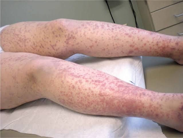
- Bleeding manifestations: Patients may present with bleeding from multiple sites, including petechiae, ecchymoses, hematuria, gastrointestinal bleeding, or bleeding from surgical wounds. This occurs due to consumption of platelets and clotting factors.
- Thrombotic events: Paradoxically, while patients may bleed, they may also experience thrombosis in small vessels leading to ischemia in organs such as kidneys (acute kidney injury), lungs (pulmonary embolism), liver (hepatic dysfunction), and skin (necrosis).
- Organ dysfunction: As microthrombi form in various organs, patients may exhibit signs of multi-organ failure. Symptoms might include altered mental status due to cerebral ischemia, respiratory distress from pulmonary involvement, or jaundice from hepatic impairment.
Complications associated with DIC include:
- Acute respiratory distress syndrome (ARDS)
- Renal failure
- Shock
- Death if not promptly recognized and treated
Laboratory Findings
Laboratory findings in DIC are critical for diagnosis and monitoring. Key laboratory tests include:
- Complete Blood Count (CBC):
- Thrombocytopenia (low platelet count) is common due to consumption during clot formation.
- Coagulation Studies:
- Prolonged prothrombin time (PT) and activated partial thromboplastin time (aPTT) indicate impaired coagulation.
- Decreased fibrinogen levels due to consumption as it is converted into fibrin for clot formation.
- Fibrinolysis Markers:
- Elevated levels of fibrin degradation products such as D-dimer are indicative of increased fibrinolysis occurring as clots are broken down.
- Peripheral Blood Smear:
- May show schistocytes (fragmented red blood cells) which are indicative of microangiopathic hemolytic anemia often seen in DIC.
These laboratory findings help differentiate DIC from other coagulopathies and guide treatment decisions.
Histopathology of Affected Organs
Histopathological examination in cases of DIC reveals characteristic changes:
- Microvascular Thrombosis:
- Small vessel occlusion with fibrin thrombi can be observed in various organs such as lungs, kidneys, liver, heart, and skin. These thrombi consist primarily of fibrin deposits along with platelets and red blood cell fragments.
- Ischemic Changes:
- Organs affected by microthrombi may show signs of ischemic necrosis or infarction due to reduced blood flow resulting from vessel occlusion.
- Hemorrhagic Areas:
- In some cases where there is significant consumption coagulopathy leading to bleeding tendencies, hemorrhagic areas may be noted alongside thrombotic changes.
- Organ-Specific Changes:
- For instance, in the lungs, there may be evidence of diffuse alveolar damage; in the kidneys, acute tubular injury; while hepatic tissues may show centrilobular necrosis due to ischemia.
These histopathological findings correlate with clinical manifestations seen in patients suffering from DIC and provide insight into the pathophysiological processes at play during this complex disorder.


