Mechanisms of Renal Concentration and Dilution of Urine
The kidneys play a crucial role in regulating the body’s fluid balance, electrolyte levels, and waste elimination through the processes of urine concentration and dilution. These processes are primarily facilitated by specialized structures within the nephron, particularly the loop of Henle, and involve two key mechanisms: countercurrent multiplication and countercurrent exchange.
Countercurrent Multipliers
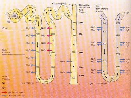
The countercurrent multiplier system is primarily located in the loop of Henle, which consists of a descending limb and an ascending limb. This mechanism is essential for creating a hyperosmotic medullary interstitium, which allows for the concentration of urine.
- Descending Limb: The descending limb of the loop of Henle is permeable to water but not to solutes. As filtrate moves down this limb, water is reabsorbed into the surrounding interstitial fluid due to osmotic gradients created by solute concentrations in the medulla. This results in an increase in osmolarity (concentration) of the filtrate as it descends.
- Ascending Limb: In contrast, the ascending limb is impermeable to water but actively transports sodium (Na+) and chloride (Cl-) ions out into the interstitial fluid. This active transport decreases the osmolarity of the filtrate as it ascends because solutes are being removed without accompanying water loss.
- Establishment of Osmotic Gradient: The combination of these two limbs creates a gradient where the deeper regions of the medulla have higher osmolarity compared to more superficial areas. This gradient is crucial for urine concentration because it allows for water reabsorption from collecting ducts later on.
- Role of Antidiuretic Hormone (ADH): When ADH is present, it increases water permeability in the collecting ducts, allowing more water to be reabsorbed back into circulation based on this established osmotic gradient. Consequently, concentrated urine is produced.
- Multiplier Effect: The term “multiplier” refers to how each cycle through this system amplifies the concentration gradient; as more Na+ and Cl- are pumped out from the ascending limb, more water can be drawn out from the descending limb, enhancing overall urine concentration.
Countercurrent Exchangers
Countercurrent exchangers refer specifically to blood flow through vasa recta—capillaries that supply blood to the renal medulla—and their interaction with tubular fluid in adjacent nephron segments.
- Vasa Recta Functionality: The vasa recta runs parallel to both limbs of the loop of Henle but in opposite directions (the descending vasa recta carries blood downwards while ascending vasa recta returns blood upwards). This arrangement helps maintain the osmotic gradient established by countercurrent multiplication.
- Osmotic Exchange Mechanism: As blood flows down into areas with high osmolarity (due to solute accumulation), it loses water and gains solutes (Na+, Cl-, urea). Conversely, as blood ascends back toward circulation through less concentrated regions, it regains some water while losing solutes back into interstitial fluid.
- Preservation of Medullary Gradient: By maintaining this exchange process without washing away solutes from the medullary interstitium, countercurrent exchangers ensure that high osmolarity remains intact for effective urine concentration when needed.
- Impact on Urine Dilution: In conditions where hydration status changes or when ADH levels drop (as seen during overhydration), less water is reabsorbed from collecting ducts leading to dilute urine production due to decreased osmotic gradients facilitated by these mechanisms.
In summary, renal concentration and dilution mechanisms rely heavily on both countercurrent multiplication within nephron loops and countercurrent exchange via vasa recta capillaries. Together they create an efficient system for regulating body fluids while ensuring waste products are effectively excreted through appropriately concentrated or diluted urine depending on physiological needs.
Role of Urea
Introduction to Urea Handling in the Kidneys
Urea is a small, polar molecule that plays a crucial role in the kidney’s ability to concentrate and dilute urine. It is produced as a byproduct of protein metabolism and is primarily excreted through the urine. The kidneys manage urea through filtration, reabsorption, and secretion processes that are essential for maintaining osmotic balance and regulating water excretion.
Filtration of Urea
The renal process begins with the filtration of blood plasma at the glomerulus. Urea is freely filtered due to its small size and lack of protein binding. Approximately 100% of urea present in plasma enters the Bowman’s capsule during glomerular filtration.
Reabsorption of Urea
After filtration, urea undergoes significant reabsorption within different segments of the nephron:
- Proximal Tubule: About 50% of filtered urea is reabsorbed here passively, primarily due to solvent drag as water is reabsorbed more than urea. This results in an increase in urea concentration within the tubular fluid.
- Loop of Henle: In the thin descending limb, water continues to be reabsorbed while urea remains concentrated due to its inability to permeate this segment effectively. As fluid moves into the thick ascending limb, some urea may diffuse back into the tubular fluid.
- Inner Medulla: The inner medullary collecting ducts play a pivotal role in concentrating urine by allowing passive reabsorption of urea via specific transporters (e.g., UT-A2). This process increases the osmolarity of the interstitial fluid, which is critical for urine concentration.
Urea’s Role in Countercurrent Multiplication
The countercurrent multiplication mechanism relies on both sodium chloride (NaCl) and urea to create an osmotic gradient within the renal medulla:
- Outer Medulla: NaCl is actively reabsorbed from the thick ascending limb, contributing significantly to creating an osmotic gradient.
- Inner Medulla: Here, both NaCl and urea contribute to increasing osmolality. The presence of high concentrations of urea enhances water reabsorption from collecting ducts under antidiuretic hormone (ADH) influence, leading to concentrated urine formation.
This interplay between NaCl and urea allows mammals to produce urine that can be significantly more concentrated than plasma—up to four times higher in humans—enabling effective water conservation during periods of dehydration or low water intake.
Dilution Mechanism Involving Urea
Conversely, when there is excess water intake, urine dilution occurs:
- Decreased ADH Levels: When hydration status improves, ADH secretion decreases, reducing water permeability in collecting ducts.
- Reduced Urea Reabsorption: With lower levels of ADH, less urea is reabsorbed back into circulation from collecting ducts; thus, more urea remains in tubular fluid.
- Increased Urine Volume: This leads to increased urinary output with lower osmolality since less solute (urea) contributes to overall osmotic pressure.
Conclusion
In summary, urea plays a dual role in renal physiology by facilitating both urine concentration and dilution processes through its complex interactions with nephron segments and hormonal regulation. Its ability to enhance osmolarity within renal interstitium underlines its importance not only as a waste product but also as a critical component for maintaining homeostasis regarding body fluids.


