Histopathologic Changes, Age Incidence, and Risk Factors for Cervical Intraepithelial Neoplasia and Its Association with Human Papillomavirus
Histopathologic Changes
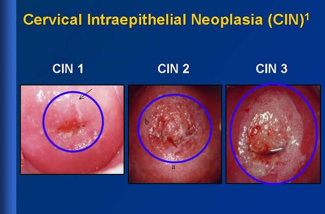
Cervical intraepithelial neoplasia (CIN) is characterized by abnormal growth of squamous cells on the surface of the cervix. The histopathologic classification of CIN is based on the degree of dysplasia observed in cervical biopsies:
- CIN I (Mild Dysplasia): This stage shows slight abnormalities in the lower third of the epithelium. The cells exhibit mild nuclear atypia, and mitotic figures may be present but are not numerous.
- CIN II (Moderate Dysplasia): In this stage, dysplastic changes involve up to two-thirds of the epithelial thickness. There is more pronounced nuclear atypia, increased mitotic activity, and irregularities in cell maturation.
- CIN III (Severe Dysplasia or Carcinoma In Situ): This represents a high-grade lesion where more than two-thirds of the epithelium is affected. The entire thickness may show severe nuclear atypia, loss of normal maturation, and a significant increase in mitotic figures. CIN III can progress to invasive carcinoma if left untreated.
The progression from normal cervical epithelium to CIN involves a series of genetic alterations often triggered by persistent infection with high-risk types of human papillomavirus (HPV), particularly HPV types 16 and 18.
Age Incidence
Cervical intraepithelial neoplasia predominantly affects younger women, with peak incidence occurring between ages 25 and 35 years. However, it can occur at any age after sexual activity begins. The risk decreases significantly after age 50 due to factors such as decreased sexual activity and improved screening practices leading to early detection and treatment.
Risk Factors
Several risk factors contribute to the development of CIN:
- Human Papillomavirus Infection: Persistent infection with high-risk HPV types is the most significant risk factor for developing CIN. HPV is sexually transmitted, and certain strains are strongly associated with cervical cancer.
- Sexual Behavior: Early onset of sexual activity, multiple sexual partners, or having a partner who has had multiple partners increases the risk of HPV infection.
- Immunosuppression: Individuals with weakened immune systems (e.g., those with HIV/AIDS) have a higher risk for persistent HPV infections leading to CIN.
- Smoking: Tobacco use has been linked to an increased risk of cervical dysplasia due to its immunosuppressive effects and potential carcinogenic properties.
- Long-term Use of Oral Contraceptives: Some studies suggest that prolonged use may increase the risk for CIN due to hormonal influences on cervical tissue.
- Co-infection with Other Sexually Transmitted Infections (STIs): Co-existing STIs can facilitate HPV transmission or exacerbate its effects on cervical cells.
- Socioeconomic Factors: Limited access to healthcare services can lead to inadequate screening and delayed diagnosis, increasing the likelihood of advanced lesions at presentation.
Association with Human Papillomavirus
The association between HPV and cervical intraepithelial neoplasia is well-established through extensive epidemiological studies demonstrating that nearly all cases of CIN are linked to persistent infection with high-risk HPV types. The oncogenic potential of these viruses arises from their ability to integrate into host cell DNA, leading to dysregulation of cellular processes that control growth and apoptosis.
High-risk HPVs express early viral proteins E6 and E7 that interfere with tumor suppressor proteins p53 and Rb respectively, promoting uncontrolled cellular proliferation characteristic of neoplastic transformation. Regular screening via Pap smears combined with HPV testing has been shown to significantly reduce incidence rates by allowing for early detection and treatment before progression occurs.
In summary, understanding histopathologic changes in CIN alongside age incidence and associated risk factors—especially those related to HPV—provides critical insights into prevention strategies aimed at reducing cervical cancer morbidity and mortality rates globally.
Squamous Cell Carcinoma Overview
Age Incidence
Squamous cell carcinoma (SCC) of the cervix is most commonly diagnosed in women between the ages of 30 and 50, with a peak incidence around 45 years. The age distribution shows that the risk increases with age, particularly after the onset of sexual activity. Women under 20 years old are rarely diagnosed with cervical SCC, while those over 65 have a higher incidence rate. This pattern reflects both the cumulative effects of risk factors over time and the natural history of human papillomavirus (HPV) infection, which is a significant precursor to cervical cancer.
Predisposing Factors
Several predisposing factors contribute to the development of squamous cell carcinoma of the cervix:
- Human Papillomavirus (HPV) Infection: The primary risk factor for cervical SCC is persistent infection with high-risk HPV types, particularly HPV-16 and HPV-18. These viruses can cause cellular changes that lead to dysplasia and eventually invasive cancer.
- Sexual Behavior: Early onset of sexual activity, multiple sexual partners, and having a partner who has had multiple partners increase the risk of HPV exposure.
- Immunosuppression: Conditions that weaken the immune system, such as HIV/AIDS or immunosuppressive therapies, can increase susceptibility to HPV infections and subsequent cervical cancer.
- Smoking: Tobacco use has been linked to an increased risk of cervical SCC due to carcinogenic substances in tobacco that may affect cervical cells.
- Long-term Use of Oral Contraceptives: Some studies suggest that prolonged use (five years or more) may be associated with an increased risk of cervical cancer.
- Socioeconomic Factors: Limited access to healthcare services, including screening programs like Pap smears and HPV vaccinations, can lead to higher rates of undiagnosed precancerous lesions and cancers.
- History of Cervical Dysplasia: Women with a history of high-grade squamous intraepithelial lesions (HSIL) are at increased risk for developing invasive cervical cancer.
Pathologic Characteristics
The pathologic characteristics of squamous cell carcinoma include:
- Histological Types: The majority are keratinizing or non-keratinizing types, with keratinizing SCC being more common in older women and associated with better prognosis compared to non-keratinizing types.
- Tumor Differentiation: Tumors can be well-differentiated (more organized structure), moderately differentiated, or poorly differentiated (less organized structure). Poorly differentiated tumors tend to have a worse prognosis due to their aggressive nature.
- Invasion Patterns: SCC typically invades locally into surrounding tissues including the stroma and may show desmoplastic reaction—a fibrous tissue response indicative of malignancy.
- Lymphovascular Invasion: Presence of tumor cells in lymphatic vessels or blood vessels indicates a higher likelihood for metastasis.
- Staging: The International Federation of Gynecology and Obstetrics (FIGO) staging system classifies cervical cancer based on depth of invasion and spread to regional lymph nodes or distant sites.
Sites of Metastases
Squamous cell carcinoma primarily metastasizes through lymphatic channels but can also spread hematogenously in advanced stages:
- Regional Lymph Nodes: The first sites for metastases are usually pelvic lymph nodes followed by para-aortic lymph nodes as the disease progresses.
- Distant Metastases: Common distant sites include:
- Lungs
- Liver
- Bones
- Abdomen
- Peritoneal Spread: Advanced cases may involve peritoneal dissemination leading to ascites or peritoneal carcinomatosis.
- Local Invasion: Direct extension into adjacent structures such as the vagina, bladder, rectum, and pelvic wall is also common in advanced disease stages.
In summary, squamous cell carcinoma of the cervix predominantly affects women aged 30-50 years old, driven largely by HPV infection along with various lifestyle and health factors. Its pathological characteristics include distinct histological types and patterns indicative of invasiveness while metastasis typically occurs first through lymphatics before spreading distantly via blood circulation or local invasion into neighboring organs.


