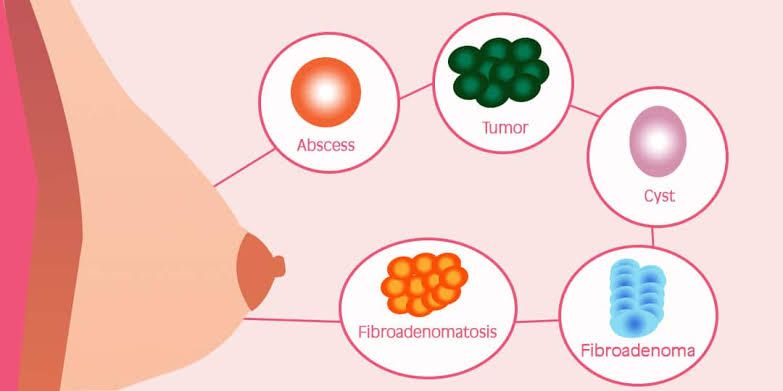
Major Types of Breast Lumps
1. Benign Breast Lumps
Benign breast lumps are non-cancerous and account for the majority of breast lumps. Approximately 80% of breast lumps that are biopsied are benign. The most common types include:
- Fibrocystic Changes: This condition affects 50 to 60 percent of women, particularly those aged 20 to 50. It is characterized by the development of fibrous lumps and/or multiple small cysts due to an exaggerated response of breast tissue to hormonal changes during the menstrual cycle. Symptoms often include increased size and tenderness of lumps before menstruation, which typically decrease afterward. Fibrocystic changes usually resolve after menopause.
- Fibroadenomas: These are solid, benign tumors made up of fibrous and glandular tissue, most commonly found in women aged 18 to 35. They are generally painless and movable when palpated, although they may become tender before menstruation.
- Papillomas: These small wart-like growths occur in the lining of the mammary ducts near the nipple and can cause clear or bloody discharge from the nipple.
2. Malignant Tumors
Malignant breast tumors are cancerous and can invade surrounding tissues if not detected early. Key characteristics include:
- Hard Lumps or Thickening: Malignant tumors often present as single, hard lumps that may be painless but can also be associated with changes in the nipple’s appearance or skin dimpling. They typically arise from mammary glands or ducts.
- Metastasis Potential: If left untreated, malignant tumors can spread to nearby lymph nodes and other parts of the body through a process called metastasis, significantly decreasing survival rates.
Breast cancer remains the most commonly diagnosed cancer among American women, with approximately one in eight women expected to develop it during their lifetime.
3. Other Benign Conditions
In addition to the major types listed above, other benign conditions can also lead to breast lumps:
- Breast Cysts: These are soft fluid-filled sacs that can form due to hormonal fluctuations related to the menstrual cycle or other physiological changes such as pregnancy or menopause.
- Other Benign Fibrocystic Masses: These masses may consist of a combination of fibrous tissue and cysts resulting from hormonal influences on breast tissue.
It is crucial for any woman who discovers a lump in her breast to consult a healthcare professional for evaluation and diagnosis, as self-diagnosis is unreliable.
Common Risk Factors for Benign Breast Disease
Benign breast disease encompasses a variety of noncancerous conditions affecting breast tissue. Understanding the risk factors associated with these conditions can help in early detection and management. Here are some common risk factors:
1. Family History of Breast Cancer
Having a family history of breast cancer significantly increases the likelihood of developing benign breast conditions. Research indicates that premenopausal women with a family history are twice as likely to develop certain types of noncancerous abnormal cell growth, such as epithelial proliferation with atypia (EPA). This condition not only raises the risk for benign issues but also has implications for future breast cancer risk.
2. Hormone Replacement Therapy (HRT)
Postmenopausal hormone therapy is linked to higher rates of benign breast conditions, including EPA and other types that do not typically increase cancer risk. Women who undergo HRT may experience changes in their breast tissue due to hormonal fluctuations, which can lead to the development of benign lumps or cysts.
3. Age
Age is a significant factor in the development of benign breast disease. As women age, particularly during their reproductive years and into menopause, they may experience more changes in breast tissue due to hormonal shifts, increasing the likelihood of benign conditions.
4. Obesity
Interestingly, obesity has been associated with lower rates of certain benign breast conditions compared to women of normal weight, except for specific types like epithelial proliferation (EP). The relationship between body weight and breast health is complex and may involve hormonal influences on breast tissue.
5. Reproductive History
Women who have not given birth tend to have different risks compared to those who have had children. Premenopausal women without children are less likely to develop EP but may be more prone to fluid-filled cysts. Conversely, having multiple pregnancies can influence the type and frequency of benign changes experienced.
6. Use of Oral Contraceptives
The use of oral contraceptives has been shown to reduce the likelihood of developing certain types of benign growths such as fibroadenomas with atypia and fibrocystic changes. This protective effect is thought to be related to how these medications regulate hormone levels.
7. Hormonal Changes Related to Menstruation or Pregnancy
Hormonal fluctuations during menstruation or pregnancy can lead to temporary changes in breast tissue, resulting in tenderness or lump formation. These changes are usually benign but highlight how sensitive breast tissue is to hormonal variations.
In summary, while many factors contribute to the development of benign breast disease, understanding these common risk factors can empower individuals to monitor their health proactively and seek medical advice when necessary.
Diagnostic Modalities and Their Sequence in the Workup of a Patient with a Breast Mass and Nipple Discharge
1. Introduction
The evaluation of a breast mass or nipple discharge involves a systematic approach to ensure accurate diagnosis and appropriate management. The workup typically includes a combination of clinical assessment, imaging studies, and possibly histological evaluation.
2. Workup for a Patient with a Breast Mass
Step 1: Clinical Evaluation
- History Taking: A thorough history is essential, including the duration of the mass, associated symptoms (pain, changes in size), personal and family history of breast cancer, and menstrual history.
- Physical Examination: A detailed examination of the breast and regional lymph nodes is performed to assess the characteristics of the mass (size, shape, consistency) and to check for any signs of lymphadenopathy.
Step 2: Imaging Studies
- Mammography: This is often the first imaging modality used in women over 30 years old. It helps identify calcifications or masses that may not be palpable.
- Ultrasound: Typically used as an adjunct to mammography, especially in younger women or when further characterization of a mass is needed. Ultrasound can differentiate between solid and cystic lesions.
Step 3: Biopsy
- Fine Needle Aspiration (FNA): This may be performed if there is suspicion of malignancy based on imaging findings. It allows for cytological analysis.
- Core Needle Biopsy: Provides more tissue than FNA and is often preferred for definitive diagnosis.
- Surgical Biopsy: In cases where FNA or core biopsy results are inconclusive or if there are complex lesions.
Step 4: Additional Imaging (if necessary)
- MRI: May be indicated for further evaluation in certain cases, such as assessing multifocal disease or in patients with dense breast tissue.
3. Workup for a Patient with Nipple Discharge
Step 1: Clinical Evaluation
- History Taking: Important factors include the nature of the discharge (spontaneous vs. elicited), color (clear, bloody), duration, associated symptoms (pain or mass), and any medications that might influence discharge.
- Physical Examination: Focuses on examining both breasts for masses or abnormalities and checking for any signs of infection or inflammation.
Step 2: Imaging Studies
- Mammography: Recommended particularly if there are concerning features such as unilateral discharge or associated masses.
- Ultrasound: Useful to evaluate underlying pathology when discharge is present; can help identify ductal abnormalities or masses.
Step 3: Ductography (Galactography)
- If imaging suggests an intraductal lesion, ductography can be performed to visualize the ducts after injecting contrast material into the affected duct.
Step 4: Biopsy
- If imaging reveals suspicious findings or if there’s concern about malignancy based on clinical evaluation, biopsy may be warranted:
- Ductal Lavage/Biopsy: Can be performed during ductography if an abnormality is seen.
- Surgical Excision: May be necessary if there’s significant concern regarding malignancy despite previous evaluations.
4. Conclusion
The diagnostic workup for both breast masses and nipple discharge follows a structured approach involving clinical assessment followed by appropriate imaging studies and biopsies as needed. The sequence may vary slightly depending on individual patient factors but generally adheres to this framework to ensure comprehensive evaluation.
Natural History of Benign Breast Disorders
The natural history of benign breast disorders encompasses a range of conditions that affect breast tissue, primarily characterized by non-cancerous changes. Understanding these disorders involves examining their prevalence, types, symptoms, diagnosis, and potential progression over time.
Prevalence and Types
Benign breast disorders are quite common among individuals with breast tissue, particularly women. It is estimated that up to 50% of women will experience some form of benign breast disease during their lifetime. The most prevalent types include:
- Fibrocystic Changes: These are characterized by lumpy or rope-like breast tissue and may cause discomfort or pain (mastalgia). They are often influenced by hormonal fluctuations associated with the menstrual cycle.
- Fibroadenomas: These are solid, non-cancerous tumors made up of glandular and connective tissue. They are most common in younger women and typically do not require treatment unless they cause discomfort or grow significantly.
- Cysts: Fluid-filled sacs that can develop in the breast tissue. They can vary in size and may be tender but are generally harmless.
- Atypical Hyperplasia: This condition involves an abnormal increase in the number of cells within the breast ducts or lobules and is associated with a higher risk of developing breast cancer later on.
- Gynecomastia: This condition occurs in men and people assigned male at birth (AMAB) when there is an imbalance between estrogen and testosterone levels, leading to enlarged breast tissue.
Symptoms
Many benign breast disorders present with noticeable symptoms such as lumps, tenderness, or changes in texture. Some individuals may also experience nipple discharge or swelling. However, many benign conditions can be asymptomatic and discovered incidentally during routine examinations or imaging studies like mammograms.
Diagnosis
Diagnosis typically involves a combination of clinical examination, imaging studies (such as ultrasound or mammography), and sometimes biopsy to differentiate between benign conditions and potential malignancies. Most benign lumps do not require invasive treatment; however, atypical hyperplasia may necessitate closer monitoring due to its association with increased cancer risk.
Natural Progression
The natural progression of benign breast disorders varies widely depending on the specific condition:
- Fibrocystic Changes: These often fluctuate with hormonal cycles but generally do not progress to cancer.
- Fibroadenomas: While they can persist for years, they usually remain stable in size; surgical removal is only recommended if they become symptomatic.
- Cysts: Many cysts resolve spontaneously without intervention; however, larger cysts may require aspiration if they cause discomfort.
- Atypical Hyperplasia: Individuals diagnosed with this condition should undergo regular monitoring due to the elevated risk for developing breast cancer over time.
Overall, while benign breast disorders can cause anxiety due to concerns about cancer risk, most cases are manageable and do not lead to serious health issues. Regular check-ups and awareness of any changes in breast tissue are essential for early detection and management.
Treatment for Fibroadenoma and Fibrocystic Diseases
Fibroadenoma Treatment
- Observation: In many cases, fibroadenomas do not require treatment, especially if they are small, asymptomatic, and confirmed to be benign through imaging or biopsy. Healthcare providers often recommend regular monitoring through physical exams and imaging tests (like ultrasound or mammograms) every three to six months to check for any changes in size or characteristics.
- Surgical Removal: If a fibroadenoma is large, painful, or shows suspicious features on imaging studies, surgical excision may be recommended. This involves removing the lump from the breast tissue. The procedure is typically performed under local anesthesia and can be done as an outpatient procedure.
- Cryoablation: This less common method involves freezing the fibroadenoma to destroy its cells. It is generally considered when surgery is not preferred by the patient or deemed necessary by the healthcare provider.
- Follow-Up Care: After treatment, follow-up appointments are crucial to monitor for any recurrence of fibroadenomas or new lumps.
Fibrocystic Disease Treatment
- Lifestyle Modifications: Many individuals with fibrocystic breast changes find relief from symptoms through lifestyle adjustments such as dietary changes (reducing caffeine and fat intake), regular exercise, and wearing supportive bras.
- Pain Management: Over-the-counter pain relievers like ibuprofen or acetaminophen can help alleviate discomfort associated with fibrocystic breasts. Some may also benefit from applying warm compresses to the affected area.
- Hormonal Treatments: In some cases, hormonal therapies may be prescribed to manage symptoms related to hormonal fluctuations that exacerbate fibrocystic changes. This could include birth control pills or other hormone-regulating medications.
- Surgical Options: If cysts become particularly large or painful, aspiration (removing fluid from cysts using a needle) may be performed for symptomatic relief. Surgical removal of cysts is rare but can be considered if they are recurrent and troublesome.
- Regular Monitoring: Similar to fibroadenomas, regular breast exams and imaging tests are important for individuals with fibrocystic disease to monitor any changes in breast tissue over time.
In summary, both conditions often involve a watchful waiting approach unless symptoms warrant intervention. Regular communication with healthcare providers is essential for managing these benign breast conditions effectively.


