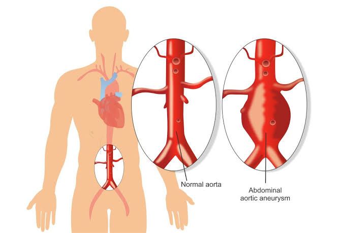
An aneurysm is a bulging, weakened area in the wall of a blood vessel, resulting in an abnormal widening or ballooning that exceeds 50% of the vessel’s normal diameter. Aneurysms can occur in any blood vessel but are most commonly found in arteries, particularly the aorta.
Common Sites and Relative Incidence of Arterial Aneurysms
Aneurysms can occur in various locations within the arterial system, with the most common sites being the aorta, popliteal artery, and carotid artery. Each site has its own relative incidence and associated risk factors.
1. Aortic Aneurysms
The aorta is the largest artery in the body and is divided into several segments: the ascending aorta, aortic arch, descending thoracic aorta, and abdominal aorta.
- Abdominal Aortic Aneurysms (AAAs): These are the most prevalent type of aortic aneurysm. The incidence of AAAs has been reported to be between 1.3% to 8.9% in men and 1.0% to 2.2% in women, with higher rates observed in older populations (typically over age 65). Risk factors include advanced age, male gender, smoking history, and family history.
- Thoracic Aortic Aneurysms (TAAs): These are less common than AAAs but still significant. The estimated incidence is about 5-10 per 100,000 person-years. TAAs can be further classified into ascending aortic aneurysms (approximately 60% of cases), descending thoracic aneurysms (about 35%), and those involving the aortic arch (less than 10%).
2. Peripheral Artery Aneurysms
Apart from the aorta, aneurysms can also occur in peripheral arteries:
- Popliteal Artery Aneurysms: These are among the most common peripheral aneurysms and typically occur behind the knee joint. They have an incidence rate that varies but is generally considered significant due to their potential for thromboembolic complications.
- Carotid Artery Aneurysms: Although less common than other types of aneurysms, carotid artery aneurysms can lead to serious complications such as stroke if they rupture or embolize.
3. Other Sites
Other less common sites for arterial aneurysms include:
- Mesenteric Arteries: These supply blood to the intestines and can develop aneurysms that may lead to ischemia.
- Renal Arteries: Renal artery aneurysms are rare but can cause hypertension or renal ischemia.
- Femoral Arteries: Similar to popliteal arteries, femoral artery aneurysms can occur but are less frequently reported.
In summary, while abdominal aortic aneurysms are the most prevalent type of arterial aneurysm overall, thoracic aortic aneurysms also represent a significant health concern due to their potential for rupture and dissection. Peripheral artery aneurysms like those found in the popliteal and carotid arteries also contribute notably to vascular complications.
Symptoms, Signs, Differential Diagnosis, Diagnostic and Management Plans for a Patient with a Rupturing Abdominal Aortic Aneurysm
1. Symptoms of Rupturing Abdominal Aortic Aneurysm (AAA)
The symptoms of a rupturing AAA can be sudden and severe. Common symptoms include:
- Severe Abdominal Pain: Patients often describe this pain as a sudden onset of severe, tearing or stabbing pain in the abdomen or back. The pain may radiate to the groin, buttocks, or legs.
- Hypotension: Due to internal bleeding, patients may experience low blood pressure leading to dizziness or fainting.
- Nausea and Vomiting: These symptoms can occur due to irritation of the peritoneum from blood leakage.
- Pulsatile Mass: In some cases, a pulsating mass may be palpable in the abdomen.
2. Signs of Rupturing AAA
Upon examination, several signs may indicate a ruptured AAA:
- Tachycardia: Increased heart rate is common as the body compensates for blood loss.
- Abdominal Tenderness: The abdomen may be tender upon palpation.
- Signs of Shock: This includes cold and clammy skin, confusion, and rapid breathing due to significant blood loss.
- Distended Abdomen: In cases where there is significant internal bleeding, abdominal distension may be observed.
3. Differential Diagnosis
When evaluating a patient suspected of having a ruptured AAA, it is crucial to consider other conditions that may present similarly:
- Acute Pancreatitis: Can cause severe abdominal pain but typically has associated nausea and vomiting without hypotension.
- Perforated Peptic Ulcer: Presents with acute abdominal pain but usually has more localized tenderness and signs of peritonitis.
- Renal Colic: Severe flank pain that radiates but usually does not cause hypotension unless there is significant bleeding.
- Mesenteric Ischemia: Can present with abdominal pain out of proportion to physical findings but typically does not have a pulsatile mass.
4. Diagnostic Plan
To confirm the diagnosis of a ruptured AAA, several diagnostic steps are taken:
- Ultrasound (US): A bedside ultrasound can quickly assess for the presence of an AAA and any free fluid indicative of rupture.
- Computed Tomography (CT) Scan: CT angiography is the gold standard for diagnosing an AAA and assessing its size and extent. It provides detailed images that help determine if there is rupture and the location of any hematoma.
- Physical Examination Findings: Noting vital signs such as hypotension or tachycardia can support the diagnosis.
5. Management Plan
Management of a ruptured AAA requires immediate intervention:
- Stabilization:
- Establish IV access for fluid resuscitation with crystalloids or blood products if necessary.
- Monitor vital signs closely for changes indicating shock.
- Surgical Intervention:
- Emergency surgical repair is required; options include open surgical repair or endovascular aneurysm repair (EVAR), depending on the patient’s condition and anatomy.
- Open repair involves resection of the aneurysm and placement of a synthetic graft; EVAR involves placing stent grafts via catheterization.
- Postoperative Care:
- Intensive monitoring in an ICU setting post-surgery for complications such as bleeding, infection, or organ dysfunction.
- Long-term management includes controlling risk factors such as hypertension and hyperlipidemia through medications and lifestyle changes.
In summary, recognizing the symptoms and signs associated with a ruptured abdominal aortic aneurysm is critical for timely diagnosis and management. Rapid assessment through imaging studies followed by urgent surgical intervention can significantly improve outcomes in affected patients.
Indications for Surgery in Chronic Asymptomatic Abdominal Aneurysms
Surgery for chronic asymptomatic abdominal aortic aneurysms (AAA) is typically indicated based on the size of the aneurysm and the risk of rupture. The general consensus among vascular surgeons is that surgical intervention should be considered when the diameter of the AAA reaches 5.5 cm or greater in men, and 5.0 cm or greater in women. This threshold is based on studies indicating that larger aneurysms have a significantly higher risk of rupture, which can lead to life-threatening complications.
In addition to size, other factors may influence the decision to proceed with surgery, including:
- Growth Rate: If an aneurysm shows rapid enlargement (greater than 0.5 cm per year), this may warrant surgical consideration even if it is below the standard size thresholds.
- Patient’s Life Expectancy: In patients with a limited life expectancy due to comorbid conditions, the risks associated with surgery may outweigh potential benefits.
- Patient Preference: Informed patient choice plays a crucial role; some patients may opt for surgery despite being asymptomatic if they are concerned about future risks.
Contraindications for Surgery in Chronic Asymptomatic Abdominal Aneurysms
Contraindications to surgical intervention primarily revolve around patient-specific factors that increase surgical risk or diminish potential benefits:
- Severe Comorbidities: Conditions such as advanced heart disease, severe pulmonary disease, or significant renal impairment can increase perioperative mortality and morbidity.
- Age Considerations: While age alone is not a strict contraindication, very elderly patients (typically over 80 years) may face higher risks during surgery.
- Anatomical Considerations: Complex anatomical features such as tortuous vessels or previous surgeries that complicate access to the aneurysm can make surgical repair more hazardous.
- Patient Refusal: If a patient declines surgery after being fully informed of their condition and treatment options, this refusal must be respected.
Risk Factors for Surgery in Chronic Asymptomatic Abdominal Aneurysms
Several risk factors must be evaluated when considering surgical intervention for chronic asymptomatic AAAs:
- Aneurysm Size and Growth Rate: Larger and rapidly growing aneurysms pose a higher risk of rupture and thus necessitate closer monitoring and possible surgical intervention.
- Gender Differences: Women tend to have smaller AAAs than men but have a higher relative risk of rupture at smaller sizes; hence gender should be considered in management strategies.
- Family History: A family history of AAA can indicate a genetic predisposition, increasing vigilance regarding monitoring and potential surgical intervention.
- Lifestyle Factors: Smoking is one of the most significant modifiable risk factors associated with AAA development and growth; cessation should be encouraged regardless of surgical plans.
- Hypertension and Hyperlipidemia: These conditions can exacerbate vascular health issues; managing these comorbidities is essential both pre- and post-surgery.
In summary, while chronic asymptomatic abdominal aneurysms often require careful monitoring rather than immediate intervention, specific indications such as size thresholds, growth rates, patient health status, and preferences guide decisions regarding surgery. Contraindications include severe comorbidities and anatomical challenges that could complicate procedures.
Prevention of Common Complications Following Aneurysm Surgery
Aneurysm surgery, whether it involves open surgical repair or endovascular techniques, is a critical intervention aimed at preventing rupture and associated morbidity and mortality. However, like any major surgical procedure, it carries risks of complications. Understanding these complications and implementing preventive measures is essential for improving patient outcomes.
Common Complications Following Aneurysm Surgery
- Infection: Surgical site infections (SSIs) can occur post-operatively, particularly in open surgeries where larger incisions are made.
- Hemorrhage: Both intraoperative and postoperative bleeding can lead to significant complications, including hematoma formation or shock.
- Thrombosis/Embolism: The risk of thromboembolic events increases after surgery due to changes in blood flow dynamics.
- Neurological Deficits: In cases involving cerebral aneurysms, there is a risk of stroke or transient ischemic attacks (TIAs).
- Organ Dysfunction: This includes renal failure or respiratory complications that may arise from the stress of surgery.
- Graft Complications: In endovascular procedures, issues such as graft migration or endoleaks can occur.
Prevention Strategies
- Infection Control
- Antibiotic Prophylaxis: Administering prophylactic antibiotics before surgery can significantly reduce the risk of SSIs.
- Sterile Technique: Maintaining strict sterile conditions during surgery is crucial to minimize infection risk.
- Postoperative Care: Proper wound care and monitoring for signs of infection are vital in the postoperative period.
- Hemorrhage Management
- Careful Surgical Technique: Surgeons must employ meticulous techniques to control bleeding during the operation.
- Monitoring Blood Loss: Continuous monitoring of blood loss during and after surgery allows for timely interventions if significant hemorrhage occurs.
- Fluid Resuscitation and Transfusion Protocols: Establishing protocols for fluid resuscitation and blood transfusion helps manage potential hypovolemia effectively.
- Thrombosis/Embolism Prevention
- Anticoagulation Therapy: Depending on the patient’s risk factors, anticoagulants may be administered postoperatively to prevent thromboembolic events.
- Early Mobilization: Encouraging early ambulation post-surgery reduces venous stasis and lowers the risk of deep vein thrombosis (DVT).
- Compression Devices: Utilizing pneumatic compression devices on the legs can also help prevent DVT.
- Neurological Monitoring
- For patients undergoing cerebral aneurysm repairs, continuous neurological assessments are necessary to detect any deficits early.
- Postoperative imaging (e.g., CT scans) may be employed to identify any acute changes indicative of complications like stroke.
- Organ Function Monitoring
- Regular assessment of renal function through serum creatinine levels and urine output monitoring helps detect early signs of renal impairment.
- Respiratory function should be monitored closely, especially in patients with pre-existing lung conditions or those who underwent thoracic surgeries.
- Graft Surveillance
- For patients who have undergone endovascular repair, regular follow-up imaging (such as ultrasound or CT angiography) is essential to check for graft integrity and detect endoleaks early.
- Patient Education
- Educating patients about signs and symptoms of potential complications allows for prompt reporting and intervention if issues arise post-discharge.
By implementing these prevention strategies comprehensively throughout the perioperative period—from preoperative planning through postoperative care—healthcare providers can significantly reduce the incidence of complications following aneurysm surgery.
Comparison of Thoracic, Abdominal, Femoral, and Popliteal Aneurysms
1. Presentation
- Thoracic Aneurysms: These often present asymptomatically until they reach a significant size or rupture. Symptoms may include chest pain, back pain, or dyspnea if the aneurysm compresses adjacent structures. In some cases, patients may experience hoarseness due to recurrent laryngeal nerve involvement.
- Abdominal Aneurysms: Abdominal aortic aneurysms (AAA) are typically asymptomatic until rupture. When symptomatic, they may present with abdominal pain that can radiate to the back or groin. Pulsatile abdominal mass may be palpable in larger aneurysms.
- Femoral Aneurysms: These can be asymptomatic but may present with a pulsatile mass in the groin area. Symptoms can include pain or discomfort in the thigh or leg due to compression of surrounding structures or thrombosis.
- Popliteal Aneurysms: Often asymptomatic initially, these aneurysms can lead to symptoms such as calf pain or claudication due to compromised blood flow. Patients may also experience acute limb ischemia if thrombosis occurs.
2. Complications
- Frequency of Dissection:
- Thoracic aneurysms have a higher risk of dissection compared to abdominal and peripheral aneurysms due to the high pressures within the thoracic aorta.
- Abdominal aneurysms can dissect but are more commonly associated with rupture.
- Femoral and popliteal aneurysms rarely dissect; however, they can lead to complications from thromboembolism.
- Rupture:
- The risk of rupture is highest in thoracic and abdominal aneurysms as they grow larger (greater than 5 cm for AAA). The mortality rate for ruptured AAA is particularly high.
- Femoral and popliteal aneurysms have lower rates of rupture but can still cause significant morbidity if they do rupture.
- Thrombosis:
- Thrombosis is common in femoral and popliteal aneurysms due to turbulent blood flow leading to clot formation.
- In thoracic and abdominal aneurysms, thrombosis is less common but can occur especially if there is significant stenosis or dissection.
- Embolization:
- Embolization is a significant concern in all types of aneurysms but is particularly notable in peripheral (femoral and popliteal) aneurysms where thrombus can break off and occlude distal vessels.
- In thoracic and abdominal cases, embolization usually results from thrombus formation within the dilated vessel itself.
3. Treatment
- Thoracic Aneurysms: Management typically involves surgical intervention for symptomatic patients or those with large (>5 cm) aneurysms. Options include open surgical repair or endovascular stent grafting depending on the anatomy and patient factors.
- Abdominal Aneurysms: Similar to thoracic aneurysms, treatment involves surgical repair for symptomatic cases or those over certain size thresholds (generally >5.5 cm). Endovascular repair has become increasingly popular due to its minimally invasive nature.
- Femoral Aneurysms: Treatment often involves surgical resection and bypass grafting if symptomatic or if there’s concern for complications like thrombosis. Endovascular techniques are less commonly used here compared to other locations.
- Popliteal Aneurysms: Surgical intervention is indicated when there are symptoms or evidence of ischemia. This typically involves resection of the aneurysm with bypass grafting; endovascular options are also being explored but are not as established as in other locations.
In summary, while all four types of aneurysms share some common features regarding presentation and potential complications, their management strategies differ significantly based on location, size, symptoms, and associated risks.


