Divisions of the Heart into Four Chambers and Their Features
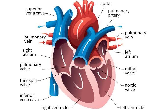
The human heart is a muscular organ that functions as the central component of the circulatory system, responsible for pumping blood throughout the body. It is divided into four chambers: two atria (upper chambers) and two ventricles (lower chambers). Each chamber has distinct internal and external features that contribute to its function.
1. Right Atrium
- External Features: The right atrium is located on the upper right side of the heart. It has a smooth wall and receives deoxygenated blood from the body through three major veins: the superior vena cava, inferior vena cava, and coronary sinus. The right atrium also features a small muscular pouch called the right auricle, which increases its capacity.
- Internal Features: Internally, the right atrium contains several important structures:
- Fossa Ovalis: A depression in the interatrial septum that is a remnant of the foramen ovale, an opening present during fetal development.
- Pectinate Muscles: These are ridged muscles found in the anterior part of the atrial wall that help with contraction.
- Tricuspid Valve: This valve separates the right atrium from the right ventricle and prevents backflow of blood when the ventricle contracts.
2. Right Ventricle
- External Features: The right ventricle is positioned below the right atrium and has a thicker muscular wall compared to the atrium because it needs to pump blood to the lungs. Its shape is somewhat crescent-like when viewed from above.
- Internal Features: Key internal structures include:
- Trabeculae Carneae: These are irregular muscular columns that line the ventricular walls, aiding in contraction.
- Pulmonary Valve: Located at the exit of the right ventricle, this valve opens into the pulmonary artery, allowing deoxygenated blood to flow towards the lungs for oxygenation.
- Chordae Tendineae and Papillary Muscles: These structures anchor and support the tricuspid valve during ventricular contraction.
3. Left Atrium
- External Features: The left atrium is situated on the upper left side of the heart and has a similar structure to that of the right atrium but typically appears smaller due to its role in receiving oxygenated blood from pulmonary circulation.
- Internal Features: Important internal components include:
- Pulmonary Veins Opening: Four pulmonary veins enter this chamber, delivering oxygen-rich blood from both lungs.
- Left Auricle: Similar to its counterpart on the right side, it serves as an extension to increase volume capacity.
- Bicuspid (Mitral) Valve: This valve allows blood to flow from left atrium into left ventricle while preventing backflow during ventricular contraction.
4. Left Ventricle
- External Features: The left ventricle has thickest walls among all four chambers due to its role in pumping oxygenated blood throughout systemic circulation. It has a conical shape which helps generate high pressure necessary for systemic circulation.
- Internal Features:
- Thick Myocardial Walls: These walls are significantly thicker than those of other chambers due to higher pressure requirements.
- Aortic Valve: This valve controls blood flow from left ventricle into ascending aorta, ensuring one-way flow into systemic circulation.
- Trabeculae Carneae and Papillary Muscles similar to those found in right ventricle assist in maintaining proper function of mitral valve during contraction.
In summary, each chamber plays a critical role in maintaining efficient circulation within different parts of cardiovascular system. The structural differences between chambers reflect their specific functions—receiving or pumping blood—and their adaptations ensure effective hemodynamics throughout life.
Conductive System of the Heart: Arrangement and Function
The conductive system of the heart is a specialized network of cells responsible for initiating and conducting electrical impulses throughout the myocardium, leading to coordinated heart contractions. This system ensures that the heart beats in a rhythmic and efficient manner, allowing for effective blood circulation. The main components of this system include the sinoatrial (SA) node, atrioventricular (AV) node, bundle of His, right and left bundle branches, and Purkinje fibers. Below is a detailed description of each part, their arrangement within the myocardium, and their specific functions.
1. Sinoatrial (SA) Node
The SA node is often referred to as the natural pacemaker of the heart. It is located in the right atrium near the entrance of the superior vena cava. The SA node generates electrical impulses that initiate each heartbeat.
- Function: The primary function of the SA node is to produce action potentials at regular intervals (typically 60-100 beats per minute in a resting adult), which spread through the atria causing them to contract and push blood into the ventricles.
- Arrangement: The SA node consists of specialized pacemaker cells that have an intrinsic ability to depolarize spontaneously due to their unique ion channel composition.
2. Atrioventricular (AV) Node
The AV node is situated at the junction between the atria and ventricles, specifically in the interatrial septum near the opening of the coronary sinus.
- Function: The AV node serves as a critical relay point for electrical impulses from the atria to the ventricles. It introduces a delay (approximately 0.1 seconds) in conduction, allowing time for ventricular filling after atrial contraction.
- Arrangement: Comprised of nodal tissue similar to that found in the SA node but with fewer pacemaker cells, it acts as both a gatekeeper and backup pacemaker if needed.
3. Bundle of His (Atrioventricular Bundle)
The Bundle of His extends from the AV node into the interventricular septum.
- Function: Its primary role is to conduct impulses from the AV node down into both ventricles via its branching pathways.
- Arrangement: This structure divides into two main branches—right and left bundle branches—that run along either side of the interventricular septum toward their respective ventricles.
4. Right and Left Bundle Branches
These branches extend from the Bundle of His into each ventricle.
- Function: They carry electrical impulses rapidly towards their respective ventricles, ensuring synchronized contraction.
- Arrangement: The right bundle branch travels along the right side of the interventricular septum while splitting into smaller Purkinje fibers that spread throughout the right ventricle; similarly, left bundle branch divides further into anterior and posterior fascicles that innervate different regions of the left ventricle.
5. Purkinje Fibers
Purkinje fibers are specialized conductive fibers located within both ventricles.
- Function: They distribute electrical impulses quickly throughout ventricular myocardium, leading to coordinated contraction from apex to base which optimizes blood ejection during systole.
- Arrangement: These fibers are spread out extensively throughout both ventricles forming a network that ensures rapid conduction across all myocardial cells.
In summary, these components work together in a highly organized manner to ensure efficient cardiac function by generating and propagating electrical signals through specific pathways within myocardial tissue. This orchestration allows for precise timing between atrial and ventricular contractions necessary for effective pumping action.
Understanding Cardiac Referred Pain
Cardiac referred pain is a phenomenon where pain originating from the heart is perceived in areas of the body that are not directly affected by the cardiac condition. This type of pain occurs due to the complex interconnections within the nervous system, particularly involving sensory nerves that transmit pain signals from both visceral organs (like the heart) and somatic tissues (like skin and muscles).
Mechanism Behind Cardiac Referred Pain
The principle underlying cardiac referred pain can be explained through several neurophysiological theories:
- Convergence-Projections Theory: This theory posits that sensory neurons from different regions converge at the same spinal cord level. For instance, nociceptive signals from the heart may travel along pathways that also carry signals from somatic structures such as the left arm or shoulder. When these signals reach higher centers in the brain, it becomes difficult for the brain to distinguish between them, leading to misinterpretation of the source of pain.
- Dermatomes and Visceral-Somatic Convergence: The body is mapped into dermatomes, which are areas of skin supplied by specific spinal nerves. The heart primarily corresponds to spinal segments T1-T5. Because these segments also innervate regions such as the left shoulder, neck, and arm, stimulation of cardiac nociceptors can lead to sensations of pain in these areas.
- Visceral Sensitivity and Somatic Pain Perception: The brain’s inability to accurately localize visceral pain contributes significantly to referred pain. Since visceral organs have fewer sensory receptors than somatic tissues, when visceral pain occurs (such as during ischemia or a heart attack), it may be perceived as somatic discomfort in adjacent areas.
Common Areas for Cardiac Referred Pain
Patients experiencing cardiac issues often report referred pain in specific locations:
- Left Arm: Many individuals feel discomfort radiating down their left arm.
- Shoulder and Neck: Pain may also manifest in the neck or shoulder region.
- Jaw and Back: Some patients experience referred pain in their jaw or upper back.
These patterns are critical for healthcare providers because they can serve as warning signs for potential cardiac events like angina or myocardial infarction (heart attack).
Clinical Implications of Cardiac Referred Pain
Recognizing cardiac referred pain is essential for timely diagnosis and treatment. Misinterpretation of this type of pain can lead patients to overlook serious conditions, delaying necessary medical intervention. For example, someone might dismiss severe chest discomfort as muscle strain when it could indicate an impending heart attack.
Healthcare professionals emphasize awareness of these symptoms during patient assessments, especially in individuals with risk factors for cardiovascular disease.
In summary, cardiac referred pain arises due to convergence of sensory pathways from visceral organs like the heart with those from somatic tissues, leading to misperceptions about where the actual source of discomfort lies.
Papillary Muscles: Locations and Importance
Papillary muscles are specialized muscles located within the ventricles of the heart. They play a crucial role in the function of the atrioventricular valves, specifically the mitral valve in the left ventricle and the tricuspid valve in the right ventricle. Their primary function is to prevent the inversion or prolapse of these valves during ventricular contraction (systole).
Locations of Papillary Muscles
In humans, there are five papillary muscles: three located in the right ventricle and two in the left ventricle.
- Left Ventricle:
- Anterolateral Papillary Muscle: This muscle typically arises from the area between the apical and middle thirds of the left ventricular wall. It is usually composed of a single body or head and provides chordae tendineae to both leaflets of the mitral valve.
- Posteromedial Papillary Muscle: Also originating from the left ventricular wall, this muscle often has two bodies or heads. It supplies chordae tendineae to both leaflets as well but is particularly vulnerable due to its blood supply being derived from a single source, primarily from either the left circumflex artery or right coronary artery depending on coronary dominance.
- Right Ventricle:
- The right ventricle contains three papillary muscles known as anterior, posterior, and septal papillary muscles. Each of these attaches via chordae tendineae to the tricuspid valve.
Importance of Papillary Muscles
The importance of papillary muscles lies in their role in maintaining proper heart function:
- Prevention of Valve Prolapse: During ventricular contraction, papillary muscles contract slightly before systole, which helps maintain tension on the chordae tendineae. This tension prevents regurgitation (backward flow) of blood into the atria by keeping the atrioventricular valves closed against high pressure generated within the ventricles.
- Mitral Valve Functionality: The anterolateral and posteromedial papillary muscles are integral to mitral valve competence. Dysfunction or rupture of these muscles can lead to severe complications such as acute mitral regurgitation, which may necessitate surgical intervention.
- Vulnerability to Injury: The posteromedial papillary muscle is particularly susceptible to ischemic injury due to its single blood supply system; occlusion of its supplying artery can lead to rupture. This highlights its clinical significance as damage can result in life-threatening conditions.
- Impact on Heart Conditions: Conditions such as myocardial infarction can lead to dysfunction or rupture of papillary muscles, resulting in worsening mitral regurgitation and potentially requiring surgical repair.
In summary, papillary muscles are essential components within cardiac anatomy that ensure effective functioning of heart valves during each heartbeat, thus playing a critical role in overall cardiovascular health.
Atrio-Ventricular Valves: Structure, Position, and Function
The atrio-ventricular (AV) valves are crucial components of the heart’s anatomy, serving as gatekeepers between the atria and ventricles. There are two primary AV valves in the human heart: the tricuspid valve and the mitral valve. Their main function is to prevent backflow of blood from the ventricles into the atria during ventricular contraction (systole).
1. Structure and Position of AV Valves
- Tricuspid Valve: Located on the right side of the heart, it separates the right atrium from the right ventricle. The tricuspid valve consists of three cusps (leaflets): anterior, posterior, and septal.
- Mitral Valve: Situated on the left side of the heart, this valve separates the left atrium from the left ventricle. The mitral valve has two cusps: anterior and posterior.
Both valves are positioned at an angle that allows them to open fully during diastole (when the heart fills with blood) and close tightly during systole.
2. Attachment of Cusps to Papillary Muscles
The cusps of both AV valves are anchored by chordae tendineae to papillary muscles located within the ventricles:
- Chordae Tendineae: These are fibrous cords that connect each cusp to papillary muscles. They play a vital role in maintaining valve closure by preventing inversion or prolapse of the cusps when ventricular pressure increases during contraction.
- Papillary Muscles: These muscular projections arise from the ventricular walls. When the ventricles contract, papillary muscles also contract, pulling on the chordae tendineae. This action stabilizes the cusps and ensures they remain closed against high pressure.
In summary:
- The tricuspid valve is connected to three papillary muscles (anterior, posterior, and septal), while two papillary muscles support the mitral valve.
3. Functional Importance of AV Valves
The functional importance of AV valves can be summarized as follows:
- Prevention of Backflow: During ventricular contraction, increased pressure forces blood into pulmonary arteries (from right ventricle) and aorta (from left ventricle). The tight closure of AV valves prevents backflow into atria.
- Coordination with Heart Cycle: The opening and closing of these valves are synchronized with cardiac cycles—opening during diastole for filling and closing during systole for ejection.
- Pressure Regulation: By ensuring unidirectional blood flow, AV valves help maintain appropriate pressure gradients necessary for effective circulation throughout systemic and pulmonary circuits.
In conclusion, atrio-ventricular valves play a vital role in cardiac function by facilitating proper blood flow direction while preventing regurgitation through their structural design and connections to papillary muscles.
Aortic and Pulmonary Semilunar Valves: Position and Functional Importance
Introduction to Semilunar Valves
The semilunar valves are crucial components of the heart’s anatomy, specifically located at the exits of the ventricles. There are two semilunar valves: the aortic semilunar valve and the pulmonary semilunar valve. Both play vital roles in maintaining unidirectional blood flow during the cardiac cycle.
Aortic Semilunar Valve
- Position: The aortic semilunar valve is situated between the left ventricle and the aorta. It is located at the base of the ascending aorta, which carries oxygenated blood away from the heart to supply the body. The valve consists of three cusps (leaflets) that open to allow blood to flow into the aorta during ventricular contraction (systole) and close to prevent backflow when the ventricle relaxes (diastole).
- Functional Importance:
- Prevention of Backflow: The primary function of the aortic semilunar valve is to prevent regurgitation of blood into the left ventricle after it has been pumped into the aorta. This ensures that oxygen-rich blood continues to circulate throughout the body efficiently.
- Pressure Regulation: During systole, as blood is ejected from the left ventricle, high pressure builds up in this chamber. The opening of the aortic valve allows this pressure to be transmitted into systemic circulation while maintaining adequate pressure levels within both chambers.
- Facilitating Blood Flow: By allowing for rapid ejection of blood into systemic circulation, it helps maintain adequate perfusion pressure necessary for organ function.
Pulmonary Semilunar Valve
- Position: The pulmonary semilunar valve is located between the right ventricle and the pulmonary artery. It sits at the base of the pulmonary trunk, which bifurcates into right and left pulmonary arteries that carry deoxygenated blood from the heart to each lung for oxygenation.
- Functional Importance:
- Prevention of Backflow: Similar to its counterpart, this valve prevents backflow of blood into the right ventricle after it has been pumped into the pulmonary artery during ventricular contraction.
- Regulating Blood Flow to Lungs: During systole, when blood is pushed from the right ventricle through this valve, it ensures that deoxygenated blood reaches the lungs efficiently for gas exchange.
- Maintaining Pressure Balance: The pulmonary semilunar valve also plays a role in maintaining appropriate pressures within both ventricles during different phases of cardiac activity.
Conclusion
Both semilunar valves are integral in ensuring efficient circulation by preventing backflow and regulating pressures within their respective circuits—systemic for the aortic valve and pulmonary for the pulmonary valve. Their proper functioning is essential for effective cardiovascular health.


