Common Types of Gallstones and Their Pathophysiology
Introduction to Gallstones
Gallstones are solid particles that form in the gallbladder, a small organ located beneath the liver that stores bile. Bile is a digestive fluid produced by the liver that helps in the digestion of fats. The formation of gallstones can lead to various complications, including cholecystitis (inflammation of the gallbladder), pancreatitis, and biliary obstruction.
Types of Gallstones
- Cholesterol Gallstones
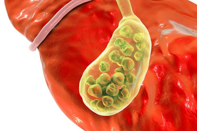
- Description: Cholesterol gallstones are the most common type, accounting for approximately 80% of all gallstone cases. They are primarily composed of hardened cholesterol and may appear yellow-green in color.
- Pathophysiology: The formation of cholesterol gallstones occurs when there is an imbalance between the components that make up bile. Factors contributing to this imbalance include:
- Supersaturation of Cholesterol: When there is too much cholesterol in relation to bile salts and phospholipids, it can precipitate out of solution and form crystals.
- Bile Salt Deficiency: Insufficient bile salts can fail to solubilize cholesterol effectively, leading to crystallization.
- Gallbladder Motility Issues: If the gallbladder does not empty properly or frequently enough, bile can become overly concentrated with cholesterol.
- Pigment Gallstones
- Description: Pigment gallstones are smaller and darker than cholesterol stones and consist mainly of bilirubin, a breakdown product of hemoglobin. They can be classified into two types: black pigment stones and brown pigment stones.
- Black Pigment Stones: These are typically associated with conditions that cause increased bilirubin production, such as hemolytic anemia.
- Brown Pigment Stones: These are often found in individuals with biliary tract infections or conditions leading to cholestasis (reduced bile flow).
- Pathophysiology:
- Increased Bilirubin Production: Conditions like hemolytic anemia lead to excess bilirubin being excreted into bile, which can precipitate as stones.
- Bacterial Infection: Infections can alter the composition of bile and promote stone formation through processes such as deconjugation of bilirubin by bacterial enzymes.
- Description: Pigment gallstones are smaller and darker than cholesterol stones and consist mainly of bilirubin, a breakdown product of hemoglobin. They can be classified into two types: black pigment stones and brown pigment stones.
- Mixed Gallstones
- Description: Mixed gallstones contain both cholesterol and bilirubin components along with other substances such as calcium salts. They represent a combination of factors leading to both cholesterol supersaturation and increased bilirubin levels.
- Pathophysiology: The formation mechanism involves:
- A combination of factors from both cholesterol and pigment stone formation pathways.
- An environment where both excess cholesterol and elevated bilirubin coexist due to metabolic or pathological changes in the body.
Conclusion
The pathophysiology behind gallstone formation is multifactorial, involving genetic predisposition, dietary factors (high fat intake), obesity, rapid weight loss, diabetes mellitus, pregnancy, age, gender (more common in females), and certain medical conditions affecting liver function or red blood cell turnover. Understanding these mechanisms is crucial for prevention strategies and management options for individuals at risk for developing gallstones.
Diseases That Predispose to Gallstones
Gallstones can form due to various underlying health conditions. Here are several diseases that are known to increase the risk of developing gallstones:
1. Obesity
Obesity is a significant risk factor for gallstones, particularly in women. Excess body weight can lead to increased cholesterol levels in bile, which contributes to the formation of cholesterol stones. The mechanism involves the liver producing more cholesterol than the bile can dissolve, leading to crystallization and stone formation.
2. Diabetes Mellitus
Individuals with diabetes often have elevated levels of triglycerides, a type of fat found in the blood. High triglyceride levels can increase the likelihood of gallstone formation by altering bile composition and promoting cholesterol supersaturation in bile.
3. Liver Cirrhosis
Cirrhosis, which is scarring of the liver due to chronic liver disease, can disrupt normal bile production and flow. This disruption may lead to an imbalance in bile salts and cholesterol, increasing the risk for pigment stones (which are typically associated with bilirubin).
4. Hemolytic Anemias
Conditions such as sickle cell anemia or thalassemia lead to increased breakdown of red blood cells (hemolysis). This process releases excess bilirubin into the bloodstream, which can then be excreted into bile and contribute to the formation of pigment stones.
5. Biliary Tract Infections
Infections within the biliary tract can alter bile composition and promote stone formation. Inflammatory processes associated with infections may lead to changes in how bile is produced or secreted, increasing the risk for both cholesterol and pigment stones.
6. Rapid Weight Loss
Rapid weight loss, often seen in individuals undergoing extreme dieting or bariatric surgery, can cause the liver to release excess cholesterol into bile as fat is metabolized quickly. This sudden change in bile composition increases the likelihood of gallstone formation.
7. Pregnancy
Pregnancy leads to hormonal changes that increase estrogen levels, which can affect gallbladder motility and reduce its ability to empty effectively. This stasis allows for greater concentration of cholesterol in bile, predisposing pregnant women to gallstones.
8. Crohn’s Disease
Crohn’s disease is an inflammatory bowel condition that affects nutrient absorption and may alter how bile acids are reabsorbed in the intestines. This malabsorption can lead to changes in bile composition that favor gallstone development.
In summary, various diseases and conditions predispose individuals to gallstone formation through mechanisms involving alterations in bile composition, impaired gallbladder function, or increased bilirubin levels.
Signs and Symptoms of Biliary Colic
Biliary colic is characterized by a specific set of signs and symptoms that arise due to the obstruction of the bile ducts, typically caused by gallstones. The key features include:
- Pain Characteristics:
- The pain associated with biliary colic is often described as a sudden onset of severe, steady pain in the right upper quadrant (RUQ) or epigastric region.
- It may radiate to the right shoulder or back.
- The pain usually lasts from 30 minutes to several hours and tends to resolve spontaneously.
- Timing and Triggers:
- Biliary colic often occurs after meals, particularly after fatty meals, as these can stimulate gallbladder contraction.
- Patients may experience episodes that recur intermittently over time.
- Associated Symptoms:
- Nausea and vomiting are common but not universal.
- There may be no fever or significant abdominal tenderness upon examination.
- Physical Examination Findings:
- On physical exam, there may be mild tenderness in the RUQ without rebound tenderness or guarding, which distinguishes it from acute cholecystitis.
Signs and Symptoms of Acute Cholecystitis
Acute cholecystitis is an inflammation of the gallbladder typically due to prolonged obstruction of the cystic duct by gallstones. Its symptoms are more severe compared to biliary colic:
- Pain Characteristics:
- The pain is similar in location (RUQ) but is usually more intense and persistent.
- It can also radiate to the right shoulder or back but tends to last longer than in biliary colic, often exceeding several hours.
- Timing and Triggers:
- Pain does not necessarily correlate with meal times; it can occur at any time.
- Patients may have a history of prior biliary colic episodes leading up to acute cholecystitis.
- Associated Symptoms:
- Fever is commonly present (often low-grade).
- Patients frequently exhibit nausea and vomiting.
- Anorexia (loss of appetite) is also common.
- Physical Examination Findings:
- On examination, there is often significant tenderness in the RUQ with possible rebound tenderness and guarding, indicating peritoneal irritation.
- A positive Murphy’s sign (pain upon palpation of the gallbladder during inspiration) is characteristic of acute cholecystitis.
- Laboratory Findings:
- Laboratory tests may show leukocytosis (elevated white blood cell count), which indicates infection or inflammation.
- Imaging studies such as ultrasound may reveal thickening of the gallbladder wall or presence of fluid around it.
Contrast Between Biliary Colic and Acute Cholecystitis
- Duration and Severity of Pain: Biliary colic pain is intermittent and resolves spontaneously within hours; acute cholecystitis involves continuous, severe pain lasting longer with associated systemic symptoms like fever.
- Physical Exam Findings: Biliary colic shows mild RUQ tenderness without guarding; acute cholecystitis presents with significant tenderness, rebound tenderness, and possibly a positive Murphy’s sign.
- Systemic Symptoms: Acute cholecystitis typically includes fever and leukocytosis, while biliary colic does not present these systemic signs.
In summary, while both conditions involve similar initial symptoms related to gallstones, their clinical presentations diverge significantly regarding pain characteristics, physical examination findings, duration of symptoms, and associated systemic manifestations.
Tests Commonly Used in the Diagnosis of Calculus Biliary Tract Disease
Calculus biliary tract disease, often referred to as gallstone disease, primarily involves the formation of stones in the gallbladder or bile ducts. The diagnosis of this condition typically involves a combination of clinical evaluation and various imaging and laboratory tests. Below is a detailed examination of the commonly used diagnostic tests, including their indications, limitations, and potential complications.
1. Ultrasound (US)
Indications:
Ultrasound is usually the first-line imaging modality for suspected biliary tract disease due to its non-invasive nature and lack of ionizing radiation. It is particularly useful for detecting gallstones in the gallbladder and assessing complications such as cholecystitis or choledocholithiasis (bile duct stones).
Limitations:
While ultrasound is effective for visualizing gallstones, it may not detect small stones or those located in the cystic duct. Additionally, patient factors such as obesity or excessive bowel gas can hinder image quality.
Potential Complications:
There are generally no complications associated with ultrasound; however, discomfort may occur during the procedure if the patient has to remain still for an extended period.
2. Computed Tomography (CT) Scan
Indications:
CT scans are utilized when there is a need for a more comprehensive assessment of abdominal pain or when complications from gallstones are suspected (e.g., pancreatitis or perforation). CT can also help visualize stones that may not be seen on ultrasound.
Limitations:
CT scans expose patients to ionizing radiation and may not be as sensitive as ultrasound for detecting small gallstones. Additionally, contrast material used in some CT scans can pose risks for patients with renal impairment.
Potential Complications:
The primary risk associated with CT scans includes allergic reactions to contrast dye and radiation exposure leading to potential long-term effects.
3. Magnetic Resonance Cholangiopancreatography (MRCP)
Indications:
MRCP is a non-invasive MRI technique specifically designed to visualize the biliary and pancreatic ducts. It is indicated when there is suspicion of bile duct obstruction or when further evaluation of complex biliary anatomy is required.
Limitations:
MRCP cannot be used in patients with certain types of metal implants or claustrophobia issues. Additionally, while MRCP can show stones within the ducts, it does not provide information about gallbladder pathology itself.
Potential Complications:
Similar to other MRI procedures, risks include allergic reactions to contrast agents (if used) and discomfort from lying still during scanning.
4. Endoscopic Retrograde Cholangiopancreatography (ERCP)
Indications:
ERCP is both diagnostic and therapeutic; it is indicated when there is a high suspicion of choledocholithiasis or when therapeutic intervention (such as stone removal) is necessary following diagnosis.
Limitations:
ERCP carries higher risks compared to non-invasive imaging techniques due to its invasive nature. It may not always successfully remove stones, necessitating additional procedures.
Potential Complications:
Complications can include pancreatitis, perforation of the duodenum, bleeding at the site of cannulation, and infections such as cholangitis.
5. Hepatobiliary Iminodiacetic Acid (HIDA) Scan
Indications:
A HIDA scan evaluates liver function and bile excretion; it can help diagnose acute cholecystitis by assessing whether the gallbladder takes up radioactive tracer material after intravenous administration.
Limitations:
This test may not directly visualize stones but rather assesses functionality; thus false negatives can occur if there’s an obstruction upstream from the gallbladder.
Potential Complications:
The risks are minimal but include allergic reactions to radioactive tracers and exposure to low levels of radiation.
In summary, each diagnostic test plays a crucial role in evaluating calculus biliary tract disease based on specific clinical scenarios. The choice among these tests depends on various factors including patient history, presenting symptoms, and initial findings from less invasive tests like ultrasound.
Natural History of Asymptomatic Gallstones in Young Patients
While many individuals with gallstones experience symptoms, a significant number remain asymptomatic, particularly young patients.
Prevalence and Risk Factors
Asymptomatic gallstones are relatively common, especially among certain demographics. In young patients, factors such as obesity, rapid weight loss, pregnancy, and certain genetic predispositions may increase the likelihood of developing gallstones. However, many young individuals may have gallstones without any noticeable symptoms.
Natural History of Asymptomatic Gallstones
- Initial Discovery:
- Many cases of asymptomatic gallstones are discovered incidentally during imaging studies for unrelated medical issues (e.g., ultrasound or CT scans).
- Long-Term Prognosis:
- Studies indicate that the majority of asymptomatic gallstone cases do not progress to symptomatic disease. Research shows that approximately 10-20% of asymptomatic individuals will develop symptoms over a period of 5-20 years.
- The risk of complications such as cholecystitis (inflammation of the gallbladder), pancreatitis (inflammation of the pancreas), or biliary colic is low in asymptomatic patients.
- Factors Influencing Progression:
- Certain factors may influence whether an asymptomatic patient will develop symptoms or complications. These include age, gender (females are at higher risk), body mass index (BMI), and lifestyle factors such as diet and physical activity levels.
- Younger patients generally have a lower risk of developing complications compared to older adults.
- Monitoring and Management:
- For most young patients with asymptomatic gallstones, active surveillance is recommended rather than immediate surgical intervention. Regular follow-up appointments can help monitor for any changes in symptoms or stone characteristics.
- Surgical removal of the gallbladder (cholecystectomy) is typically reserved for those who become symptomatic or develop complications.
- Lifestyle Considerations:
- Maintaining a healthy lifestyle through balanced nutrition and regular exercise can potentially reduce the risk of developing symptoms associated with gallstones.
- Avoiding rapid weight loss diets is also advisable since this can precipitate stone formation.
- Conclusion on Natural History:
- Overall, the natural history for young patients with asymptomatic gallstones tends to be favorable, with many remaining symptom-free throughout their lives. Continuous monitoring allows for timely intervention if symptoms arise.
In summary, while there is a possibility that some young patients with asymptomatic gallstones may eventually develop symptoms or complications over time, most will remain asymptomatic without requiring surgical treatment.
Complications of Biliary Calculi
Biliary calculi, commonly known as gallstones, can lead to several complications that may require medical intervention. The most significant complications include:
- Cholecystitis
- History: Patients often report a sudden onset of right upper quadrant pain, which may be associated with nausea and vomiting. The pain can be exacerbated by fatty meals.
- Physical Examination: On examination, there may be tenderness in the right upper quadrant, and signs such as Murphy’s sign (pain on inspiration when the examiner palpates the gallbladder) may be positive. Fever and tachycardia can also be present.
- Laboratory Findings: Laboratory tests typically show leukocytosis (elevated white blood cell count), elevated liver enzymes (AST, ALT), and possibly elevated bilirubin levels if there is bile duct involvement.
- Choledocholithiasis
- History: Patients may experience intermittent episodes of jaundice, dark urine, and pale stools. They might also report abdominal pain similar to that seen in cholecystitis.
- Physical Examination: Jaundice is often evident; abdominal examination may reveal tenderness in the right upper quadrant or epigastric region. There could also be signs of biliary obstruction.
- Laboratory Findings: Elevated bilirubin levels (both direct and indirect), alkaline phosphatase, and gamma-glutamyl transferase (GGT) are common findings. Liver function tests may show transaminitis if there is liver involvement.
- Pancreatitis
- History: Patients typically present with severe epigastric pain that radiates to the back, often accompanied by nausea and vomiting. The pain may worsen after eating.
- Physical Examination: Abdominal tenderness is usually present, and patients may exhibit signs of peritoneal irritation or guarding.
- Laboratory Findings: Serum amylase and lipase levels are usually significantly elevated. Other laboratory findings might include leukocytosis and elevated liver enzymes if there is concurrent biliary obstruction.
- Cholangitis
- History: This condition presents with the classic triad of symptoms known as Charcot’s triad: fever, jaundice, and right upper quadrant pain. Patients might also report chills.
- Physical Examination: Fever is typically present along with jaundice; abdominal examination reveals tenderness in the right upper quadrant.
- Laboratory Findings: Blood tests often show leukocytosis with a left shift (indicating infection), elevated bilirubin levels (predominantly direct), and increased alkaline phosphatase.
- Gallbladder Perforation
- History: Patients may have a history of prolonged cholecystitis symptoms followed by sudden worsening of abdominal pain, which can become diffuse.
- Physical Examination: There will likely be signs of peritonitis such as rebound tenderness or rigidity in the abdomen.
- Laboratory Findings: Laboratory tests may show leukocytosis and possibly metabolic acidosis due to sepsis or perforation.
- Fistula Formation
- History: Patients might report recurrent episodes of biliary colic or chronic diarrhea if a fistula has formed between the gallbladder and intestines.
- Physical Examination: Physical examination findings can vary but might include signs related to bowel obstruction or malabsorption depending on the location of the fistula.
- Laboratory Findings: Laboratory tests could reveal electrolyte imbalances or nutritional deficiencies depending on how long the fistula has been present.
In summary, biliary calculi can lead to various complications that manifest through specific histories, physical examinations, and laboratory findings indicative of each condition.
Medical and Surgical Management of Acute Cholecystitis
Acute cholecystitis is an inflammation of the gallbladder, typically caused by obstruction of the cystic duct, most commonly due to gallstones. The management of acute cholecystitis involves both medical and surgical approaches, which can vary based on the severity of the condition, the patient’s overall health, and any complications that may arise.
Medical Management
- Initial Assessment and Stabilization
- History and Physical Examination: Evaluate symptoms such as right upper quadrant pain, fever, nausea, and vomiting.
- Laboratory Tests: Complete blood count (CBC) to check for leukocytosis; liver function tests (LFTs) to assess for biliary obstruction; amylase/lipase levels to rule out pancreatitis.
- Imaging Studies: Ultrasound is the first-line imaging modality to confirm gallbladder inflammation and identify gallstones. A HIDA scan may be used if ultrasound results are inconclusive.
- Supportive Care
- Fluid Resuscitation: Administer intravenous fluids to maintain hydration.
- Electrolyte Management: Monitor and correct electrolyte imbalances.
- Pain Control: Use analgesics such as NSAIDs or opioids for pain management.
- Antibiotics: Initiate broad-spectrum intravenous antibiotics (e.g., piperacillin-tazobactam or ceftriaxone plus metronidazole) to cover potential bacterial infections.
- Nutritional Support
- Initially withhold oral intake (NPO status) until the patient is stable. Once stable, a gradual reintroduction of a low-fat diet may be considered.
- Monitoring for Complications
- Watch for signs of perforation or abscess formation, which may necessitate more urgent intervention.
Surgical Management
- Timing of Surgery
- The timing of surgery depends on the patient’s clinical status:
- Early laparoscopic cholecystectomy (within 24-48 hours) is preferred in uncomplicated cases.
- Delayed surgery may be necessary in patients with severe illness or those who are not surgical candidates initially.
- The timing of surgery depends on the patient’s clinical status:
- Surgical Techniques
- Laparoscopic Cholecystectomy: This minimally invasive procedure is the gold standard for treating acute cholecystitis when feasible. It involves:
- Creation of small incisions in the abdomen.
- Insertion of a camera and instruments to remove the gallbladder.
- Advantages include reduced postoperative pain, shorter recovery time, and lower complication rates compared to open surgery.
- Open Cholecystectomy: This approach may be required in cases where laparoscopic techniques are not possible due to complications like severe inflammation or anatomical difficulties.
- Laparoscopic Cholecystectomy: This minimally invasive procedure is the gold standard for treating acute cholecystitis when feasible. It involves:
- Postoperative Care
- Monitor for complications such as bleeding, infection at incision sites, or bile leaks.
- Provide pain management and encourage early mobilization to prevent thromboembolic events.
- Follow-Up Care
- Schedule follow-up appointments to monitor recovery progress and manage any long-term effects related to gallbladder removal.
Conclusion
The management of acute cholecystitis requires a comprehensive approach that includes initial medical stabilization followed by timely surgical intervention when indicated. Early recognition and appropriate treatment can significantly reduce morbidity associated with this condition.
Signs and Symptoms of Choledocholithiasis
Choledocholithiasis, the presence of gallstones in the common bile duct, can manifest through a variety of signs and symptoms. These may vary in intensity and duration depending on whether the stones cause a blockage or remain asymptomatic. The primary signs and symptoms include:
- Abdominal Pain: Patients often experience pain in the right upper or middle upper abdomen. This pain can be intermittent (biliary colic) or constant, and it may range from mild to severe.
- Jaundice: A yellowing of the skin and eyes occurs when bile flow is obstructed, leading to an accumulation of bilirubin in the bloodstream.
- Fever: An elevated body temperature may indicate an infection associated with bile duct obstruction.
- Loss of Appetite: Patients may experience a decrease in appetite due to discomfort or nausea.
- Nausea and Vomiting: These symptoms are common when there is a blockage affecting digestion.
- Clay-Colored Stools: Stools may appear pale or clay-colored due to a lack of bile reaching the intestines.
These symptoms arise primarily when a gallstone obstructs the common bile duct, leading to complications such as cholangitis (infection), pancreatitis, or biliary cirrhosis if left untreated.
Management of Choledocholithiasis
The management of choledocholithiasis typically involves both diagnostic and therapeutic approaches aimed at relieving obstruction and preventing complications. The steps involved in management include:
- Diagnosis:
- Blood Tests: Blood tests can reveal elevated liver enzymes, bilirubin levels, and signs of infection.
- Imaging Studies: Techniques such as ultrasound, CT scans, or MRCP (Magnetic Resonance Cholangiopancreatography) are used to visualize gallstones within the bile ducts.
- Endoscopic Retrograde Cholangiopancreatography (ERCP):
- This is often the first-line treatment for choledocholithiasis. During ERCP, a gastroenterologist uses an endoscope to access the duodenum and inject contrast dye into the bile ducts for imaging. If stones are found, they can often be removed during this procedure using specialized tools.
- Surgical Intervention:
- If ERCP is unsuccessful or if there are complications such as pancreatitis or recurrent stones, surgical options may be considered. This could involve laparoscopic cholecystectomy (removal of the gallbladder) along with exploration of the common bile duct to remove any remaining stones.
- Post-Treatment Care:
- After successful removal of stones, patients may require monitoring for potential complications such as infections or recurrence of stones. Lifestyle modifications may also be recommended to reduce future risks.
- Follow-Up Care:
- Regular follow-up appointments with healthcare providers are essential for monitoring liver function tests and ensuring that no further complications arise from previous conditions.
In summary, choledocholithiasis requires prompt diagnosis and management to alleviate symptoms and prevent serious complications associated with bile duct obstruction.
Diagnostic and Management Plan for Acute Right Upper Quadrant Pain
1. Initial Assessment
The first step in managing a patient with acute right upper quadrant (RUQ) pain is to conduct a thorough initial assessment. This includes:
- History Taking: Gather detailed information about the onset, duration, character, and severity of the pain. Inquire about associated symptoms such as nausea, vomiting, fever, jaundice, changes in bowel habits, or weight loss. Important past medical history includes previous episodes of similar pain, gallbladder disease, liver disease, or any recent trauma.
- Physical Examination: Perform a focused physical examination emphasizing the abdomen. Look for signs of tenderness in the RUQ, guarding, rebound tenderness, and any palpable masses. Assess for Murphy’s sign (pain on inspiration when palpating the gallbladder) which may indicate cholecystitis.
2. Differential Diagnosis
Based on the history and physical examination findings, consider a differential diagnosis that may include:
- Cholecystitis
- Biliary colic
- Hepatitis
- Pancreatitis
- Peptic ulcer disease
- Renal colic (right kidney stone)
- Pneumonia (lower lobe)
- Appendicitis (atypical presentation)
3. Diagnostic Testing
To confirm the diagnosis and rule out other conditions, order appropriate diagnostic tests:
- Laboratory Tests:
- Complete blood count (CBC) to check for leukocytosis indicating infection.
- Liver function tests (LFTs) to assess liver health and biliary obstruction.
- Amylase and lipase levels to evaluate for pancreatitis.
- Imaging Studies:
- Ultrasound of the abdomen: This is often the first imaging modality used to evaluate RUQ pain due to its effectiveness in diagnosing gallbladder disease.
- If ultrasound results are inconclusive or if there is suspicion of other conditions (e.g., pancreatitis), consider a CT scan of the abdomen.
4. Management Plan
Once a diagnosis is established based on clinical findings and test results, initiate an appropriate management plan:
- Cholecystitis: If diagnosed with acute cholecystitis:
- Initiate IV fluids and NPO status.
- Start broad-spectrum antibiotics.
- Consult surgery for possible laparoscopic cholecystectomy.
- Biliary Colic: For biliary colic without signs of infection:
- Manage pain with analgesics.
- Discuss elective cholecystectomy if recurrent episodes occur.
- Pancreatitis: If pancreatitis is confirmed:
- Admit to hospital for supportive care including IV fluids and pain management.
- Monitor laboratory values closely.
- Hepatitis or Liver Disease: If hepatitis is suspected:
- Provide supportive care and monitor liver function tests.
- Consider referral to a hepatologist if necessary.
- Other Diagnoses: Tailor management according to specific diagnoses such as treating renal stones with hydration or surgical intervention if indicated.
5. Follow-Up Care
After initial treatment:
- Schedule follow-up appointments to monitor recovery.
- Educate patients about lifestyle modifications relevant to their condition (e.g., dietary changes post-cholecystectomy).
Ensure that patients understand warning signs that would necessitate immediate medical attention such as worsening pain, fever, or jaundice.
In summary, managing acute RUQ pain involves a systematic approach starting from assessment through diagnosis to tailored treatment based on identified conditions.
Diagnostic Evaluation and Management of a Patient with Fever, Chills, and Jaundice
Fever, chills, and jaundice are clinical symptoms that can indicate a variety of underlying conditions. The combination of these symptoms often suggests an infectious process or liver dysfunction. A systematic approach to diagnosis and management is essential to identify the underlying cause and initiate appropriate treatment.
Step 1: Initial Assessment
The first step in evaluating a patient presenting with fever, chills, and jaundice involves a thorough history and physical examination:
- History Taking: Key elements include:
- Duration of symptoms
- Recent travel history
- Exposure to sick contacts
- Alcohol consumption
- Medication history (including over-the-counter drugs)
- History of liver disease or other chronic illnesses
- Physical Examination: Focus on:
- Vital signs (to assess for sepsis)
- Abdominal examination (to check for tenderness, hepatomegaly, or splenomegaly)
- Skin examination (to evaluate the extent of jaundice)
Step 2: Laboratory Investigations
Following the initial assessment, laboratory tests are crucial for diagnosis:
- Complete Blood Count (CBC): To evaluate for leukocytosis or thrombocytopenia.
- Liver Function Tests (LFTs): To assess levels of bilirubin, alkaline phosphatase, AST, and ALT. Elevated bilirubin levels confirm jaundice.
- Coagulation Profile: Prothrombin time (PT) may be prolonged in liver dysfunction.
- Blood Cultures: To identify any bacterial infections.
- Urinalysis: To check for bilirubinuria or urobilinogen.
Additional tests may include:
- Hepatitis Panel: To rule out viral hepatitis.
- Serology for specific infections: Such as leptospirosis or malaria if indicated by history.
- Imaging Studies: Ultrasound or CT scan may be warranted to evaluate the biliary tree and liver structure.
Step 3: Differential Diagnosis
Based on the findings from the history, physical exam, and laboratory tests, several differential diagnoses should be considered:
- Acute Viral Hepatitis
- Cholestatic Liver Disease
- Sepsis with Hepatic Involvement
- Hemolytic Anemia
- Biliary Obstruction (e.g., gallstones)
- Leptospirosis or other zoonotic infections
Each condition has specific implications for management.
Step 4: Management Strategies
Management will depend on the underlying cause identified through diagnostic evaluation:
- Supportive Care:
- Hydration and electrolyte management are critical in all cases.
- Antipyretics such as acetaminophen can be used to manage fever.
- Specific Treatments Based on Diagnosis:
- For viral hepatitis, supportive care is often sufficient; antiviral therapy may be indicated in severe cases.
- If cholestasis is due to gallstones causing obstruction, surgical intervention may be necessary.
- In cases of sepsis with hepatic involvement, broad-spectrum intravenous antibiotics should be initiated promptly based on local guidelines until culture results are available.
- Monitoring and Follow-Up:
- Continuous monitoring of vital signs and laboratory parameters is essential to assess response to treatment.
- Regular follow-up appointments should be scheduled to monitor liver function recovery.
- Referral Considerations:
- Referral to a gastroenterologist or infectious disease specialist may be necessary depending on the complexity of the case.
- Patient Education:
- Educating patients about signs of worsening symptoms that require immediate medical attention is crucial.
In summary, managing a patient with fever, chills, and jaundice requires a structured approach involving thorough assessment, targeted diagnostics, identification of underlying causes, appropriate treatment strategies tailored to those causes, ongoing monitoring, and patient education.
Murphy’s Sign, Courvoisier’s Sign and T Tube
1. Murphy’s Sign
Murphy’s sign is a clinical test used to assess for gallbladder inflammation, typically in the context of acute cholecystitis. The test is performed by palpating the right upper quadrant of the abdomen while asking the patient to take a deep breath. A positive Murphy’s sign occurs when the patient experiences pain and abruptly stops inhaling due to discomfort upon palpation of the gallbladder area. This sign indicates that there may be an inflamed gallbladder, often associated with gallstones obstructing the cystic duct.
2. Courvoisier’s Sign
Courvoisier’s sign refers to the clinical finding of an enlarged gallbladder in the presence of jaundice. This sign suggests that there is an obstruction in the bile duct, commonly due to a malignancy (such as pancreatic cancer) or a stone in the common bile duct (choledocholithiasis). In normal circumstances, if jaundice is present but the gallbladder is not palpable, it typically indicates that there is no obstruction and that liver disease or hemolysis may be responsible for the jaundice.
3. T Tube
A T tube (or T-tube drain) is a type of drainage device used primarily after surgical procedures involving the biliary system, such as cholecystectomy or exploration of bile ducts. The T tube allows for drainage of bile from the common bile duct into a collection bag outside of the body. It serves several purposes: it helps prevent bile leakage into surrounding tissues post-surgery, allows for monitoring of bile output, and can facilitate further imaging studies (like cholangiography) if needed. The T tube is typically left in place for several days to weeks depending on individual patient circumstances and recovery progress.
Gallstone Ileus
Gallstone ileus is a condition where a gallstone passes through a fistula (an abnormal connection) between the biliary tract and gastrointestinal tract, usually following chronic inflammation from gallstones. This can lead to intestinal obstruction when a large gallstone lodges in the bowel lumen, most commonly at sites like the ileocecal valve. Symptoms include abdominal pain, vomiting, distension, and constipation. Diagnosis often involves imaging studies such as X-rays or CT scans showing air within the biliary tree or evidence of obstruction.
The management of gallstone ileus typically requires surgical intervention to remove both the obstructing stone and any affected bowel segments if necessary.
Carcinomas of the Gallbladder, Bile Duct, and Ampulla of Vater: Survival and Presenting Symptoms
Carcinomas affecting the biliary tract, including the gallbladder, bile duct, and ampulla of Vater, present significant clinical challenges due to their often late diagnosis and variable prognosis. Understanding the differences in survival rates and presenting symptoms among these cancers is crucial for effective management and patient counseling.
1. Carcinoma of the Gallbladder
Presenting Symptoms: Gallbladder carcinoma often presents with nonspecific symptoms that can mimic other gastrointestinal disorders. Common presenting symptoms include:
- Abdominal pain (often in the right upper quadrant)
- Nausea and vomiting
- Weight loss
- Jaundice (less common at presentation)
- Anorexia
Due to its location and the nature of its growth, gallbladder cancer may not be diagnosed until it has reached an advanced stage. Many patients are asymptomatic until late in the disease process.
Survival Rates: The prognosis for gallbladder carcinoma is generally poor. The overall 5-year survival rate is approximately 5% to 15%, depending on the stage at diagnosis. Early-stage disease (localized) has a better prognosis with a 5-year survival rate of about 30% to 40%. However, most cases are diagnosed at an advanced stage when surgical resection is not feasible.
2. Carcinoma of the Bile Duct (Cholangiocarcinoma)
Presenting Symptoms: Cholangiocarcinoma can arise in any part of the bile duct system and typically presents with:
- Jaundice (often one of the first symptoms)
- Pruritus (itching)
- Dark urine
- Pale stools
- Abdominal pain
- Weight loss
Jaundice occurs due to obstruction of bile flow, which is a hallmark symptom that often leads to earlier diagnosis compared to gallbladder cancer.
Survival Rates: The prognosis for cholangiocarcinoma varies significantly based on tumor location (intrahepatic vs. extrahepatic) and resectability. The overall 5-year survival rate ranges from 10% to 30%. Intrahepatic cholangiocarcinomas tend to have a slightly better prognosis than extrahepatic forms if detected early enough for surgical intervention.
3. Carcinoma of the Ampulla of Vater
Presenting Symptoms: Ampullary carcinoma often presents with:
- Jaundice (due to bile duct obstruction)
- Abdominal pain
- Gastrointestinal bleeding
- Changes in bowel habits (such as diarrhea or constipation)
Patients may experience symptoms related to both biliary obstruction and gastrointestinal involvement, leading to earlier detection compared to other biliary tract cancers.
Survival Rates: The prognosis for ampullary carcinoma is generally more favorable than that for gallbladder or bile duct cancers. The overall 5-year survival rate can range from 20% to over 50%, particularly if diagnosed early when surgical resection is possible. Factors such as tumor size, lymph node involvement, and margin status after surgery significantly influence outcomes.
Conclusion
In summary, while all three types of carcinomas present challenges in terms of diagnosis and treatment, they differ markedly in their presenting symptoms and survival rates. Gallbladder carcinoma tends to have poorer outcomes due to late presentation; cholangiocarcinoma varies widely based on location; while ampullary carcinoma generally offers a better prognosis when detected early.


