Pathogenesis of Various Forms of Reactive Erythemas
Reactive erythemas are skin conditions characterized by redness and inflammation, often resulting from various underlying mechanisms. Here, we will explore the pathogenesis of four specific forms: urticarial, erythema multiforme, erythema nodosum, and vasculitis.
Urticarial Erythema
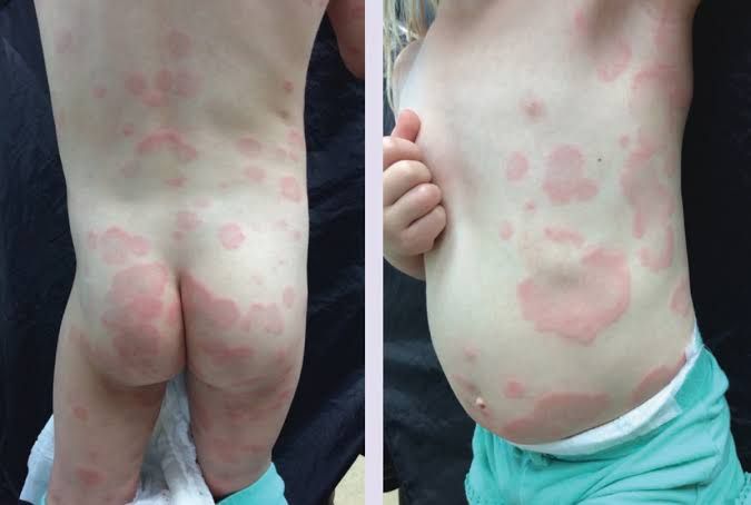
Urticaria, commonly known as hives, is characterized by raised, itchy welts on the skin. The pathogenesis involves:
- Mast Cell Activation: Urticaria is primarily driven by the activation of mast cells in the dermis. This can occur due to various triggers such as allergens (food, medications), physical stimuli (pressure, temperature), or infections.
- Histamine Release: Upon activation, mast cells degranulate and release histamine and other inflammatory mediators. Histamine causes vasodilation and increased vascular permeability, leading to the characteristic swelling and redness.
- Immune Response: In some cases, urticaria may involve an autoimmune component where antibodies target mast cells or their mediators. Chronic urticaria can be particularly complex with a significant immune response involved.
- Neuropeptides: Additionally, neuropeptides released from sensory nerves can contribute to inflammation and itchiness associated with urticaria.
Erythema Multiforme
Erythema multiforme (EM) is an acute condition often triggered by infections or medications. Its pathogenesis includes:
- Immune-Mediated Mechanism: EM is believed to be a hypersensitivity reaction that occurs in response to infections (most commonly herpes simplex virus) or drugs.
- Keratinocyte Damage: The immune system targets keratinocytes in the epidermis leading to cell death and subsequent formation of target lesions—characteristic red macules with central vesicles.
- Cytotoxic T Cells: CD8+ cytotoxic T lymphocytes play a crucial role in this process by recognizing infected or damaged keratinocytes and inducing apoptosis.
- Inflammatory Mediators: The release of cytokines such as TNF-alpha and IL-6 further amplifies the inflammatory response contributing to the clinical manifestations of EM.
Erythema Nodosum
Erythema nodosum (EN) presents as painful nodules typically located on the lower extremities and has distinct pathophysiological features:
- Panniculitis: EN is classified as a form of panniculitis involving inflammation of the subcutaneous fat tissue rather than the dermis itself.
- Delayed-Type Hypersensitivity Reaction: It is often associated with systemic conditions such as infections (e.g., streptococcal infection), sarcoidosis, or drug reactions that provoke a delayed-type hypersensitivity reaction.
- Inflammatory Cell Infiltration: The condition involves infiltration of neutrophils followed by lymphocytes into the subcutaneous fat leading to painful erythematous nodules.
- Cytokine Release: Similar to other reactive erythemas, cytokines play a significant role in EN’s pathogenesis; they promote inflammation and attract more immune cells to the site of injury.
Vasculitis
Vasculitis refers to inflammation of blood vessels which can lead to various skin manifestations including purpura or ulcers:
- Immune Complex Deposition: Vasculitis often results from immune complex deposition within vessel walls triggering an inflammatory response that damages endothelial cells lining blood vessels.
- Types of Vasculitis: There are several types including small-vessel vasculitis (e.g., IgA vasculitis) which affects capillaries and venules; medium-vessel vasculitis (e.g., polyarteritis nodosa); and large-vessel vasculitis (e.g., giant cell arteritis).
- Inflammatory Cell Recruitment: Neutrophils are typically among the first responders at sites of vascular injury followed by monocytes/macrophages which perpetuate inflammation through cytokine production.
- Clinical Manifestations: Depending on the type affected, symptoms may include palpable purpura (due to leakage from inflamed vessels), ulcers, or even systemic symptoms if larger vessels are involved leading to ischemia in affected tissues.
In summary, while each form of reactive erythema has unique triggers and pathological processes—ranging from mast cell activation in urticaria to immune-mediated damage in erythema multiforme—the common thread among them is an aberrant immune response that leads to inflammation manifesting as skin changes.
Relevance of Knowing Particular Types of Reactive Erythemas
- Diagnosis: Different types of reactive erythemas can indicate specific dermatological or systemic conditions. For instance, conditions such as urticaria (hives), contact dermatitis, and drug reactions present with erythema but have distinct etiologies and treatment protocols. Accurate identification helps healthcare providers to differentiate between these conditions and avoid misdiagnosis.
- Underlying Causes: Reactive erythemas can be symptomatic of broader systemic issues. For example, erythema multiforme may be linked to infections or drug reactions, while other forms might indicate autoimmune disorders or malignancies. Recognizing the type of erythema can lead to further investigations into these underlying causes.
- Treatment Implications: The management strategies for different types of reactive erythemas vary significantly. For instance, corticosteroids may be effective for inflammatory causes like eczema but not for infectious causes like cellulitis. Understanding the type allows clinicians to tailor treatments appropriately.
- Prognostic Value: Certain types of reactive erythemas have prognostic implications regarding the severity and duration of the condition. For example, persistent erythema associated with systemic lupus erythematosus may indicate a more severe disease course compared to transient hives due to an allergic reaction.
- Patient Education and Management: Educating patients about their specific type of reactive erythema can empower them in managing their condition effectively. It also aids in recognizing triggers that could exacerbate their symptoms, leading to better self-management strategies.
Conclusion
In summary, understanding the various types of reactive erythemas is essential not only for clinical practice but also for enhancing patient outcomes through informed decision-making and education.
Importance of Performing Dermatologic History, Examination, and Additional Diascopy in the Evaluation of Patients with Reactive Erythemas
1. Dermatologic History
A comprehensive dermatologic history is crucial for identifying potential triggers and understanding the context of the patient’s symptoms. Key components include:
- Onset and Duration: Knowing when the symptoms began and how long they have persisted helps differentiate between acute and chronic conditions.
- Associated Symptoms: Inquiring about systemic symptoms (fever, malaise) or other skin manifestations can provide clues to underlying systemic diseases.
- Triggers: Identifying potential allergens, medications, infections, or environmental factors is essential for conditions like urticaria or erythema multiforme.
- Past Medical History: A history of autoimmune diseases or previous episodes of similar skin reactions can inform the diagnosis.
2. Dermatologic Examination
The physical examination focuses on the characteristics of the lesions:
- Morphology: The appearance (macules, papules, plaques) and distribution (localized vs. generalized) help narrow down differential diagnoses.
- Color Changes: Observing color variations can indicate vascular involvement; for instance, purpura in vasculitis versus wheals in urticaria.
- Palpation: Assessing tenderness or induration can differentiate between inflammatory processes (like erythema nodosum) and non-inflammatory lesions.
3. Additional Diascopy
Diascopy is a technique used to evaluate vascular lesions by applying pressure to the skin with a glass plate or similar device. This method helps determine whether a lesion is vascular in nature:
- Vascular Lesions: If blanching occurs under pressure, it indicates that the redness is due to dilated blood vessels (as seen in urticaria).
- Non-Vascular Lesions: If there is no blanching (as seen in purpura associated with vasculitis), it suggests extravasation of blood into the dermis.
This distinction is critical because it influences management strategies; for example, non-blanching lesions may require further investigation for underlying systemic issues.
4. Differential Diagnosis
Each reactive erythema has specific diagnostic criteria:
- Urticaria: Characterized by transient wheals that are often itchy; history may reveal recent exposure to allergens.
- Erythema Multiforme: Typically presents with target lesions; associated with infections like herpes simplex virus.
- Erythema Nodosum: Presents as painful nodules on extensor surfaces; often linked to infections or medications.
- Vasculitis: May present with palpable purpura; requires consideration of systemic involvement and possible biopsy for confirmation.
5. Conclusion
Performing a detailed dermatologic history, thorough examination, and additional diascopy plays an integral role in accurately diagnosing reactive erythemas. These steps not only aid in distinguishing between different conditions but also guide appropriate management strategies tailored to individual patient needs.
Diagnostic Tests for Reactive Erythemas
Diagnosing the underlying cause of reactive erythemas is crucial for effective treatment. The diagnostic approach typically involves a combination of clinical evaluation and specific tests tailored to the suspected etiology. Below are the most important diagnostic tests required for patients with various forms of reactive erythemas.
1. Clinical History and Physical Examination
The first step in diagnosing reactive erythema is a thorough clinical history and physical examination. This includes:
- Patient History: Gathering information about the onset, duration, and characteristics of the rash, associated symptoms (such as itching or burning), potential triggers (like medications, foods, or environmental factors), and any previous skin conditions.
- Physical Examination: Assessing the distribution, morphology, and pattern of the erythema. This can help differentiate between various types of reactive erythemas such as drug eruptions, infections, or autoimmune conditions.
2. Patch Testing
For cases suspected to be related to contact dermatitis (a common form of reactive erythema), patch testing is essential. This test helps identify specific allergens that may be causing an allergic reaction:
- Procedure: Small amounts of potential allergens are applied to the skin using patches. After 48 hours, the patches are removed, and reactions are assessed at 48 hours and again at 72-96 hours.
- Indications: Particularly useful for patients with localized erythema that may suggest contact dermatitis.
3. Skin Biopsy
In certain cases where the diagnosis remains unclear after initial evaluation, a skin biopsy may be warranted:
- Purpose: A biopsy allows for histopathological examination of skin tissue to identify inflammatory cells or other changes indicative of specific dermatological conditions.
- Indications: Useful in differentiating between various forms of dermatitis (e.g., psoriasis vs. eczema) or ruling out malignancies in atypical presentations.
4. Laboratory Tests
Depending on clinical suspicion, several laboratory tests may be performed:
- Complete Blood Count (CBC): To check for signs of infection or systemic involvement (e.g., leukocytosis).
- Erythrocyte Sedimentation Rate (ESR) / C-reactive Protein (CRP): These tests assess inflammation levels in the body and can indicate systemic inflammatory processes.
- Serological Tests: Specific serologies may be ordered based on clinical suspicion (e.g., ANA for lupus erythematosus if an autoimmune process is suspected).
5. Microbiological Cultures
If there is suspicion of an infectious etiology contributing to the reactive erythema:
- Bacterial Cultures: Swabs from lesions can help identify bacterial infections such as cellulitis or impetigo.
- Fungal Cultures: If fungal infection is suspected based on clinical presentation (e.g., tinea), scraping from lesions can be cultured.
- Viral Testing: In cases where herpes simplex virus or other viral infections are suspected, PCR testing from vesicular lesions may be indicated.
6. Imaging Studies
In rare cases where deeper structures might be involved or when systemic disease is suspected:
- Ultrasound or MRI: These imaging modalities can help evaluate deeper tissue involvement if there’s concern about underlying pathology beyond superficial skin layers.
In summary, diagnosing reactive erythemas requires a multifaceted approach that begins with a detailed patient history and physical examination followed by targeted diagnostic tests such as patch testing, skin biopsy, laboratory evaluations, microbiological cultures, and possibly imaging studies depending on individual case presentations.
Principles of Management of Various Forms of Reactive Erythemas
Reactive erythemas encompass a variety of dermatological conditions characterized by redness of the skin due to inflammation or vascular changes. The management principles for these conditions—urticaria, erythema multiforme, erythema nodosum, and vasculitis—vary based on their underlying causes, symptoms, and severity. Below is a detailed examination of each condition and its management.
Urticaria
Urticaria, commonly known as hives, is characterized by raised, itchy welts on the skin. It can be acute or chronic and may result from various triggers including allergens, medications, infections, or stress.
- Identification and Avoidance of Triggers: The first step in managing urticaria is identifying potential triggers through patient history and allergy testing. Avoiding known triggers can significantly reduce episodes.
- Antihistamines: Second-generation antihistamines (e.g., cetirizine, loratadine) are the first-line treatment for urticaria. They help alleviate itching and reduce the size of hives.
- Corticosteroids: For severe cases or acute exacerbations that do not respond to antihistamines, short courses of oral corticosteroids may be prescribed to reduce inflammation.
- Other Treatments: In chronic cases unresponsive to standard treatments, options such as omalizumab (a monoclonal antibody) or immunosuppressants may be considered.
Erythema Multiforme
Erythema multiforme is an acute condition often triggered by infections (most commonly herpes simplex virus) or medications. It presents with target-like lesions on the skin.
- Identifying Triggers: Similar to urticaria, identifying any underlying infection or medication is crucial for effective management.
- Symptomatic Treatment: Mild cases may require symptomatic treatment with antihistamines for itching and topical corticosteroids for localized lesions.
- Systemic Corticosteroids: In more severe cases (especially if mucosal involvement occurs), systemic corticosteroids may be indicated to control inflammation.
- Antiviral Therapy: If herpes simplex virus is identified as a trigger, antiviral medications such as acyclovir may be used to prevent recurrences.
- Supportive Care: Ensuring adequate hydration and pain management is also important in managing this condition.
Erythema Nodosum
Erythema nodosum presents as painful red nodules typically located on the lower legs and can be associated with infections, medications, or systemic diseases like sarcoidosis.
- Identifying Underlying Causes: A thorough evaluation including history-taking and laboratory tests is essential to identify any underlying conditions that may need treatment.
- Rest and Supportive Care: Patients are advised to rest and elevate affected limbs to reduce swelling and discomfort.
- Nonsteroidal Anti-Inflammatory Drugs (NSAIDs): NSAIDs such as ibuprofen can help relieve pain and inflammation associated with erythema nodosum.
- Corticosteroids: In persistent cases where symptoms are severe or do not improve with NSAIDs alone, systemic corticosteroids may be warranted.
- Treating Underlying Conditions: If an underlying cause such as an infection is identified (e.g., streptococcal infection), appropriate treatment should be initiated.
Vasculitis
Vasculitis refers to a group of disorders characterized by inflammation of blood vessels which can lead to various skin manifestations including purpura or ulcers depending on the type involved.
- Diagnosis Confirmation: Accurate diagnosis often requires biopsy of affected tissue along with laboratory tests to determine specific types of vasculitis (e.g., ANCA-associated vasculitis).
- Immunosuppressive Therapy: Management typically involves immunosuppressive agents such as corticosteroids combined with other agents like azathioprine or cyclophosphamide depending on severity and type of vasculitis.
- Symptomatic Treatment: Pain relief through NSAIDs may also be part of the management plan alongside addressing any systemic symptoms present (fever, malaise).
- Monitoring for Complications: Regular follow-up is necessary due to potential complications arising from both the disease itself and its treatment (e.g., infections due to immunosuppression).
- Addressing Underlying Conditions: If secondary vasculitis arises from another condition (like lupus), treating that primary condition becomes crucial in managing vasculitis effectively.
In conclusion, while each form of reactive erythema has distinct characteristics requiring tailored approaches for management, common principles include identifying triggers when possible, symptomatic relief through medications like antihistamines or NSAIDs, use of corticosteroids in more severe cases, supportive care measures, and addressing any underlying conditions contributing to the erythematous response.


