Formation of Dorsal Aorta
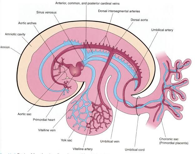
The dorsal aorta is a crucial structure in the embryonic development of vertebrates, serving as the first functional intra-embryonic blood vessel. Its formation involves a series of dynamic processes that include the emergence of bilateral vessels, their migration, and eventual fusion into a single vessel.
Embryological Origins
The dorsal aorta arises from paired embryological structures known as the dorsal aortae, which develop from the aortic arches originating from the aortic sac. Initially, these vessels are positioned laterally in relation to the developing embryo.
Lateral-to-Medial Migration
As development progresses, the paired dorsal aortae undergo significant morphological changes. They migrate from their initial lateral positions toward the midline of the embryo. This lateral-to-medial translocation is essential for their eventual fusion. The movement is guided by various molecular signals and cellular interactions that orchestrate this remodeling process.
Fusion into Descending Aorta
Once the dorsal aortae have migrated medially, they fuse at the midline to form a single descending aorta. This fusion marks an important transition in vascular development, allowing for more efficient blood circulation within the embryo. The newly formed descending aorta will continue to develop and give rise to other major arteries as well as branches that supply different regions of the body.
Branching and Functionality
After fusion, the dorsal aorta begins to branch out and provide blood supply to various embryonic structures. It gives rise to vital arteries that supply oxygenated blood to tissues and organs during early development. Additionally, it plays an essential role in hematopoiesis (the formation of blood cells), particularly generating hematopoietic stem cells that are crucial for later stages of development.
Conclusion on Dorsal Aorta Formation
In summary, the formation of the dorsal aorta is characterized by its embryological origins as paired vessels, their lateral-to-medial migration leading to fusion at the midline, and subsequent branching that supports both vascularization and hematopoiesis within developing embryos.
Formation of Aortic Arches and Their Fate
The aortic arches, also known as pharyngeal arch arteries, are a series of six paired embryological structures that develop during the early stages of human embryogenesis. They arise from the aortic sac and are located ventral to the dorsal aorta. The formation of these arches is crucial for the development of major arteries in the head and neck region.
Developmental Timeline
The formation of the aortic arches begins around the third week of gestation. Initially, these structures appear symmetrical on both sides of the embryo but undergo significant remodeling to create an asymmetrical arrangement in the mature circulatory system. Each arch forms sequentially within the context of pharyngeal arches, which are transient structures that contribute to various anatomical features in the developing embryo.
Details of Each Aortic Arch
- Arch 1 and Arch 2:
- The first and second aortic arches regress early in development.
- A remnant of the first arch contributes to the maxillary artery, which is a branch of the external carotid artery.
- The second arch gives rise to several vessels: its ventral end develops into the ascending pharyngeal artery, while its dorsal end typically forms the stapedial artery, which usually regresses but may persist in some mammals.
- Additionally, remnants from this arch contribute to the hyoid artery.
- Arch 3:
- This arch is critical as it forms part of what is known as the carotid arch.
- It gives rise to both common carotid arteries bilaterally and contributes to proximal segments of internal carotid arteries.
- Arch 4:
- Known as the systemic arch, this structure has different fates on either side:
- The right fourth arch develops into part of the right subclavian artery.
- The left fourth arch contributes to forming a segment of the aorta between where the left common carotid and left subclavian arteries originate.
- Known as the systemic arch, this structure has different fates on either side:
- Arch 5:
- This arch is unique because it either fails to form or forms incompletely before regressing entirely.
- Arch 6:
- The sixth arches have distinct roles:
- The right sixth arch persists partially as part of the right pulmonary artery while its distal section degenerates.
- The left sixth arch gives rise to both pulmonary arteries and forms the ductus arteriosus, which connects pulmonary circulation with systemic circulation during fetal life.
- After birth, increased oxygen levels cause this ductus arteriosus to constrict and eventually become obliterated within weeks after delivery, transforming into ligamentum arteriosum.
- The sixth arches have distinct roles:
Clinical Significance
The fate of these aortic arches is clinically significant because abnormalities can lead to congenital heart defects or vascular anomalies. For instance:
- Persistence or regression errors can result in conditions such as aberrant subclavian arteries or double aortic arches, which can create vascular rings around vital structures like trachea and esophagus.
In summary, understanding how each aortic arch develops and what they ultimately become is essential for recognizing potential clinical implications associated with their malformations.
Transformation of Fetal into Adult Circulation
The transition from fetal to adult circulation is a critical process that occurs shortly after birth. This transformation involves several physiological changes that adapt the circulatory system to function independently from the placenta. Below, I will outline the major changes and processes involved in this transformation step by step.
1. Fetal Circulation Overview
In fetal life, the circulatory system is designed to support the developing fetus while it is still in utero. Key features of fetal circulation include:
- Placental Circulation: The placenta serves as the organ for gas exchange, nutrient transfer, and waste removal. Oxygenated blood from the mother flows into the placenta and then enters the fetus through the umbilical vein.
- Shunts: To bypass non-functional organs (like the lungs and liver), fetuses have several shunts:
- Ductus Venosus: This vessel allows most of the oxygenated blood from the umbilical vein to bypass the liver and flow directly into the inferior vena cava.
- Foramen Ovale: This opening between the right and left atria allows blood to flow directly from the right atrium to the left atrium, bypassing pulmonary circulation.
- Ductus Arteriosus: This connection between the pulmonary artery and aorta allows blood to flow from the pulmonary artery into systemic circulation, further bypassing non-functional lungs.
2. Initiation of Breathing
Upon birth, several immediate changes occur:
- First Breath: The newborn takes its first breath, which expands the lungs and decreases pulmonary vascular resistance due to increased oxygen levels. This change leads to increased blood flow through pulmonary arteries.
- Increased Blood Flow to Lungs: As air fills the lungs, oxygen-rich blood returns via pulmonary veins to the left atrium.
3. Closure of Shunts
As a result of these changes, several key structures undergo closure:
- Foramen Ovale Closure: The increase in pressure in the left atrium (due to increased return from lungs) causes functional closure of this shunt within minutes after birth. Over time (weeks to months), it becomes anatomically sealed.
- Ductus Arteriosus Closure: Increased oxygen levels and decreased prostaglandin E1 levels lead to contraction of smooth muscle in this ductus, causing it to close within hours after birth. It eventually forms a fibrous remnant known as the ligamentum arteriosum.
- Ductus Venosus Closure: With cessation of placental blood flow, this vessel constricts and becomes a fibrous cord called the ligamentum venosum.
4. Establishment of Adult Circulation
With these closures complete, adult circulation is established:
- Blood now flows from systemic circulation into the right atrium via superior and inferior vena cavae.
- From there, it moves into the right ventricle and is pumped into pulmonary arteries for oxygenation in lungs.
- Oxygenated blood returns via pulmonary veins into left atrium, moves into left ventricle, and is pumped out through aorta for systemic distribution.
This transition marks a significant shift in how blood circulates within an individual—moving from reliance on placental structures to independent functioning organs.
In summary, at birth, significant physiological changes occur that transform fetal circulation into adult circulation through initiation of breathing leading to increased lung perfusion, closure of shunts (foramen ovale, ductus arteriosus), and establishment of normal pathways for systemic and pulmonary circulation.
Congenital Malformations During Cardiovascular Development
Congenital malformations of the cardiovascular system can occur at various stages of development, often leading to significant clinical implications. These malformations arise due to disruptions in the complex processes that govern heart and blood vessel formation during embryonic development. Below, we will explore several key congenital malformations categorized by their developmental stage.
1. Early Cardiac Development (Weeks 3-4)
During the early stages of embryogenesis, specifically between the third and fourth weeks, critical events occur that lay the foundation for normal cardiac structure. Disruptions during this period can lead to:
- Atrial Septal Defect (ASD): This defect involves an abnormal opening in the atrial septum, allowing blood to flow between the left and right atria. It can result from improper fusion of septal tissue or failure of the foramen ovale to close after birth.
- Ventricular Septal Defect (VSD): A VSD is characterized by a defect in the ventricular septum, which separates the left and right ventricles. This condition arises from incomplete closure of the interventricular septum during development.
- Transposition of the Great Arteries (TGA): In TGA, the aorta and pulmonary artery are switched, leading to two separate circulatory systems that do not communicate effectively. This occurs due to errors in the rotation and alignment of great vessels during embryonic development.
2. Formation of Outflow Tracts (Weeks 5-7)
The outflow tracts develop as part of heart morphogenesis around weeks five to seven. Congenital malformations occurring during this phase include:
- Tetralogy of Fallot (ToF): This condition encompasses four defects: VSD, pulmonary stenosis, overriding aorta, and right ventricular hypertrophy. The malformation results from abnormal neural crest cell migration affecting outflow tract formation.
- Pulmonary Stenosis: This refers to narrowing at or near the valve that obstructs blood flow from the right ventricle into the pulmonary artery. It may arise from abnormal valve formation or fusion of valve leaflets.
3. Development of Valves (Weeks 6-8)
The formation and maturation of heart valves occur primarily between weeks six and eight. Malformations during this period include:
- Aortic Stenosis: Aortic stenosis is characterized by narrowing at or below the aortic valve, which can be caused by congenital fusion of valve cusps or abnormal leaflet development.
- Mitral Valve Prolapse: This condition occurs when one or both mitral valve leaflets bulge back into the left atrium during contraction. It may result from improper connective tissue development affecting valve structure.
4. Late Cardiac Development (Weeks 8-12)
As development progresses into later stages, further structural refinements take place:
- Coarctation of the Aorta: This is a narrowing of a segment of the aorta that typically occurs just distal to where the ductus arteriosus inserts into the aorta. It can result from abnormal remodeling during fetal life.
- Single Ventricle Defects: Conditions such as hypoplastic left heart syndrome arise when one side of the heart does not develop properly, resulting in inadequate systemic circulation.
Conclusion
Congenital cardiovascular malformations represent a diverse group of conditions arising from disruptions at various stages throughout cardiac development. Understanding these defects is crucial for diagnosis and management strategies in affected individuals.


