Initial Anatomic Location of Deep Vein Thrombosis
Deep vein thrombosis (DVT) primarily occurs in the deep veins of the lower extremities, with the most common initial anatomic locations being the popliteal vein, femoral vein, and iliac veins. The popliteal vein is located behind the knee and is often where thrombi first form due to its anatomical position and blood flow dynamics. Following this, thrombi may extend into the femoral vein and potentially into the iliac veins as well. In some cases, DVT can also occur in other areas such as the upper extremities, particularly in patients with certain risk factors or conditions.
Clinical Factors Leading to Increased Incidence of Venous Thrombosis
Several clinical factors contribute to an increased incidence of venous thrombosis, commonly summarized by Virchow’s triad: stasis of blood flow, endothelial injury, and hypercoagulability.
- Stasis of Blood Flow:
- Prolonged immobility is a significant contributor to stasis. This can occur during long flights or car rides, post-surgery recovery periods, or in patients who are bedridden due to illness.
- Conditions that limit mobility, such as obesity or neurological disorders (e.g., stroke), also increase the risk by promoting stagnant blood flow in the veins.
- Endothelial Injury:
- Trauma or surgery can damage the endothelial lining of blood vessels, leading to thrombosis. Orthopedic surgeries (especially hip and knee replacements) are particularly associated with a higher risk.
- Other factors include previous venous thrombosis episodes and conditions like varicose veins that can cause chronic endothelial damage.
- Hypercoagulability:
- Certain medical conditions predispose individuals to a hypercoagulable state. These include genetic disorders such as Factor V Leiden mutation or prothrombin gene mutation.
- Acquired conditions like cancer (particularly pancreatic and lung cancers), pregnancy, hormone replacement therapy, and oral contraceptive use significantly increase clotting risks due to changes in coagulation factors.
- Age and Comorbidities:
- Older age is a well-established risk factor for DVT due to both increased prevalence of comorbidities and natural changes in vascular health.
- Comorbid conditions such as heart failure, chronic obstructive pulmonary disease (COPD), or inflammatory diseases can further exacerbate risks through mechanisms involving reduced mobility or altered hemodynamics.
- Lifestyle Factors:
- Smoking has been shown to affect coagulation pathways negatively and contributes to endothelial dysfunction.
- Dehydration can lead to increased blood viscosity which may promote thrombus formation.
In summary, DVT typically begins in the deep veins of the legs—most commonly in the popliteal vein—and is influenced by multiple clinical factors including stasis of blood flow due to immobility, endothelial injury from trauma or surgery, hypercoagulable states from genetic or acquired conditions, age-related changes, comorbidities, and lifestyle choices such as smoking.
Noninvasive and Invasive Testing Procedures for Diagnosing Venous Valvular Incompetence and Deep Vein Thrombosis
Venous valvular incompetence (VVI) and deep vein thrombosis (DVT) are significant vascular conditions that can lead to serious complications, including chronic venous insufficiency and pulmonary embolism. Accurate diagnosis is crucial for effective management. Various testing procedures, both noninvasive and invasive, are employed to diagnose these conditions.
Noninvasive Testing Procedures
- Ultrasound (Doppler Ultrasound):
- Description: This is the primary noninvasive method used to assess venous flow and detect abnormalities such as DVT or valvular incompetence. It utilizes high-frequency sound waves to create images of blood vessels.
- Application: For DVT, a Doppler ultrasound evaluates blood flow in the veins of the legs. It can identify clots by showing reduced or absent blood flow. For VVI, it assesses the function of venous valves by measuring reflux during maneuvers like leg elevation or compression.
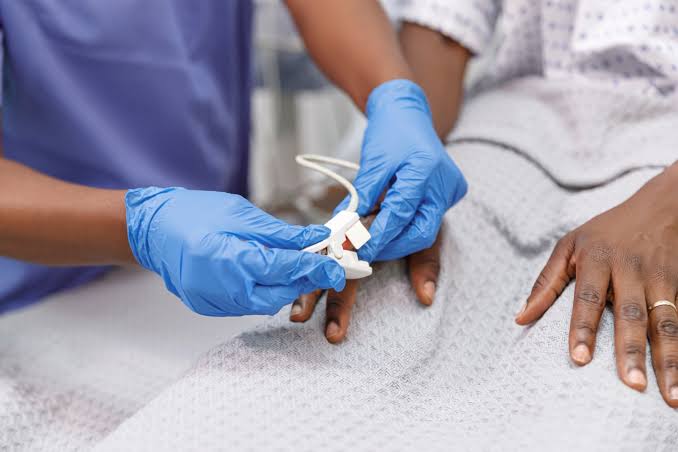
- Photoplethysmography (PPG):
- Description: PPG is a simple, noninvasive technique that measures changes in blood volume in the microvascular bed of tissue.
- Application: It is used to evaluate venous refill time and can help identify venous insufficiency by assessing how quickly blood returns to the veins after being emptied.
- Air Plethysmography (APG):
- Description: This method measures changes in volume within a limb using air-filled cuffs.
- Application: APG provides quantitative data on venous function, including parameters like ejection fraction and residual volume, which are useful for diagnosing VVI.
- Magnetic Resonance Venography (MRV):
- Description: MRV uses magnetic resonance imaging (MRI) techniques specifically tailored for visualizing veins.
- Application: It can provide detailed images of venous anatomy and help detect thrombus presence in cases where ultrasound results are inconclusive.
- Computed Tomography Venography (CTV):
- Description: CTV involves the use of CT scans with contrast material to visualize veins.
- Application: This technique is particularly useful for detecting DVT in complex cases or when other imaging modalities are not definitive.
Invasive Testing Procedures
- Venography:
- Description: This is an invasive procedure where a contrast dye is injected into a vein, followed by X-ray imaging.
- Application: Venography provides direct visualization of the venous system and can confirm the presence of thrombus or assess valve function directly.
- Endovenous Laser Therapy (EVLT):
- Description: While primarily a treatment modality, EVLT involves catheterization of veins under ultrasound guidance.
- Application: During this procedure, laser energy is applied to treat incompetent veins; however, it also allows for assessment of venous anatomy and valve function prior to treatment.
- Phlebography with Catheterization:
- Description: Similar to traditional venography but performed with catheterization techniques that allow for selective visualization of specific veins.
- Application: This method can be used when more detailed information about specific segments of the venous system is required.
- Direct Measurement Techniques (e.g., Pressure Measurements):
- Description: In some cases, direct measurement of pressure within veins may be performed through catheterization.
- Application: These measurements can help assess hemodynamic changes associated with valvular incompetence or thrombosis.
- Surgical Exploration (Rarely Used):
- In rare cases where other diagnostic methods fail or when there’s a need for immediate intervention due to severe symptoms, surgical exploration may be considered.
Conclusion
The choice between noninvasive and invasive testing procedures depends on various factors including clinical presentation, availability of resources, patient safety considerations, and specific diagnostic needs. Noninvasive methods like Doppler ultrasound remain first-line approaches due to their safety profile and effectiveness in diagnosing both DVT and VVI.
Differential Diagnosis of Acute Edema Associated with Leg Pain
Acute edema in the legs, particularly when accompanied by pain, can arise from various underlying conditions. A systematic approach to the differential diagnosis is essential for effective management. Below are the main categories and specific conditions to consider:
1. Vascular Causes
- Deep Venous Thrombosis (DVT): This is a common cause of unilateral leg swelling and pain, characterized by sudden onset edema, tenderness, and sometimes warmth over the affected area.
- Venous Insufficiency: Chronic venous insufficiency can lead to acute exacerbations of swelling and pain due to increased venous pressure.
- Superficial Thrombophlebitis: Inflammation of superficial veins can cause localized swelling and pain.
2. Infectious Causes
- Cellulitis: A bacterial skin infection that presents with redness, swelling, warmth, and pain in the affected leg.
- Abscess Formation: Localized collections of pus can lead to significant swelling and tenderness.
3. Traumatic Causes
- Acute Compartment Syndrome: This condition occurs when there is increased pressure within a muscle compartment, leading to severe pain, swelling, and potentially permanent muscle damage if not treated promptly.
- Fractures or Sprains: Trauma to the leg can result in localized edema and pain.
4. Inflammatory Conditions
- Gout or Pseudogout: These conditions can cause acute joint inflammation leading to swelling and pain in the lower extremities.
- Rheumatoid Arthritis Flare-Up: Inflammatory arthritis may present with acute swelling and discomfort in the joints of the legs.
5. Systemic Conditions
- Congestive Heart Failure (CHF): Can lead to bilateral leg edema; however, acute exacerbations may present with unilateral symptoms if there is a specific vascular issue.
- Kidney Disease: Acute kidney injury or nephrotic syndrome can lead to fluid retention manifesting as leg edema.
6. Lymphatic Causes
- Lymphedema: Although typically chronic, acute exacerbations can occur due to infections or trauma affecting lymphatic drainage.
In evaluating a patient with acute edema associated with leg pain, it is crucial to obtain a thorough history including timing of symptoms, associated features (e.g., fever), medication history (e.g., recent initiation of calcium channel blockers), and any relevant past medical history that could suggest systemic disease. Physical examination should focus on assessing for signs of DVT (e.g., Homan’s sign), infection (e.g., erythema), or signs indicative of compartment syndrome (e.g., tense compartments).
The diagnostic workup may include imaging studies such as Doppler ultrasound for DVT evaluation or CT scans for suspected abscesses or fractures.
Five Modalities for Preventing the Development of Venous Thrombosis in Surgical Patients
Effective prevention strategies are essential to reduce the risk of thromboembolic complications. Here are five key modalities for preventing venous thrombosis in surgical patients:
1. Pharmacologic Prophylaxis
Pharmacologic prophylaxis involves the use of anticoagulant medications to reduce the risk of thrombus formation. Common agents include:
- Low Molecular Weight Heparins (LMWH): Medications like enoxaparin and dalteparin are frequently used due to their efficacy in reducing DVT incidence.
- Unfractionated Heparin (UFH): Administered subcutaneously or intravenously, UFH is often used in high-risk patients or those with renal impairment.
- Direct Oral Anticoagulants (DOACs): Agents such as rivaroxaban and apixaban are increasingly utilized for their ease of administration and predictable pharmacokinetics.
The choice of agent depends on patient-specific factors, including the type of surgery, risk factors for thrombosis, and potential drug interactions.
2. Mechanical Prophylaxis
Mechanical methods aim to enhance venous return and prevent stasis through physical means. Key techniques include:
- Compression Stockings: Graduated compression stockings apply pressure to the legs, promoting venous blood flow and reducing stasis.
- Intermittent Pneumatic Compression Devices (IPC): These devices inflate and deflate cuffs around the legs, mimicking muscle contractions that facilitate venous return.
Mechanical prophylaxis is particularly useful for patients who may not tolerate anticoagulation therapy due to bleeding risks or other contraindications.
3. Early Mobilization
Encouraging early mobilization post-surgery is a critical non-pharmacological strategy. This approach includes:
- Ambulation: Patients should be encouraged to get out of bed and walk as soon as it is safe post-operatively.
- Range-of-Motion Exercises: Simple exercises can be performed while still in bed to stimulate circulation in the lower extremities.
Early mobilization helps combat immobility-related venous stasis, significantly lowering DVT risk.
4. Risk Assessment Protocols
Implementing standardized risk assessment protocols allows healthcare providers to identify patients at higher risk for venous thromboembolism (VTE). Tools such as the Caprini Risk Assessment Model help stratify patients based on various factors including:
- Surgical type
- Patient history (e.g., previous VTE events)
- Comorbid conditions (e.g., obesity, cancer)
By identifying high-risk individuals, tailored prevention strategies can be employed more effectively.
5. Education and Awareness
Educating both healthcare providers and patients about VTE risks and prevention strategies is crucial. This includes:
- Patient Education: Informing patients about the importance of mobility, recognizing symptoms of DVT, and adhering to prescribed prophylactic measures.
- Staff Training: Ensuring that all surgical team members understand VTE prevention protocols enhances compliance with preventive measures.
Awareness initiatives can lead to improved outcomes by fostering a culture of vigilance regarding thromboembolic risks.
In conclusion, preventing venous thrombosis in surgical patients requires a multifaceted approach that combines pharmacologic interventions, mechanical devices, early mobilization strategies, thorough risk assessments, and education efforts aimed at both healthcare providers and patients.
Anticoagulant and Thrombolytic Administration, Evaluation, and Contraindications
1. Anticoagulant Administration
Anticoagulants are medications that prevent blood clot formation. They can be administered through various routes depending on the specific drug and clinical situation:
- Oral Anticoagulants: Medications such as warfarin, rivaroxaban, apixaban, and dabigatran are commonly used. Warfarin is typically initiated with a loading dose followed by maintenance doses adjusted based on INR (International Normalized Ratio) monitoring. Direct oral anticoagulants (DOACs) like rivaroxaban and apixaban do not require routine monitoring but should be taken consistently to maintain therapeutic levels.
- Parenteral Anticoagulants: These include unfractionated heparin (UFH), low molecular weight heparins (LMWH) like enoxaparin, and fondaparinux. UFH is usually administered intravenously or subcutaneously, with continuous infusion for rapid effect and frequent aPTT (activated partial thromboplastin time) monitoring to adjust dosing. LMWH is typically given via subcutaneous injection with fixed dosing based on body weight, requiring less frequent monitoring than UFH.
2. Thrombolytic Administration
Thrombolytics are agents that dissolve blood clots and are primarily used in acute settings such as myocardial infarction or pulmonary embolism:
- Intravenous Administration: Thrombolytics like alteplase (tPA), reteplase, and tenecteplase are administered intravenously. The administration can be done as a bolus or continuous infusion depending on the specific agent used. For instance, alteplase is often given as a bolus followed by an infusion over several hours.
- Intra-arterial Administration: In certain cases, thrombolytics may be delivered directly into the artery affected by the clot (e.g., during catheter-directed thrombolysis). This method allows for higher local concentrations of the drug while minimizing systemic exposure.
3. Evaluation of Adequacy of Therapy
Evaluating the adequacy of anticoagulation therapy involves regular monitoring of specific laboratory parameters:
- For Anticoagulants:
- Warfarin: The INR is monitored regularly to ensure it remains within the therapeutic range (typically 2.0 to 3.0 for most indications).
- LMWH/UFH: Anti-Xa levels may be measured for LMWH to ensure appropriate dosing; aPTT is monitored for UFH.
- For Thrombolytics:
- Clinical response is assessed through symptom relief (e.g., chest pain resolution in myocardial infarction) and imaging studies (e.g., echocardiography or angiography) to evaluate perfusion restoration.
- Monitoring for bleeding complications is crucial during thrombolytic therapy.
4. Contraindications to Therapy
Both anticoagulants and thrombolytics have specific contraindications that must be considered before initiation:
- Anticoagulant Contraindications:
- Active bleeding disorders or conditions that predispose to bleeding (e.g., recent surgery, trauma).
- Severe liver disease affecting coagulation factors.
- Pregnancy (specific anticoagulants vary in safety).
- Thrombolytic Contraindications:
- History of intracranial hemorrhage or known structural cerebral vascular lesions.
- Recent major surgery or trauma within the past few weeks.
- Active bleeding or significant hypertension at presentation.
In both cases, careful patient selection and risk assessment are essential to minimize adverse outcomes associated with these therapies.
Clinical Syndrome of Pulmonary Embolus
Pulmonary embolism (PE) is a critical condition that arises when a blood clot, typically originating from the deep veins of the legs (deep vein thrombosis or DVT), travels to the lungs and obstructs one or more pulmonary arteries. This obstruction can lead to significant morbidity and mortality, making prompt recognition and management essential.
Symptoms and Signs
The clinical presentation of PE can vary widely, but common symptoms include:
- Dyspnea: Sudden onset of shortness of breath is the most frequent symptom reported by patients.
- Chest Pain: This may be pleuritic in nature (worsening with deep breaths) or may present as a feeling of tightness or pressure.
- Cough: Patients may experience a dry cough or cough up blood (hemoptysis).
- Tachycardia: An increased heart rate is often noted due to the body’s response to decreased oxygenation.
- Hypoxia: Low oxygen saturation levels can occur, leading to cyanosis in severe cases.
- Anxiety: Patients may exhibit signs of anxiety or restlessness due to hypoxia.
Physical examination findings may include tachypnea, crackles on lung auscultation, and signs of DVT such as unilateral leg swelling and tenderness.
Diagnosis
The diagnosis of PE involves several steps prioritized based on clinical suspicion:
- Clinical Assessment: Initial evaluation includes assessing risk factors for thromboembolism (e.g., recent surgery, immobility, cancer) and using clinical prediction rules like the Wells score or the Geneva score to estimate pre-test probability.
- D-dimer Testing: A negative D-dimer test can help rule out PE in low-risk patients; however, elevated levels are not specific and can occur in other conditions.
- Imaging Studies:
- CT Pulmonary Angiography (CTPA): This is the gold standard for diagnosing PE as it provides direct visualization of pulmonary arteries.
- Ventilation-Perfusion (V/Q) Scan: This is an alternative imaging modality used especially in patients who cannot undergo CTPA due to contrast allergies or renal impairment.
- Ultrasound for DVT Detection: In cases where there is suspicion of DVT contributing to PE, ultrasound imaging of the legs may be performed.
- Additional Tests: In some cases, echocardiography may be utilized to assess right heart strain or dysfunction secondary to PE.
Management Priorities
Once a diagnosis of life-threatening pulmonary embolism is established, immediate management priorities include:
- Stabilization:
- Ensure airway patency and provide supplemental oxygen if hypoxia is present.
- Establish intravenous access for medication administration.
- Anticoagulation Therapy:
- Initiate anticoagulation promptly with agents such as unfractionated heparin or low molecular weight heparin (LMWH). The choice depends on patient-specific factors including renal function and bleeding risk.
- Thrombolytic Therapy:
- For patients with massive PE causing hemodynamic instability (e.g., hypotension), thrombolytics such as alteplase may be indicated to dissolve clots rapidly.
- Supportive Care:
- Monitor vital signs closely; manage fluid status carefully to avoid volume overload while ensuring adequate perfusion.
- Consider advanced therapies such as mechanical ventilation if respiratory failure occurs.
- Long-term Management:
- After stabilization, evaluate for long-term anticoagulation therapy based on individual risk factors for recurrence versus bleeding complications.
In summary, recognizing the clinical syndrome associated with pulmonary embolism involves understanding its symptoms and signs while prioritizing rapid diagnosis through clinical assessment and imaging studies followed by immediate stabilization and treatment interventions tailored to the severity of the condition.
Indications for Surgical Intervention in Venous Thrombosis and Pulmonary Embolus
(a) Indications for Surgical Intervention in Venous Thrombosis
- Severe Symptoms or Complications:
- Patients experiencing severe pain, swelling, or skin changes due to DVT may require surgical intervention if conservative measures fail. This is particularly true if there is a risk of limb loss or significant disability.
- Phlegmasia Cerulea Dolens:
- This is a severe form of DVT characterized by extensive venous occlusion leading to limb ischemia. Surgical options such as thrombectomy may be indicated to restore blood flow and prevent tissue necrosis.
- Failure of Anticoagulation:
- If a patient does not respond to anticoagulation therapy or has recurrent thromboembolic events despite treatment, surgical options such as catheter-directed thrombolysis or mechanical thrombectomy may be considered.
- Chronic Venous Insufficiency:
- In cases where DVT has led to chronic venous insufficiency causing significant morbidity, surgical interventions like venous valve reconstruction or bypass procedures might be warranted.
- Massive Thrombus Burden:
- When there is a large thrombus burden that poses a risk of embolization or severe complications, surgical removal of the clot may be necessary.
(b) Indications for Surgical Intervention in Pulmonary Embolus
- Massive Pulmonary Embolism:
- Patients presenting with massive PE (e.g., hemodynamic instability, shock) often require urgent surgical intervention such as embolectomy when they do not respond to thrombolytic therapy or when thrombolytics are contraindicated.
- Submassive Pulmonary Embolism:
- In cases of submassive PE where patients have right ventricular dysfunction but are stable otherwise, surgical embolectomy may be indicated if there is evidence of deterioration despite medical management.
- Recurrent PE Despite Anticoagulation:
- For patients who experience recurrent pulmonary emboli while on anticoagulation therapy, surgical options like inferior vena cava (IVC) filter placement may be considered to prevent further embolic events.
- Contraindications to Thrombolysis:
- If a patient has contraindications to systemic thrombolysis (e.g., recent surgery, bleeding disorders), surgical embolectomy can provide an alternative means of removing the clot from the pulmonary circulation.
- Chronic Thromboembolic Pulmonary Hypertension (CTEPH):
- In patients who develop CTEPH following an episode of PE, pulmonary endarterectomy may be indicated to remove organized clots from the pulmonary arteries and improve hemodynamics.
Conclusion Surgical intervention in venous thrombosis and pulmonary embolism is reserved for specific clinical scenarios where conservative management fails or poses unacceptable risks. The decision for surgery must consider the patient’s overall health status, the extent of disease, and potential benefits versus risks associated with the procedure.
Diagnostic, Operative, and Nonoperative Management of Venous Ulcers and Varicose Veins
1. Diagnostic Management
The diagnosis of venous ulcers and varicose veins involves a comprehensive assessment that includes:
- Clinical Evaluation: A detailed history and physical examination are essential. Patients typically present with symptoms such as leg swelling, pain, heaviness, and skin changes. The presence of varicosities or ulcers is noted during the examination.
- Ankle-Brachial Index (ABI): This test compares the blood pressure in the patient’s ankle with the blood pressure in the arm to assess for peripheral arterial disease (PAD). An ABI of less than 0.8 may indicate significant arterial disease, which can complicate venous ulcer management.
- Duplex Ultrasound: This non-invasive imaging technique is crucial for evaluating venous anatomy and function. It helps identify reflux in the superficial or deep venous systems and assesses the presence of thrombosis.
- Venography: Although less commonly used today due to advancements in ultrasound technology, venography can be employed to visualize veins directly if necessary.
- Skin Assessment: Evaluating the ulcer’s characteristics (size, depth, exudate) and surrounding skin condition helps determine treatment strategies.
2. Operative Management
Operative management is indicated when conservative measures fail or when there are significant underlying venous issues contributing to ulcer formation or varicosities. Surgical options include:
- Saphenous Vein Stripping: This procedure involves removing the great saphenous vein to eliminate reflux. It is effective for treating symptomatic varicose veins.
- Endovenous Laser Treatment (EVLT): A minimally invasive procedure where laser energy is used to close off varicose veins. It has gained popularity due to its effectiveness and reduced recovery time compared to traditional surgery.
- Radiofrequency Ablation (RFA): Similar to EVLT but uses radiofrequency energy instead of laser energy to achieve vein closure. RFA is also minimally invasive with a quick recovery period.
- Ultrasound-Guided Sclerotherapy: Involves injecting a sclerosant into affected veins under ultrasound guidance, leading to vein closure. This method is particularly useful for smaller varicosities.
- Debridement and Skin Grafting for Ulcers: In cases where ulcers are chronic or infected, surgical debridement may be necessary followed by skin grafting if there is significant tissue loss.
3. Nonoperative Management
Nonoperative management focuses on conservative treatment strategies aimed at improving symptoms and promoting healing of venous ulcers:
- Compression Therapy: The cornerstone of treatment for both varicose veins and venous ulcers. Compression stockings help reduce edema, improve venous return, and promote ulcer healing by applying graduated pressure on the legs.
- Wound Care Management: Proper care of venous ulcers includes cleaning, dressing changes using appropriate materials (e.g., hydrocolloids), and maintaining a moist wound environment to facilitate healing.
- Leg Elevation: Encouraging patients to elevate their legs periodically can help reduce swelling and improve circulation.
- Exercise Programs: Regular physical activity enhances calf muscle pump function which aids in venous return; walking programs are often recommended.
- Pharmacological Interventions: Medications such as pentoxifylline may be prescribed to enhance microcirculation in chronic wounds; however, their efficacy remains debated.
- Patient Education: Educating patients about lifestyle modifications such as weight management, avoiding prolonged standing or sitting, and adherence to compression therapy plays a vital role in managing symptoms effectively.
In summary, effective management of venous ulcers and varicose veins requires a multifaceted approach involving accurate diagnosis through clinical evaluation and imaging techniques followed by tailored operative or nonoperative treatments based on individual patient needs.
An overview of Lymphedema
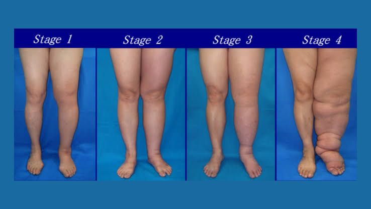
Lymphedema is a condition characterized by the accumulation of lymphatic fluid in the interstitial tissues, leading to swelling, most commonly in the limbs. It can be classified into two main categories: primary lymphedema and secondary lymphedema. Within these categories, there are specific types such as lymphedema praecox and lymphedema tarda.
Primary Lymphedema
Primary lymphedema is caused by developmental abnormalities in the lymphatic system. It can occur without any identifiable cause and is often hereditary. This type of lymphedema can be further divided into:
- Lymphedema Praecox: This form typically manifests before the age of 35, often during puberty or pregnancy. It is usually bilateral (affecting both sides) but can also be unilateral (affecting one side). The swelling may start in the feet or legs and can progress over time if not managed properly. Genetic mutations affecting lymphatic development have been identified in some cases.
- Lymphedema Tarda: This type appears after the age of 35 and is less common than praecox. It may develop due to various factors including obesity, trauma, or infections that affect lymphatic drainage over time. Unlike praecox, it does not have a clear genetic basis but may still have underlying anatomical issues with lymphatic vessels.
Secondary Lymphedema
Secondary lymphedema occurs as a result of damage to the lymphatic system due to external factors rather than congenital issues. Common causes include:
- Surgery: Particularly surgeries involving lymph node removal (e.g., cancer treatments).
- Radiation Therapy: Used for cancer treatment which can damage lymphatic vessels.
- Infections: Such as cellulitis or filariasis that can lead to scarring and obstruction of lymphatics.
- Trauma: Injuries that disrupt normal lymphatic flow.
- Obesity: Excess fat tissue can compress lymphatics and impair drainage.
Secondary lymphedema can develop at any age and often presents with localized swelling that may worsen over time if not treated.
Diagnosis and Management
Diagnosis of lymphedema involves clinical evaluation, patient history, and sometimes imaging studies like MRI or ultrasound to assess lymphatic function. Treatment options vary based on severity but typically include:
- Compression Therapy: Use of compression garments to reduce swelling.
- Manual Lymph Drainage (MLD): A specialized massage technique aimed at promoting lymph flow.
- Exercise: Tailored physical activity programs to enhance circulation.
- Skin Care: To prevent infections which are more likely in swollen areas.
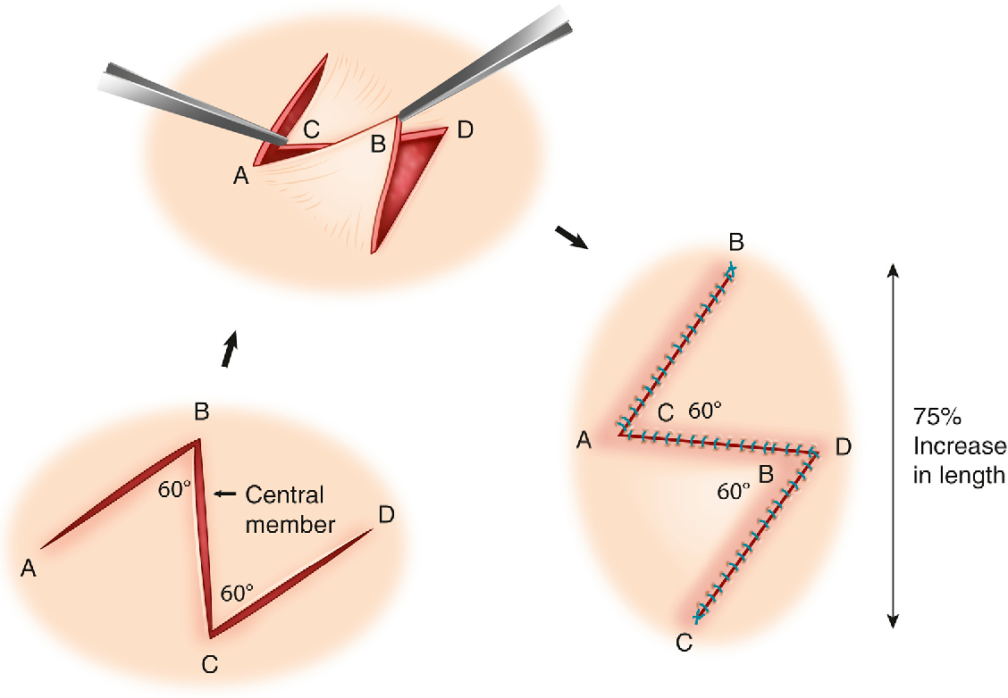
- Surgical Options: In severe cases where conservative measures fail, surgical interventions such as lymphovenous bypass or debulking procedures may be considered.
Overall management requires a multidisciplinary approach involving healthcare providers specializing in lymphedema care.
In conclusion, understanding the distinctions between primary forms like lymphedema praecox and tarda as well as secondary causes is crucial for effective diagnosis and treatment strategies for individuals affected by this condition.
Pathophysiology of Lymphedema explained
Lymphedema is primarily characterized by the accumulation of lymphatic fluid in the interstitial spaces due to impaired lymphatic drainage. The lymphatic system plays a crucial role in maintaining fluid balance, immune function, and fat absorption. When this system is compromised, it can lead to significant clinical manifestations.
- Impaired Lymphatic Transport: The pathophysiology begins with an impairment in the lymphatic transport system. This can occur due to congenital malformations (primary lymphedema) or as a result of damage from surgery, radiation, trauma, or infections (secondary lymphedema). In both cases, the lymphatic vessels are unable to effectively transport excess interstitial fluid back into the bloodstream.
- Fluid Accumulation: As lymphatic drainage becomes obstructed or insufficient, proteins and lipids accumulate in the interstitial space. This accumulation leads to increased osmotic pressure within the tissues, causing further fluid retention and swelling.
- Tissue Changes: Over time, chronic lymphedema results in progressive architectural changes within the affected tissues. These changes include:
- Adipose Tissue Deposition: The body may respond to chronic swelling by increasing fat deposition in the affected area.
- Fibrosis: There is also an increase in collagen deposition leading to fibrosis, which stiffens and thickens the tissue.
- Inflammation: Chronic inflammation may develop as a response to prolonged fluid accumulation and tissue damage.
- Complications: The impaired lymphatic system not only causes physical symptoms but also increases susceptibility to infections due to skin integrity issues and altered immune responses. In severe cases, there is a risk of developing lymphangiosarcoma, a rare form of cancer associated with long-standing lymphedema.
Treatment of Lymphedema
While there is currently no cure for lymphedema, various treatment strategies aim to manage symptoms and improve quality of life:
- Conservative Management:
- Compression Therapy: Use of compression garments or bandaging helps maintain reduced swelling by applying external pressure on affected limbs.
- Manual Lymphatic Drainage (MLD): A specialized form of massage therapy that encourages lymph flow through manual manipulation.
- Exercise: Regular physical activity can promote lymph circulation and reduce swelling.
- Physical Therapy: Physical therapists trained in lymphedema management can provide tailored exercise programs and techniques that help facilitate lymph drainage.
- Medication:
- Antibiotics may be prescribed for infections that arise due to skin breakdown.
- Pain management medications can help alleviate discomfort associated with swelling.
- Surgical Options:
- Surgical interventions are considered when conservative treatments fail or if there is significant functional impairment due to severe lymphedema.
- Procedures may include:
- Lymphovenous Bypass: Connecting lymphatic vessels directly to nearby veins.
- Liposuction for Lymphedema: Removal of excess fatty tissue that has accumulated over time.
- Debulking Surgery: Reducing the volume of swollen tissue.
- Lifestyle Modifications:
- Patients are encouraged to adopt preventive measures such as skin care routines to protect against infection and maintaining a healthy weight through diet and exercise.
In summary, understanding the pathophysiology of lymphedema highlights its complexity involving fluid dynamics and tissue responses while emphasizing that effective treatment requires a multidisciplinary approach tailored to individual patient needs.


