Basis for the Antithrombotic Role of Normal Endothelium
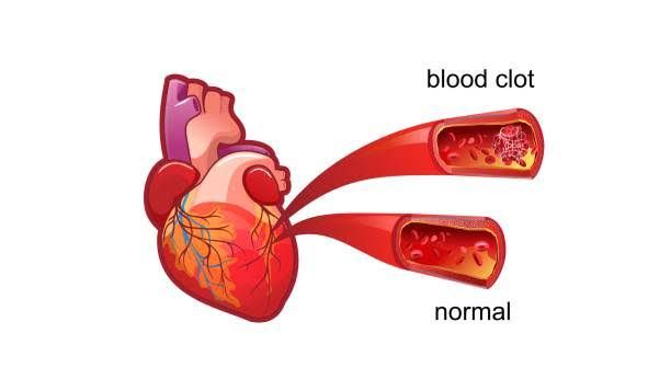
The endothelium, a thin layer of cells lining the blood vessels, plays a crucial role in maintaining vascular health and preventing thrombosis. Its antithrombotic properties are essential for regulating hemostasis and ensuring that blood flows smoothly without unnecessary clot formation. The mechanisms underlying the antithrombotic role of normal endothelium can be understood through several key functions and interactions:
1. Production of Anticoagulant Factors
Normal endothelial cells produce various anticoagulant substances that inhibit coagulation processes. These include:
- Nitric Oxide (NO): This potent vasodilator is synthesized by endothelial nitric oxide synthase (eNOS). NO inhibits platelet activation and aggregation by preventing their adherence to the vessel wall. It also promotes vasodilation, which enhances blood flow and reduces shear stress on the endothelium.
- Prostacyclin (PGI2): Another important product of endothelial cells, prostacyclin is derived from arachidonic acid via cyclooxygenase enzymes. It acts similarly to nitric oxide by inhibiting platelet aggregation and promoting vasodilation.
- Tissue Factor Pathway Inhibitor (TFPI): This protein inhibits the tissue factor/factor VIIa complex, which is crucial for initiating the extrinsic pathway of coagulation. By limiting this pathway, TFPI helps prevent excessive clot formation.
- Antithrombin III: Endothelial cells also facilitate the action of antithrombin III, a plasma protein that inactivates thrombin and other serine proteases involved in coagulation.
2. Regulation of Platelet Activity
Endothelial cells express surface receptors that modulate platelet function:
- Receptors for Adenosine Diphosphate (ADP) and Thromboxane A2: Under normal conditions, endothelial cells release nucleotides such as ATP and ADP that can inhibit platelet activation through specific receptors (e.g., P2Y12). This balance prevents inappropriate platelet aggregation.
- Expression of Cell Adhesion Molecules: Healthy endothelium maintains low levels of cell adhesion molecules such as P-selectin and von Willebrand factor (vWF), which are involved in platelet adhesion. When activated or injured, endothelial cells increase expression of these molecules to promote hemostasis when necessary but keep them suppressed under normal conditions to avoid unwanted clotting.
3. Fibrinolytic Activity
Endothelial cells contribute to fibrinolysis—the process by which clots are dissolved—by producing tissue-type plasminogen activator (tPA):
- Tissue-Type Plasminogen Activator (tPA): tPA converts plasminogen into plasmin, an enzyme that breaks down fibrin clots. The release of tPA from healthy endothelial cells ensures that once a clot has served its purpose in sealing a wound, it can be effectively removed from circulation.
4. Barrier Function
The integrity of the endothelial barrier is vital for its antithrombotic function:
- Prevention of Leukocyte Adhesion: Under normal conditions, healthy endothelium restricts leukocyte adhesion to prevent inflammation-induced thrombosis. Inflammatory cytokines can activate endothelial cells, leading to increased expression of adhesion molecules; however, under homeostatic conditions, this expression is minimized.
- Fluid Exchange Regulation: The endothelium regulates the exchange of fluids and solutes between blood and tissues while maintaining a selective barrier against procoagulant factors entering the bloodstream.
5. Response to Shear Stress
Endothelial cells adapt their functions based on mechanical forces exerted by flowing blood:
- Mechanotransduction: Endothelial cells sense shear stress from blood flow through specialized structures called mechanoreceptors. This sensing leads to changes in gene expression that enhance their antithrombotic properties under normal physiological conditions while preparing them for rapid response during injury or inflammation.
In summary, the antithrombotic role of normal endothelium is multifaceted, involving the production of anticoagulants, regulation of platelet activity, promotion of fibrinolysis, maintenance of barrier integrity, and adaptation to shear stress. These mechanisms work together to ensure proper hemostasis while preventing pathological thrombus formation.
Antithrombin III (ATIII), Protein C, and Protein S: Actions and Mechanisms
1. Antithrombin III (ATIII)
Antithrombin III is a glycoprotein produced primarily in the liver that plays a crucial role in regulating blood coagulation. Its primary function is to inhibit serine proteases involved in the coagulation cascade, particularly thrombin (factor IIa) and factor Xa. ATIII binds to these activated coagulation factors and neutralizes their activity, thereby preventing excessive clot formation.
The action of ATIII is significantly enhanced by heparin, a naturally occurring anticoagulant found in the body. Heparin accelerates the binding of ATIII to thrombin and factor Xa, increasing its inhibitory effect. This mechanism is vital for maintaining hemostatic balance and preventing thromboembolic disorders.
2. Protein C
Protein C is another critical component of the anticoagulation system, synthesized in the liver as an inactive zymogen. Upon activation (to activated protein C or APC) by thrombin bound to thrombomodulin on endothelial cells, it exerts its anticoagulant effects primarily through two mechanisms:
- Inhibition of Factors Va and VIIIa: Activated protein C works in conjunction with protein S to degrade factor Va and factor VIIIa, which are essential cofactors for prothrombinase and tenase complexes respectively. By inhibiting these factors, APC reduces thrombin generation and limits clot formation.
- Anti-inflammatory Effects: In addition to its anticoagulant properties, activated protein C has anti-inflammatory effects that help modulate the inflammatory response during vascular injury.
3. Protein S
Protein S serves as a cofactor for activated protein C in its role as an anticoagulant. It enhances the ability of APC to inactivate factors Va and VIIIa effectively. Protein S exists in two forms: free (active) form and bound form (complexed with complement component C4b). Only free protein S can facilitate the action of APC.
Deficiencies or dysfunctions in either protein C or protein S can lead to increased risk of thrombosis due to impaired regulation of coagulation processes. Both proteins are essential for maintaining normal hemostasis and preventing thromboembolic diseases.
In summary, Antithrombin III inhibits key coagulation factors such as thrombin and factor Xa; Protein C, when activated by thrombin, inhibits factors Va and VIIIa; while Protein S acts as a cofactor for activated Protein C, enhancing its anticoagulant effect.
Pathogenesis of Activated Protein C (APC) Resistance
Activated protein C (APC) resistance is a condition characterized by the body’s inability to effectively utilize activated protein C, which plays a crucial role in regulating blood coagulation. Normally, APC, in conjunction with its cofactor protein S, functions to degrade coagulation factors Va and VIIIa. This degradation process is essential for maintaining hemostatic balance and preventing excessive clotting.
Mechanism of Action of APC
Under normal physiological conditions, when thrombin is generated during the coagulation cascade, it activates protein C. Once activated, APC binds to protein S and together they cleave factor Va and factor VIIIa. This action reduces thrombin generation and limits clot formation. The effective functioning of this pathway is vital for preventing venous thrombosis.
Genetic Basis of APC Resistance
The most common hereditary cause of APC resistance is the Factor V Leiden mutation (R506Q), which occurs due to a single nucleotide substitution in the F5 gene. This mutation alters the structure of factor V, making it resistant to cleavage by activated protein C. As a result, factor Va remains active longer than it should, leading to increased thrombin generation and a hypercoagulable state.
In addition to Factor V Leiden, other genetic variants such as Factor V Cambridge and certain haplotypes can also contribute to APC resistance but are less prevalent. These mutations similarly impair the ability of APC to inactivate factor Va.
Acquired Forms of APC Resistance
While hereditary forms are significant, acquired forms of APC resistance can also occur due to various conditions that elevate factor VIII levels or alter coagulation dynamics. For instance:
- Pregnancy: Increased levels of estrogen lead to higher concentrations of clotting factors.
- Hormonal Therapy: Use of oral contraceptives containing estrogen can induce a hypercoagulable state.
- Certain Diseases: Conditions like antiphospholipid syndrome can also lead to acquired resistance.
These acquired factors may exacerbate the risk associated with inherited mutations like Factor V Leiden.
Clinical Implications
Patients with APC resistance have an increased risk for venous thromboembolism (VTE), including deep vein thrombosis (DVT) and pulmonary embolism (PE). The presence of additional risk factors—such as surgery, prolonged immobility, or other genetic predispositions—can further elevate this risk.
Diagnosis typically involves specific tests that measure the response to activated protein C; these include both aPTT-based tests and endogenous thrombin potential assays. Treatment strategies may vary based on whether individuals are symptomatic or asymptomatic and often involve anticoagulation therapy in high-risk situations.
In summary, the pathogenesis of activated protein C resistance primarily revolves around genetic mutations that hinder the normal function of coagulation regulation mechanisms, leading to an increased propensity for thrombosis under certain conditions.
Common Conditions Associated with Primary (Genetic) and Acquired Thrombosis
Thrombosis refers to the formation of a blood clot within a blood vessel, which can lead to serious health complications. It can be classified into two main categories: primary (genetic) thrombosis and acquired thrombosis. Each category has its own set of associated conditions.
Primary (Genetic) Thrombosis
Primary thrombosis is often linked to inherited genetic disorders that predispose individuals to abnormal clotting. The most common conditions associated with primary thrombosis include:
- Factor V Leiden Mutation: This is a mutation in the Factor V gene that makes it resistant to inactivation by activated protein C, leading to an increased risk of venous thromboembolism (VTE).
- Prothrombin Gene Mutation (G20210A): This mutation results in elevated levels of prothrombin, a protein essential for blood clotting, thereby increasing the risk of thrombosis.
- Antithrombin III Deficiency: Antithrombin III is a protein that helps regulate blood coagulation. A deficiency can lead to an increased risk of developing clots.
- Protein C Deficiency: Protein C plays a crucial role in controlling blood clotting; its deficiency can lead to an increased tendency for thrombosis.
- Protein S Deficiency: Similar to Protein C, Protein S is involved in regulating coagulation. Its deficiency can also increase the risk of thrombotic events.
- Hyperhomocysteinemia: Elevated levels of homocysteine, an amino acid, are associated with an increased risk of arterial and venous thrombosis.
- Inherited Thrombophilia Syndromes: These include various combinations of the above genetic factors that collectively increase the risk for thrombotic events.
Acquired Thrombosis
Acquired thrombosis occurs due to external factors or conditions that increase the likelihood of clot formation. Common conditions associated with acquired thrombosis include:
- Prolonged Immobility: Situations such as long flights or bed rest after surgery can lead to venous stasis, increasing the risk for deep vein thrombosis (DVT).
- Cancer: Certain cancers and their treatments can promote a hypercoagulable state, significantly increasing the risk for thrombotic events.
- Hormonal Factors: Use of oral contraceptives or hormone replacement therapy has been linked to an increased risk of venous thromboembolism due to hormonal changes affecting coagulation pathways.
- Pregnancy and Postpartum Period: Pregnancy induces physiological changes that increase coagulability; thus, women are at higher risk during pregnancy and shortly after childbirth.
- Obesity: Excess body weight is associated with chronic inflammation and altered hemostatic function, contributing to a higher incidence of thrombotic events.
- Chronic Inflammatory Diseases: Conditions like rheumatoid arthritis or inflammatory bowel disease can create a pro-inflammatory state that increases thrombus formation.
- Cardiovascular Diseases: Atrial fibrillation and other heart conditions can lead to stasis and turbulence in blood flow, predisposing individuals to clot formation.
- Surgery or Trauma: Surgical procedures or significant injuries can trigger clotting mechanisms as part of the body’s response to injury, particularly orthopedic surgeries involving lower limbs.
- Central Venous Catheters: The presence of catheters can irritate blood vessels and promote thrombus formation at the site of insertion or along the catheter path.
In summary, both primary (genetic) and acquired forms of thrombosis have distinct but sometimes overlapping sets of conditions that contribute to their development.


