Maturational Sequence of Erythroid Cells in the Bone Marrow
The process of erythropoiesis, or the formation of red blood cells (RBCs), occurs primarily in the bone marrow and involves a series of distinct cell types that mature through specific stages. The key stages in this maturation sequence include proerythroblast, erythroblast, normoblast, and reticulocyte. Each stage is characterized by unique morphological features and developmental changes.
1. Proerythroblast
The proerythroblast is the earliest recognizable precursor in the erythroid lineage. It is a large cell with a diameter of approximately 14-20 micrometers. The nucleus is round and occupies most of the cell volume, exhibiting a fine chromatin pattern and one or more nucleoli. The cytoplasm is basophilic due to the presence of ribosomes and RNA, indicating active protein synthesis. Proerythroblasts are committed to the erythroid lineage and respond to erythropoietin (EPO), a hormone that stimulates red blood cell production.
2. Erythroblast
As proerythroblasts mature, they develop into erythroblasts, which can be further classified into three sub-stages: basophilic erythroblast, polychromatic erythroblast, and orthochromatic erythroblast.
- Basophilic Erythroblast: This stage features intense basophilia due to abundant ribosomal RNA. The nucleus remains large with a coarse chromatin structure.
- Polychromatic Erythroblast: In this stage, hemoglobin synthesis begins, leading to a change in cytoplasmic color from blue (basophilic) to a mix of blue and pink (polychromatic). The nucleus starts to condense as the cell prepares for further maturation.
- Orthochromatic Erythroblast (Normoblast): At this point, hemoglobin production is at its peak. The cytoplasm appears more eosinophilic (pink) due to high hemoglobin content. The nucleus becomes smaller and more condensed until it is eventually extruded from the cell.
3. Normoblast
The term “normoblast” refers specifically to the orthochromatic erythroblast just before enucleation. Normoblasts are characterized by their dense nuclei that are about to be expelled from the cell as part of normal maturation. Once enucleation occurs, these cells lose their nuclei but retain some organelles.
4. Reticulocyte
After enucleation, the resulting cell is called a reticulocyte. Reticulocytes are slightly larger than mature red blood cells and still contain remnants of organelles such as ribosomes and endoplasmic reticulum, which can be visualized using special stains (e.g., methylene blue). They typically account for about 0.5% to 1% of circulating red blood cells in healthy adults and enter circulation from the bone marrow within 1-2 days after their formation.
Reticulocytes mature into fully functional erythrocytes within 1-2 days after entering circulation when they lose their remaining organelles.
In summary, the sequence from proerythroblast through reticulocyte represents critical stages in RBC development characterized by distinct morphological changes and functional adaptations necessary for effective oxygen transport throughout the body.
Aplastic Anemia: Etiology, Diagnostic Criteria, Clinical Features, and Management
Etiology
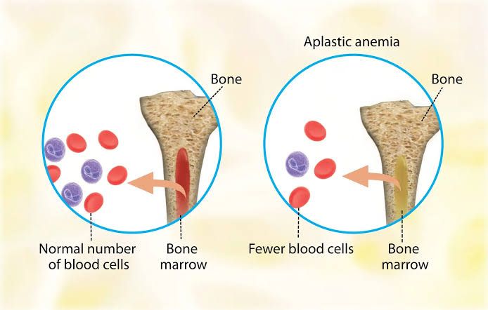
Aplastic anemia is a rare but serious hematological condition characterized by the failure of the bone marrow to produce adequate amounts of blood cells, leading to pancytopenia (a reduction in red blood cells, white blood cells, and platelets). The etiology of aplastic anemia can be broadly categorized into two main types: acquired and inherited.
- Acquired Aplastic Anemia: This form accounts for the majority of cases and can result from various factors:
- Autoimmune Disorders: Conditions such as systemic lupus erythematosus (SLE) can trigger an immune response that attacks bone marrow stem cells.
- Exposure to Chemicals: Certain chemicals like benzene, pesticides, and some medications (e.g., chloramphenicol, nonsteroidal anti-inflammatory drugs) are known to cause aplastic anemia.
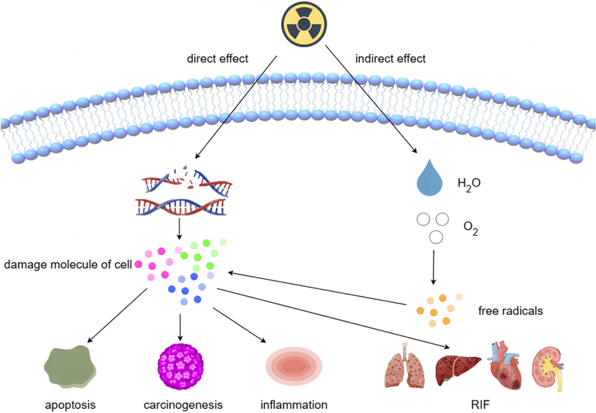
- Radiation Exposure: High doses of radiation can damage bone marrow.
- Viral Infections: Infections with viruses such as hepatitis viruses (especially hepatitis A and E), Epstein-Barr virus (EBV), cytomegalovirus (CMV), and human immunodeficiency virus (HIV) have been implicated.
- Idiopathic Cases: In many instances, no specific cause is identified.
- Inherited Aplastic Anemia: This less common form includes genetic syndromes such as:
- Fanconi Anemia: A genetic disorder that affects DNA repair mechanisms.
- Diamond-Blackfan Anemia: Primarily affects red blood cell production.
- Other rare genetic conditions may also predispose individuals to aplastic anemia.
Diagnostic Criteria
The diagnosis of aplastic anemia typically involves a combination of clinical evaluation and laboratory tests:
- Clinical History and Physical Examination: Patients often present with symptoms related to low blood counts such as fatigue, pallor, easy bruising or bleeding, recurrent infections due to leukopenia, and other signs associated with anemia.
- Complete Blood Count (CBC): The CBC will show:
- Low hemoglobin levels indicating anemia.
- Low platelet count indicating thrombocytopenia.
- Low white blood cell count indicating leukopenia.
- Bone Marrow Biopsy: This is a critical diagnostic tool. In aplastic anemia, the bone marrow is typically hypocellular (showing reduced cellularity) with a significant decrease in hematopoietic cells while retaining fat spaces.
- Additional Tests:
- Reticulocyte count may be low due to inadequate production of new red blood cells.
- Peripheral blood smear may show features consistent with pancytopenia.
- Tests for viral infections or autoimmune markers may be conducted based on clinical suspicion.
- Severity Classification: Aplastic anemia can be classified based on severity into mild, moderate, or severe forms according to specific criteria involving blood counts and clinical symptoms.
Clinical Features
The clinical manifestations of aplastic anemia arise primarily from the deficiency in red blood cells, white blood cells, and platelets:
- Anemia Symptoms: Fatigue, weakness, pallor due to reduced oxygen-carrying capacity.
- Thrombocytopenia Symptoms: Easy bruising, petechiae (small red spots on the skin), prolonged bleeding from cuts or dental work.
- Leukopenia Symptoms: Increased susceptibility to infections; patients may experience recurrent fevers or infections due to low white blood cell counts.
Complications can include severe infections or bleeding episodes which may require urgent medical intervention.
Management
The management of aplastic anemia depends on its severity and underlying cause:
- Supportive Care:
- Blood transfusions may be necessary for symptomatic patients experiencing severe anemia or significant bleeding episodes.
- Platelet transfusions can help manage thrombocytopenia-related bleeding risks.
- Immunosuppressive Therapy (IST):
- For patients with severe aplastic anemia without a matched sibling donor for transplantation, IST using agents like antithymocyte globulin (ATG) combined with cyclosporine is standard treatment aimed at suppressing the immune system’s attack on bone marrow stem cells.
- Bone Marrow Transplantation (BMT):
- Allogeneic stem cell transplantation is considered curative for younger patients with severe aplastic anemia who have a suitable donor. This involves replacing the defective bone marrow with healthy stem cells from a donor.
- Androgens & Growth Factors:
- Androgens like danazol may stimulate erythropoiesis in some cases; however, their use has declined due to side effects and limited efficacy compared to other treatments.
- Erythropoietin-stimulating agents might also be used in select cases where there’s evidence of ineffective erythropoiesis.
- Monitoring & Follow-Up Care:
- Regular monitoring through complete blood counts is essential for assessing treatment response and managing complications promptly.
In conclusion, aplastic anemia requires a multidisciplinary approach for effective management tailored to individual patient needs based on severity and underlying causes.
Classification of Myelophthisic Anemias
Myelophthisic anemia is a type of anemia that occurs due to the replacement of bone marrow tissue with abnormal cells or fibrous tissue, leading to impaired hematopoiesis (the formation of blood cells). This condition can arise from various underlying causes, including malignancies, infections, and other diseases that affect the bone marrow. Myelophthisic anemia is characterized by a reduction in red blood cell production, which results in symptoms such as fatigue, pallor, and weakness.
Myelophthisic anemias can be classified based on their underlying causes and the nature of the infiltrating material. The primary classifications include:
- Malignant Infiltrative Disorders:
- Leukemia: Acute or chronic leukemias can infiltrate the bone marrow and disrupt normal hematopoiesis.
- Lymphoma: Certain types of lymphoma can invade the bone marrow space.
- Metastatic Carcinoma: Cancers from other sites (e.g., breast, prostate) may metastasize to the bone marrow.
- Non-Malignant Infiltrative Disorders:
- Fibrosis: Conditions like primary myelofibrosis lead to excessive fibrous tissue formation within the marrow.
- Granulomatous Diseases: Diseases such as sarcoidosis or tuberculosis can cause granuloma formation in the bone marrow.
- Infectious Processes: Certain infections (e.g., viral infections) may lead to infiltration and suppression of normal hematopoiesis.
- Other Causes:
- Aplastic Anemia: Although primarily characterized by a failure of hematopoiesis rather than infiltration, it may overlap with myelophthisic processes when there is concurrent infiltration.
- Nutritional Deficiencies: Severe deficiencies in vitamins (like B12 or folate) may mimic myelophthisic anemia but are not classified under this category strictly.
Symptoms and Diagnosis
Patients with myelophthisic anemia often present with symptoms related to anemia itself—such as fatigue and pallor—as well as signs related to thrombocytopenia (low platelet count) or leukopenia (low white blood cell count), depending on the extent of bone marrow involvement. Diagnosis typically involves blood tests showing normocytic or macrocytic anemia, reticulocytopenia, and possibly a bone marrow biopsy revealing abnormal cellularity or fibrosis.
Treatment
Management strategies for myelophthisic anemia focus on treating the underlying cause. For instance, chemotherapy may be indicated for malignancies, while supportive care such as transfusions might be necessary for symptomatic relief.
The Role of Erythropoietin in Hematopoiesis
Erythropoietin (EPO) is a glycoprotein hormone that plays a crucial role in the regulation of erythropoiesis, which is the process of producing red blood cells (RBCs) from hematopoietic stem cells in the bone marrow. Understanding its function involves examining its site of production, mechanism of action, and target cells.
Site of Production
Erythropoietin is primarily produced in the kidneys. Specifically, it is synthesized by interstitial fibroblasts located in the renal cortex and outer medulla. Under normal physiological conditions, these cells secrete EPO in response to hypoxia, or low oxygen levels in the blood. This response is mediated by hypoxia-inducible factors (HIFs), which are transcription factors that become stabilized under low oxygen conditions. When oxygen levels drop, HIFs activate the transcription of the EPO gene, leading to increased production and secretion of erythropoietin into the bloodstream.
In addition to the kidneys, small amounts of EPO can also be produced by other tissues such as the liver and brain; however, these contributions are minimal compared to renal production.
Mechanism of Action
Once released into circulation, erythropoietin travels to the bone marrow where it exerts its effects on hematopoiesis. The primary mechanism through which EPO influences red blood cell production involves binding to specific receptors on erythroid progenitor cells within the bone marrow. These progenitor cells include burst-forming unit-erythroid (BFU-E) and colony-forming unit-erythroid (CFU-E) cells.
Upon binding to its receptor (the erythropoietin receptor or EPOR), several intracellular signaling pathways are activated, most notably the JAK2/STAT5 pathway. This activation leads to:
- Cell Proliferation: EPO stimulates the proliferation of erythroid progenitor cells.
- Differentiation: It promotes differentiation into mature red blood cells.
- Survival: EPO enhances cell survival by inhibiting apoptosis (programmed cell death) in erythroid progenitors.
These actions collectively increase the number of red blood cells produced in response to increased demand for oxygen transport due to conditions such as anemia or high altitudes.
Target Cells
The primary target cells for erythropoietin are those within the erythroid lineage in the bone marrow. Specifically:
- BFU-E Cells: These are early progenitor cells that respond to EPO stimulation by proliferating and differentiating into more committed progenitor cells.
- CFU-E Cells: These are more mature progenitor cells that further differentiate into reticulocytes—the immediate precursors to mature red blood cells.
In summary, erythropoietin plays a vital role in hematopoiesis by promoting red blood cell formation through its action on specific progenitor cells in the bone marrow, with its production primarily occurring in response to hypoxic conditions within the kidneys.
Classification of Anemias According to Pathophysiologic Criteria
Anemia can be classified based on its underlying pathophysiology, which refers to the mechanisms that lead to the development of anemia. This classification is essential for understanding the causes and guiding appropriate treatment. The two primary categories of anemia according to pathophysiologic criteria are regenerative and hyporegenerative anemias.
Regenerative Anemia
Regenerative anemia occurs when the bone marrow responds adequately to anemia by increasing red blood cell (RBC) production. This response is typically seen in situations where there is a loss of red blood cells due to bleeding or hemolysis. The key characteristics include:
- Increased Reticulocyte Count: In regenerative anemia, the reticulocyte count is elevated as the body attempts to compensate for the loss of mature erythrocytes.
- Causes:
- Acute Blood Loss: Such as from trauma or surgery, leading to a rapid decrease in RBCs.
- Hemolytic Anemia: Conditions where RBCs are destroyed prematurely, such as autoimmune hemolytic anemia, sickle cell disease, or hereditary spherocytosis.
- Response to Treatment: For example, after iron supplementation in iron deficiency anemia, if there is sufficient iron available for erythropoiesis.
Hyporegenerative Anemia
Hyporegenerative anemia occurs when the bone marrow fails to produce enough red blood cells despite adequate stimulation. This can result from various factors affecting erythropoiesis. Key characteristics include:
- Decreased Reticulocyte Count: In hyporegenerative anemias, reticulocyte counts are low because the bone marrow cannot produce sufficient new RBCs.
- Causes:
- Nutritional Deficiencies: Such as iron deficiency, vitamin B12 deficiency, or folate deficiency that impair RBC production.
- Bone Marrow Disorders: Conditions like aplastic anemia or myelodysplastic syndromes where the bone marrow’s ability to generate blood cells is compromised.
- Chronic Diseases: Chronic inflammatory states (e.g., chronic kidney disease) can lead to decreased erythropoietin production and impaired response of the bone marrow.
Integration of Classifications
The integration of these classifications helps clinicians determine not only the type of anemia but also its potential causes and implications for treatment. For instance:
- A patient with high reticulocyte counts may require investigation into recent bleeding or hemolysis.
- Conversely, a patient with low reticulocyte counts may need evaluation for nutritional deficiencies or bone marrow pathology.
Understanding these classifications allows healthcare providers to tailor their diagnostic approach and management strategies effectively.
Classification of Anemias According to Mean Corpuscular Volume (MCV)
Anemia can be classified based on the mean corpuscular volume (MCV), which is a measure of the average volume of red blood cells. The MCV helps in categorizing anemia into three main types: microcytic, normocytic, and macrocytic anemia. Each type has distinct causes and characteristics.
1. Microcytic Anemia
Microcytic anemia is characterized by a low MCV, typically less than 80 femtoliters (fL). This type of anemia often indicates a deficiency in hemoglobin synthesis or iron availability.
Examples of Microcytic Anemia:
- Iron Deficiency Anemia: This is the most common type of microcytic anemia, resulting from insufficient iron intake, chronic blood loss, or increased demand during pregnancy.
- Thalassemia: A genetic disorder that affects hemoglobin production, leading to smaller than normal red blood cells.
- Anemia of Chronic Disease: Often associated with chronic infections, inflammatory diseases, or malignancies that can lead to impaired iron utilization.
2. Normocytic Anemia
Normocytic anemia is characterized by a normal MCV range (80-100 fL) but presents with a reduced number of red blood cells. This type may indicate underlying systemic issues rather than problems with red blood cell production.
Examples of Normocytic Anemia:
- Aplastic Anemia: A condition where the bone marrow fails to produce adequate amounts of blood cells due to autoimmune diseases, toxins, or infections.
- Anemia of Chronic Kidney Disease: Impaired erythropoietin production due to kidney dysfunction leads to decreased red blood cell production.
- Hemolytic Anemia: Conditions where red blood cells are destroyed faster than they can be produced; this can occur due to autoimmune disorders or inherited conditions like sickle cell disease.
3. Macrocytic Anemia
Macrocytic anemia is characterized by an elevated MCV (greater than 100 fL). This type usually indicates problems with DNA synthesis in red blood cell precursors.
Examples of Macrocytic Anemia:
- Vitamin B12 Deficiency: Often caused by pernicious anemia or malabsorption syndromes leading to inadequate vitamin B12 levels necessary for proper red blood cell formation.
- Folate Deficiency: Similar to vitamin B12 deficiency but specifically related to insufficient folate intake or absorption; commonly seen in individuals with poor diet or certain medical conditions.
- Myelodysplastic Syndromes: A group of disorders caused by poorly formed or dysfunctional blood cells due to abnormalities in the bone marrow.
In summary, classifying anemias according to MCV provides valuable insights into their underlying causes and guides appropriate treatment strategies. Understanding these classifications aids healthcare professionals in diagnosing and managing various types of anemia effectively.
Reticulocyte Count and Corrected Reticulocyte Count
The reticulocyte count is a laboratory test that measures the number of reticulocytes, which are immature red blood cells (RBCs), in the bloodstream. This count is an important indicator of bone marrow activity and erythropoiesis (the production of red blood cells). Reticulocytes are typically released into the bloodstream from the bone marrow and mature into fully functional red blood cells within 1-2 days.
Normal Values and Interpretation
In healthy adults, the normal reticulocyte count ranges from approximately 0.5% to 2.5% of total red blood cell count. However, these values can vary based on factors such as age, sex, and altitude.
Corrected Reticulocyte Count
The corrected reticulocyte count adjusts the raw reticulocyte percentage based on the patient’s hematocrit (Hct) or hemoglobin level. This correction is crucial because it provides a more accurate assessment of bone marrow response to anemia or other conditions affecting red blood cell production. The formula for calculating the corrected reticulocyte count is:

Where:
- Normal Hct is typically around 45% for males and 40% for females.
This correction helps to determine whether the bone marrow is adequately responding to anemia or other stimuli.
Diseases Associated with High Reticulocyte Counts
A high reticulocyte count indicates increased erythropoiesis, which can occur in several clinical scenarios:
- Hemolytic Anemia: Conditions where red blood cells are destroyed prematurely (e.g., autoimmune hemolytic anemia, sickle cell disease).
- Acute Blood Loss: Following significant hemorrhage (e.g., trauma, gastrointestinal bleeding), the body compensates by increasing RBC production.
- Response to Treatment: After treatment for iron deficiency anemia or vitamin B12 deficiency, a rebound increase in reticulocytes may be observed.
- Hypoxia: Chronic hypoxic conditions (e.g., chronic obstructive pulmonary disease [COPD], living at high altitudes) stimulate erythropoietin production leading to increased reticulocytes.
Diseases Associated with Low Reticulocyte Counts
Conversely, a low reticulocyte count suggests inadequate bone marrow response or decreased RBC production:
- Aplastic Anemia: A condition where the bone marrow fails to produce sufficient blood cells due to damage from toxins, radiation, or autoimmune diseases.
- Iron Deficiency Anemia: Inadequate iron levels lead to reduced hemoglobin synthesis and subsequently lower reticulocyte counts.
- Chronic Kidney Disease: Reduced erythropoietin production due to kidney dysfunction leads to decreased stimulation of red blood cell production.
- Bone Marrow Disorders: Conditions such as myelodysplastic syndromes or leukemia can impair normal hematopoiesis resulting in low reticulocyte counts.
In summary, both high and low reticulocyte counts provide valuable insights into a patient’s hematologic status and underlying pathologies related to red blood cell production.


