Structural Abnormalities of Alpha and Beta Thalassemia
Thalassemia is a group of inherited blood disorders characterized by reduced or absent synthesis of globin chains, which are essential components of hemoglobin. The two main types of thalassemia are alpha thalassemia and beta thalassemia, each associated with specific structural abnormalities in the globin chains.
Alpha Thalassemia
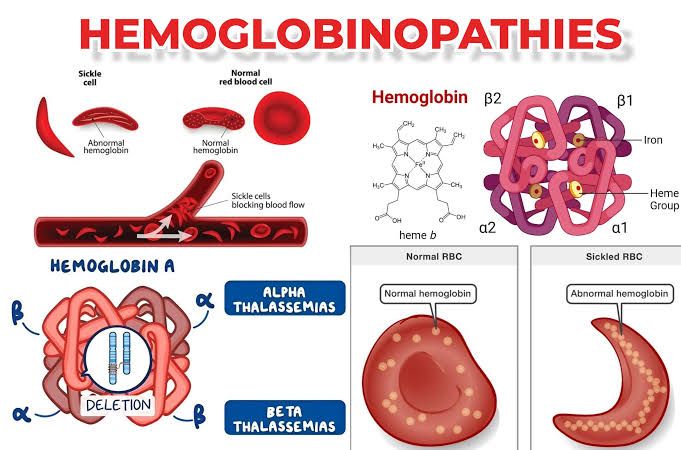
Alpha thalassemia results from mutations in the HBA1 and HBA2 genes located on chromosome 16, which encode the alpha globin chains. The structural abnormalities can be categorized based on the number of affected alpha globin genes:
- Silent Carrier State (1 gene deletion): Individuals have one normal gene and three mutated genes. They typically exhibit no clinical symptoms but may show slight microcytic anemia.
- Alpha Thalassemia Trait (2 gene deletions): This condition leads to mild anemia due to reduced alpha chain production, resulting in an imbalance between alpha and beta chains. The excess beta chains form unstable tetramers known as hemoglobin H (β4), which can precipitate within red blood cells.
- Hemoglobin H Disease (3 gene deletions): Characterized by moderate to severe anemia, this condition results from a significant deficiency of alpha chains, leading to an excess of beta chains that form hemoglobin H. Patients may experience splenomegaly and other complications due to hemolysis.
- Alpha Thalassemia Major (4 gene deletions): Also known as hydrops fetalis, this severe form occurs when all four alpha genes are deleted. It leads to fetal death or severe anemia at birth due to the absence of functional hemoglobin.
Beta Thalassemia
Beta thalassemia arises from mutations in the HBB gene located on chromosome 11, responsible for encoding beta globin chains. The severity depends on whether the mutation is classified as “beta-thalassemia major” (homozygous) or “beta-thalassemia trait” (heterozygous):
- Beta Thalassemia Minor (Trait): Individuals have one normal beta globin gene and one mutated gene, leading to mild microcytic anemia without significant clinical symptoms.
- Beta Thalassemia Intermedia: This form presents with moderate anemia due to partial production of beta globin chains; patients may require occasional blood transfusions but generally maintain better health than those with major forms.
- Beta Thalassemia Major (Cooley’s Anemia): Characterized by severe anemia requiring regular blood transfusions for survival, this condition results from a complete lack of functional beta globin production. The imbalance leads to excess alpha chains that precipitate within erythroid precursors, causing ineffective erythropoiesis and extramedullary hematopoiesis.
Summary
In summary, both alpha and beta thalassemias involve structural abnormalities related to the synthesis of their respective globin chains, leading to various clinical manifestations depending on the severity of the genetic mutations involved.
Structural Abnormalities of Sickle Cell Anemia, Hemoglobin C Disease, and Hemoglobin SC Disease
1. Sickle Cell Anemia (HbS)
Sickle cell anemia is primarily caused by a mutation in the beta-globin gene (HBB) on chromosome 11. This mutation results in the substitution of valine for glutamic acid at the sixth position of the beta-globin chain. The structural abnormality leads to the formation of hemoglobin S (HbS), which polymerizes under low oxygen conditions, causing red blood cells to deform into a characteristic sickle shape. These sickle-shaped cells are rigid and can obstruct blood flow in small vessels, leading to vaso-occlusive crises, pain, and potential organ damage.
The sickling process is influenced by several factors including dehydration, acidosis, and hypoxia. The sickled cells have a shorter lifespan than normal red blood cells (approximately 10-20 days compared to 120 days), leading to chronic hemolytic anemia. Additionally, HbS has a lower solubility than normal hemoglobin (HbA), particularly at low oxygen tensions.
2. Hemoglobin C Disease (HbC)
Hemoglobin C disease arises from another mutation in the HBB gene where lysine replaces glutamic acid at the sixth position of the beta-globin chain. This results in the production of hemoglobin C (HbC). Structurally, HbC is less soluble than HbA but more soluble than HbS. When present in homozygous form (HbCC), individuals may experience mild hemolytic anemia due to the increased tendency of red blood cells to undergo lysis.
The red blood cells containing HbC can also become distorted but typically do not sickle like those with HbS. Instead, they may form crystals within red blood cells under certain conditions, contributing to their premature destruction and leading to splenomegaly and other complications associated with chronic hemolysis.
3. Hemoglobin SC Disease (HbSC)
Hemoglobin SC disease occurs when an individual inherits one gene for hemoglobin S and one gene for hemoglobin C. The structural abnormalities here involve both types of abnormal hemoglobins: HbS and HbC coexist within red blood cells. The presence of both types leads to a mixed phenotype where some red blood cells may sickle while others may crystallize due to HbC.
Patients with HbSC disease often experience symptoms that are intermediate between those seen in sickle cell anemia and those seen in hemoglobin C disease alone. They may have episodes of pain due to vaso-occlusion similar to those experienced by patients with sickle cell anemia but generally have milder symptoms compared to homozygous sickle cell disease.
In summary:
- Sickle Cell Anemia (HbS): Caused by a single amino acid substitution leading to polymerization under low oxygen.
- Hemoglobin C Disease (HbC): Involves a different substitution resulting in less soluble hemoglobin that can cause mild anemia.
- Hemoglobin SC Disease (HbSC): A combination of both mutations leading to mixed symptoms from both disorders.
Principle Behind Hemoglobin Electrophoresis as a Diagnostic Tool for Hemoglobinopathies
Hemoglobin electrophoresis is a laboratory technique used to separate and identify different types of hemoglobin present in a blood sample. This method is particularly valuable in diagnosing hemoglobinopathies, which are disorders caused by abnormalities in the structure or production of hemoglobin. The principle behind hemoglobin electrophoresis involves several key steps and concepts:
1. Understanding Hemoglobin Structure
Hemoglobin is a protein found in red blood cells responsible for transporting oxygen throughout the body. It consists of four polypeptide chains (two alpha and two beta chains) that can vary based on genetic mutations. These variations can lead to different types of hemoglobin, such as normal adult hemoglobin (HbA), fetal hemoglobin (HbF), and various abnormal forms like HbS (sickle cell hemoglobin) or HbC.
2. Sample Preparation
To perform electrophoresis, a blood sample is collected from the patient, typically through venipuncture. The red blood cells are then lysed to release hemoglobin into solution. This step may involve using a buffer solution that maintains an appropriate pH for optimal separation during electrophoresis.
3. Application of Electric Field
Once the hemolysate (the solution containing free hemoglobin) is prepared, it is placed in an agarose gel or cellulose acetate medium within an electrophoresis chamber. An electric current is applied across the gel, causing the charged molecules of hemoglobin to migrate towards the electrode with the opposite charge (anode for negatively charged molecules). The rate at which each type of hemoglobin moves depends on its size and charge.
4. Separation Based on Charge and Size
Different types of hemoglobins have distinct charges due to their amino acid compositions, leading them to migrate at different rates through the gel matrix. For instance, HbS has a different charge compared to HbA due to a single amino acid substitution (valine instead of glutamic acid at position 6 of the beta chain). As a result, when subjected to an electric field, these variants will separate into distinct bands on the gel.
5. Visualization and Interpretation
After electrophoresis is complete, the gel is stained with specific dyes that bind to proteins, allowing visualization of the separated bands under UV light or other imaging techniques. Each band corresponds to a specific type of hemoglobin, which can be quantified by measuring its intensity relative to normal controls.
The pattern observed on the gel provides critical diagnostic information:
- Normal individuals typically show prominent bands for HbA and small amounts of HbF.
- Individuals with sickle cell disease will show significant amounts of HbS.
- Those with thalassemia may exhibit reduced levels of certain types while having increased levels of others.
6. Clinical Relevance
The results from hemoglobin electrophoresis help clinicians diagnose various conditions related to abnormal hemoglobins:
- Sickle cell disease
- Thalassemias
- Other rare variants like HbC or HbE
By identifying these conditions early, appropriate management strategies can be implemented to reduce complications associated with these disorders.
In summary, hemoglobin electrophoresis serves as an essential diagnostic tool by leveraging differences in charge and size among various forms of hemoglobin under an electric field, enabling healthcare providers to diagnose and manage hematological disorders effectively.
Molecular Diagnostic Techniques for Hemoglobin Disorders
Hemoglobin disorders, including thalassemias and sickle cell disease, are among the most prevalent genetic conditions worldwide. Molecular diagnostic techniques play a crucial role in identifying these disorders, particularly when initial protein-based methods are inconclusive. Here’s a detailed discussion of the various molecular diagnostic techniques used in the diagnosis of hemoglobin disorders.
1. Overview of Hemoglobin Disorders
Hemoglobinopathies are characterized by either a reduced synthesis of one or more globin chains (as seen in thalassemias) or the production of structurally abnormal hemoglobins. The complexity of these disorders necessitates precise diagnostic methods to inform treatment and genetic counseling.
2. Initial Diagnostic Approaches
Before delving into molecular techniques, it is essential to note that initial diagnoses often utilize protein-based methods such as:
- Hemoglobin Electrophoresis: This technique separates different types of hemoglobin based on their charge and size, allowing for the identification of abnormal hemoglobins.
- High-Performance Liquid Chromatography (HPLC): HPLC provides a more quantitative analysis of hemoglobin variants and can detect minor components that electrophoresis might miss.
These methods are effective for preliminary screening but may not provide definitive diagnoses in complex cases.
3. Molecular Diagnostic Techniques
When protein-based diagnostics yield uncertain results or when there is a need for genetic counseling, molecular diagnostic techniques become essential. The following are key molecular approaches:
- Polymerase Chain Reaction (PCR): PCR is a cornerstone technique that amplifies specific DNA sequences associated with hemoglobin disorders. It allows for the detection of known mutations in globin genes, which can confirm diagnoses such as β-thalassemia or sickle cell disease.
- Reverse Transcription PCR (RT-PCR): This variant is used to analyze mRNA levels, providing insights into gene expression related to hemoglobin synthesis. It is particularly useful in assessing conditions where gene expression may be altered.
- Sequencing Technologies:
- Sanger Sequencing: This traditional method is still widely used for confirming specific mutations identified through PCR.
- Next-Generation Sequencing (NGS): NGS allows for comprehensive analysis of multiple genes simultaneously, making it invaluable for identifying rare mutations and complex genotypes associated with various hemoglobinopathies.
- Multiplex Ligation-dependent Probe Amplification (MLPA): MLPA can detect large deletions or duplications within the globin gene clusters that standard PCR may not identify. This technique is particularly important for diagnosing α-thalassemia caused by deletions.
- Fluorescence In Situ Hybridization (FISH): FISH is utilized to visualize specific DNA sequences on chromosomes, helping to identify chromosomal abnormalities associated with certain hemoglobin disorders.
- Array Comparative Genomic Hybridization (aCGH): This technique assesses copy number variations across the genome and can identify genomic imbalances linked to hemoglobinopathies.
4. Role in Prenatal Diagnosis
Molecular diagnostics also play a significant role in prenatal testing for hemoglobin disorders. Techniques such as non-invasive prenatal testing (NIPT) using maternal blood samples can identify fetal DNA mutations associated with severe forms of thalassemia or sickle cell disease early in pregnancy.
5. Importance of Genetic Counseling
The integration of molecular diagnostics into clinical practice enhances genetic counseling efforts by providing accurate risk assessments for couples considering having children. Understanding specific mutations helps healthcare providers offer tailored advice regarding reproductive options and management strategies.
In summary, molecular diagnostic techniques have revolutionized the approach to diagnosing hemoglobin disorders by enabling precise identification of genetic mutations and variations that underlie these conditions. As technology advances, these methods will continue to evolve, improving both diagnosis and patient outcomes.


