Anatomy of the Female Pelvis
The female pelvis is a complex structure that plays a crucial role in supporting various organs and facilitating reproductive functions. It consists of bones, muscles, ligaments, and organs that work together to maintain bodily functions and support childbirth.
1. Structure of the Female Pelvis
The pelvis is located at the lower part of the torso, between the abdomen and the legs. It provides structural support for the intestines and houses important organs such as the bladder and reproductive structures. The female pelvis differs from the male pelvis primarily in its shape and size, designed to accommodate childbirth. The female pelvis is generally broader and wider than that of males.
2. Bones of the Female Pelvis
- Hip Bones: There are two hip bones (one on each side), which form the pelvic girdle. Each hip bone consists of three fused components:
- Ilium: The largest part, broad and fan-shaped.
- Pubis: Connects with its counterpart at the pubic symphysis.
- Ischium: Supports body weight when sitting.
- Sacrum: Composed of five fused vertebrae, it connects to the hip bones and supports body weight.
- Coccyx: Also known as the tailbone, it consists of four fused vertebrae forming a triangular shape at the base of the sacrum.
3. Muscles of the Female Pelvis
- Levator Ani Muscles: This group includes three muscles that support pelvic organs:
- Puborectalis: Helps control bowel movements.
- Pubococcygeus: Provides major support for pelvic structures.
- Iliococcygeus: Assists in lifting pelvic floor structures.
- Coccygeus Muscle: A smaller muscle that connects from the ischium to both sacrum and coccyx.
4. Organs within the Female Pelvis
- Uterus: A thick-walled organ where fetal development occurs during pregnancy; it sheds its lining monthly during menstruation if no pregnancy occurs.
- Ovaries: Two almond-shaped organs located on either side of the uterus; they produce eggs and hormones like estrogen and progesterone.
- Fallopian Tubes: These tubes connect each ovary to the uterus, facilitating egg transport through cilia-lined channels.
- Cervix: The narrow lower part of the uterus that opens into the vagina; it allows sperm entry into the uterus while also producing mucus to protect against infections.
- Vagina: The muscular canal connecting cervix to external genitalia; it serves as a passageway for menstrual fluid, sexual intercourse, and childbirth.
- Rectum & Bladder:
- The rectum is where feces collect before exiting through the anus.
- The bladder stores urine until it’s expelled through a shorter urethra compared to males.
5. Ligaments Supporting Pelvic Structures
- Broad Ligament: Supports reproductive organs by extending from both sides of the pelvic wall; it has subdivisions:
- Mesometrium (supports uterus)
- Mesovarium (supports ovaries)
- Mesosalpinx (supports fallopian tubes)
- Uterine Ligaments: These include round ligaments, cardinal ligaments, pubocervical ligaments, and uterosacral ligaments which provide additional support for uterine positioning.
- Ovarian Ligaments: These consist of ovarian ligament and suspensory ligament which anchor ovaries in place.
6. Health Considerations Related to Female Pelvis
Various conditions can affect pelvic health including:
- Pelvic inflammatory disease (PID)
- Endometriosis
- Pelvic organ prolapse
Symptoms may include lower abdominal pain, unusual discharge, painful intercourse, or changes in bowel habits. Regular gynecological check-ups are essential for maintaining pelvic health.
In summary, understanding female pelvic anatomy is vital for recognizing its functions related to reproduction as well as identifying potential health issues that may arise within this complex system.
Types of Female Pelvic Cavity
The female pelvic cavity can be categorized into four main types based on the shape of the pelvic inlet, which is crucial for childbirth. Each type has distinct characteristics that can influence the ease or difficulty of vaginal delivery.
1. Gynecoid Pelvis
The gynecoid pelvis is the most common type found in females, estimated to be present in about 50% of individuals assigned female at birth (AFAB). This pelvis shape is characterized by a round, wide, and shallow structure, which provides ample space for a baby to pass through during vaginal delivery. The gynecoid pelvis is considered the most favorable for childbirth due to its accommodating dimensions.
2. Android Pelvis
The android pelvis resembles the male pelvis more closely than other female types. It is narrower and has a heart or wedge-like shape. This configuration can make labor more challenging because the narrower birth canal may slow down the baby’s descent during delivery. Women with an android pelvis may have a higher likelihood of requiring a cesarean section (C-section).
3. Anthropoid Pelvis
The anthropoid pelvis has an elongated shape that resembles an upright egg or oval. It is deeper from front to back compared to the android pelvis but still narrower than the gynecoid type. While some women with an anthropoid pelvis can deliver vaginally, they may experience longer labor durations due to the less spacious nature of this pelvic shape.
4. Platypelloid Pelvis
The platypelloid pelvis, also known as a flat pelvis, is characterized by its wide but shallow structure, resembling an egg lying on its side. This type is the least common among females and can pose significant challenges during vaginal birth because it may hinder the baby’s ability to pass through the pelvic inlet effectively. Many women with a platypelloid pelvis often require a C-section for safe delivery.
In summary, these four types—gynecoid, android, anthropoid, and platypelloid—represent different anatomical configurations of the female pelvic cavity that can significantly impact childbirth experiences.
Mechanism of Labor in the First and Second Stages of Labor
First Stage of Labor
The first stage of labor is characterized by progressive changes in the cervix and uterine contractions that facilitate the birth process. This stage can be divided into three phases: latent, active, and transition.
- Latent Phase:
- During this initial phase, contractions begin to occur but are typically mild and irregular. The cervix starts to efface (thin out) and dilate (open) to about 3-4 centimeters. Women may not recognize they are in labor due to the minimal discomfort experienced.
- Active Phase:
- In this phase, contractions become more intense, longer (lasting about 40-60 seconds), and more frequent (occurring every 3-5 minutes). The cervix dilates from 4 to 7 centimeters. The increased strength and frequency of contractions help push the fetus down into the birth canal.
- Transition Phase:
- This is the final part of the first stage where the cervix dilates from 8 to 10 centimeters. Contractions are very strong, lasting up to 90 seconds and occurring every two to three minutes. Most women feel a strong urge to push as the fetus descends further into the birth canal.
The mechanism during this stage involves coordinated uterine contractions that help in cervical dilation and effacement, allowing for fetal descent.
The second stage begins when the cervix is fully dilated at 10 centimeters and ends with the delivery of the baby. This stage is often referred to as the “pushing” stage.
- Pushing Phase:
- During this phase, women actively participate by pushing with each contraction. The pressure from contractions helps move the baby down through the birth canal.
- As the baby’s head descends, it rotates into an optimal position for delivery (usually facing downward). When crowning occurs, it indicates that part of the baby’s head is visible at the vaginal opening.
- The healthcare provider may guide or assist in delivering the baby as it emerges through the vagina.
The mechanism during this stage relies on effective maternal effort combined with powerful uterine contractions that facilitate fetal expulsion from the uterus through coordinated movements within the birth canal.
In summary, both stages involve complex physiological processes driven by uterine contractions that promote cervical changes and ultimately lead to childbirth.
Interpretation of Antenatal and Intrapartum CTG
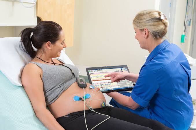
Cardiotocography (CTG) is a method used to monitor fetal heart rate and uterine contractions during pregnancy and labor. It provides continuous data that can help assess fetal well-being, particularly in pregnancies at increased risk of complications. The interpretation of CTG involves analyzing the patterns of fetal heart rate (FHR) and uterine activity to identify any signs of fetal distress or abnormality.
Antenatal CTG Interpretation
Antenatal CTG is typically performed in high-risk pregnancies to monitor the fetus’s condition before labor begins. The key components assessed during antenatal CTG include:
- Baseline Fetal Heart Rate (FHR): This is the average heart rate over a 10-minute period, measured in beats per minute (bpm). A normal baseline FHR ranges from 110 to 160 bpm. Deviations from this range may indicate potential issues:
- Tachycardia: FHR >160 bpm may suggest fetal hypoxia or infection.
- Bradycardia: FHR <110 bpm may indicate umbilical cord compression or other complications.
- Variability: This refers to fluctuations in the FHR that are indicative of fetal autonomic nervous system function. Variability is categorized as:
- Absent variability: No fluctuations; concerning for fetal distress.
- Minimal variability: Fluctuations <5 bpm; may require further evaluation.
- Moderate variability: Fluctuations 6-25 bpm; considered reassuring.
- Marked variability: Fluctuations >25 bpm; could indicate stress or stimulation.
- Accelerations: These are temporary increases in FHR, usually associated with fetal movement and are considered a positive sign of fetal well-being.
- Decelerations: These are decreases in FHR and can be classified into three types:
- Early decelerations: Gradual decrease coinciding with contractions; typically benign.
- Variable decelerations: Abrupt decreases not consistently related to contractions; often due to umbilical cord compression.
- Late decelerations: Gradual decrease occurring after the peak of contractions; concerning for uteroplacental insufficiency.
- Overall Assessment: The overall interpretation combines these factors to determine if the fetus is experiencing distress or if further intervention is needed.
Intrapartum CTG Interpretation
Intrapartum CTG monitoring occurs during labor and focuses on real-time assessment of both maternal contractions and fetal heart rate patterns:
- Continuous Monitoring: Intrapartum CTG allows for continuous observation, which helps detect changes promptly during labor.
- Assessment Similarities with Antenatal CTG:
- Baseline FHR, variability, accelerations, and decelerations are evaluated similarly as in antenatal assessments.
- Contraction Patterns: The frequency, duration, and intensity of uterine contractions are also monitored:
- Normal contraction patterns should allow for adequate rest periods between contractions for optimal fetal oxygenation.
- Intervention Criteria:
- If abnormal patterns such as persistent late decelerations or significant variable decelerations occur, immediate interventions may be warranted, including repositioning the mother, administering oxygen, or preparing for possible cesarean delivery if necessary.
- Documentation and Communication: Accurate documentation of findings is crucial for ongoing care decisions and communication among healthcare providers regarding the status of both mother and fetus.
- Clinical Decision-Making Based on Findings:
- Decisions regarding interventions during labor should be based on a comprehensive analysis of both maternal condition and CTG findings.
In summary, effective interpretation of both antenatal and intrapartum CTGs requires an understanding of normal versus abnormal patterns in FHR and uterine activity, enabling timely interventions that can improve outcomes for mothers and babies.
Causes of Abnormal CTG in Labor
1. Fetal Hypoxia
Fetal hypoxia, or a lack of oxygen to the fetus, is one of the primary concerns during labor and can lead to abnormal CTG patterns. This condition may arise from various factors, including uterine contractions that are too frequent or too strong (hyperstimulation), which can compress the umbilical cord and reduce blood flow to the fetus. Additionally, maternal conditions such as anemia or respiratory issues can contribute to fetal hypoxia.
2. Uteroplacental Insufficiency
This condition occurs when the placenta fails to provide adequate blood flow and nutrients to the fetus. Causes may include maternal hypertension, diabetes, or placental abruption (where the placenta detaches from the uterus prematurely). Uteroplacental insufficiency often results in decreased fetal heart rate variability and late decelerations on a CTG.
3. Maternal Medical Conditions
Certain maternal health issues can affect fetal well-being during labor. Conditions such as gestational diabetes, preeclampsia, and chronic hypertension can lead to abnormal CTG readings by impacting placental function and fetal oxygenation. These conditions may also increase the risk of complications during delivery.
4. Cord Compression
Umbilical cord compression can occur due to various reasons, including oligohydramnios (low amniotic fluid levels) or abnormal fetal positioning (such as a transverse lie). This compression can lead to variable decelerations in the fetal heart rate observed on a CTG strip.
5. Infection
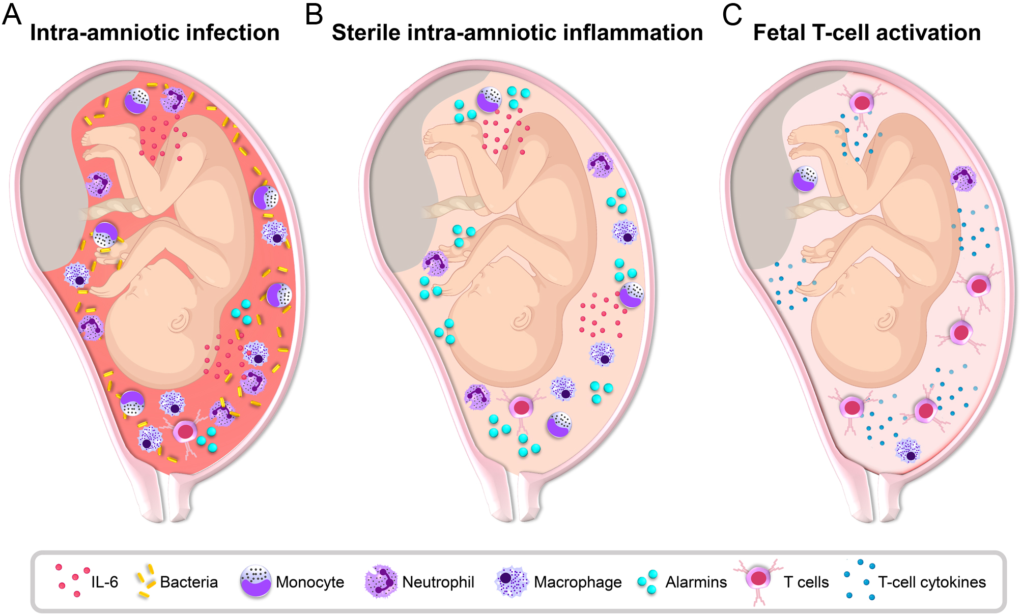
Intrauterine infections, such as chorioamnionitis (infection of the amniotic sac), can cause changes in fetal heart rate patterns due to inflammatory responses affecting both maternal and fetal physiology. This may manifest as tachycardia (increased heart rate) in the fetus on a CTG.
6. Medications
Certain medications administered during labor, such as oxytocin for induction or augmentation of labor, can influence uterine activity and potentially lead to abnormal CTG patterns if they cause excessive uterine contractions or hyperstimulation.
7. Fetal Distress
Fetal distress refers to signs that indicate the fetus is not coping well with labor. This could be due to any combination of factors mentioned above but typically presents with significant changes in heart rate patterns—either bradycardia (decreased heart rate) or tachycardia—on a CTG monitor.
8. Maternal Positioning
The position of the mother during labor can impact fetal heart rate monitoring outcomes. Certain positions may exacerbate cord compression or reduce uteroplacental perfusion, leading to abnormal readings on a CTG.
In summary, abnormal cardiotocography readings during labor can result from a variety of causes related to both maternal health and fetal conditions. Identifying these causes is crucial for timely intervention and ensuring optimal outcomes for both mother and baby.


