Microscopic Structure of the Upper Respiratory Passage
The upper respiratory passage includes structures such as the nasal cavity, pharynx, and larynx. The microscopic structure of these areas is primarily characterized by the presence of respiratory mucosa, which plays a crucial role in protecting the respiratory system and facilitating gas exchange.
1. Respiratory Mucosa Composition
The respiratory mucosa is a specialized epithelial lining that covers most of the upper respiratory tract. It consists of several key components:
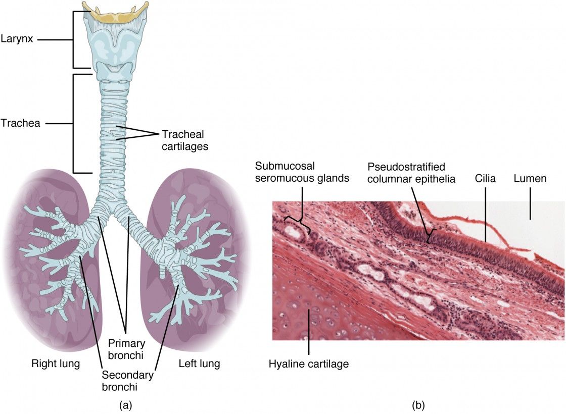
- Epithelium: The epithelium in the upper respiratory tract is primarily pseudostratified ciliated columnar epithelium. This type of epithelium appears to have multiple layers due to varying cell heights but is actually a single layer with all cells attached to the basement membrane. The cilia on these epithelial cells beat in a coordinated manner to move mucus and trapped particles out of the airways.
- Goblet Cells: Interspersed among the ciliated cells are goblet cells, which are responsible for producing mucus. This mucus traps dust, pathogens, and other particulates inhaled with air, preventing them from entering deeper parts of the respiratory system.
- Basal Cells: These are stem cells located at the base of the epithelium that can differentiate into other types of epithelial cells, helping maintain and repair the mucosal surface.
2. Lamina Propria
Beneath the epithelial layer lies the lamina propria, a connective tissue layer rich in blood vessels, immune cells, and glands:
- Blood Vessels: The extensive vascular network helps warm and humidify incoming air. The close proximity of blood vessels to the epithelium facilitates efficient heat exchange.
- Mucous Glands: In addition to goblet cells, there are also seromucous glands within the lamina propria that secrete mucus and serous fluid. This secretion further aids in trapping particles and pathogens while also providing moisture to inspired air.
- Immune Cells: The lamina propria contains various immune cells such as lymphocytes and macrophages that play a role in defending against inhaled pathogens.
3. Olfactory Epithelium
In specific regions like the superior nasal concha, there exists olfactory epithelium which is distinct from respiratory mucosa:
- Olfactory Receptor Neurons: These specialized neurons detect odor molecules and send signals to the brain for smell perception.
- Supporting Cells: Similar to glial cells in function, supporting cells provide structural support and nourishment to olfactory receptor neurons.
4. Overall Functionality
The combination of these structures allows for several critical functions:
- Air Filtration: The mucus traps airborne particles while cilia help expel them from the airway.
- Humidification and Warming: As air passes through this region, it is warmed by blood vessels and moistened by mucus secretions before reaching lower parts of the respiratory tract.
- Defense Mechanism: The presence of immune cells within the mucosa provides an immediate response mechanism against pathogens.
In summary, the upper respiratory passage’s microscopic structure is intricately designed for optimal functioning in filtering, humidifying, warming air, and protecting against infections through its specialized epithelial composition known as respiratory mucosa.
Correlating the Structure and Expected Function of the Different Components of the Nose and Trachea
1. Overview of the Nose and Trachea
The nose and trachea are integral components of the respiratory system, each serving distinct yet complementary roles in facilitating respiration. The nose primarily functions as an entry point for air, while the trachea serves as a conduit to transport air to the lungs. Understanding their structures helps elucidate their functions.
2. Structure of the Nose
The nose consists of several key components:
- External Nose: The visible part includes the nasal bridge, tip, and nostrils (nares). It is made up of cartilage and skin.
- Nasal Cavity: Inside, it is divided into two halves by the nasal septum. The cavity is lined with mucous membranes that contain cilia and goblet cells.
- Turbinates (Conchae): These are bony structures within the nasal cavity that increase surface area, helping to warm and humidify incoming air.
- Olfactory Epithelium: Located at the roof of the nasal cavity, this specialized tissue contains sensory receptors for smell.
3. Function of the Nose
The primary functions of the nose include:
- Air Filtration: The mucous membranes trap dust, pathogens, and other particles from inhaled air.
- Humidification: As air passes through the nasal cavity, it is moistened by mucus secreted by goblet cells.
- Temperature Regulation: The rich blood supply in the nasal tissues warms incoming air to body temperature.
- Olfaction: The olfactory epithelium allows for detection of odors, which is crucial for taste and environmental awareness.
4. Structure of the Trachea
The trachea is a tubular structure that extends from the larynx to the bronchi. Its key structural features include:
- Cartilaginous Rings: C-shaped rings made of hyaline cartilage provide structural support while allowing flexibility during breathing.
- Mucosal Lining: Similar to that in the nose, it contains ciliated epithelial cells and goblet cells that produce mucus.
- Smooth Muscle Layer (Trachealis Muscle): This muscle connects ends of cartilaginous rings and allows constriction or dilation during breathing.
5. Function of the Trachea
The trachea’s functions include:
- Air Passageway: It serves as a direct route for air to travel between the larynx and lungs.
- Protection Against Aspiration: The ciliated lining helps move mucus and trapped particles upward toward the throat for expulsion or swallowing.
- Regulation of Airflow: The smooth muscle can adjust diameter based on airflow needs (e.g., during exercise).
6. Correlation Between Structure and Function
The correlation between structure and function in both organs can be summarized as follows:
- In both structures, mucous membranes with cilia serve critical roles in filtering out debris from inhaled air.
- The presence of cartilage in both structures provides necessary rigidity while allowing flexibility; this is particularly important in maintaining an open airway during various activities such as speaking or exercising.
- Turbinates in the nose enhance its ability to condition air (warming/humidifying), which prepares it for optimal gas exchange once it reaches alveoli in lungs.
- The olfactory epithelium’s specialized structure allows for effective detection of airborne chemicals, linking sensory perception directly with respiratory function.
In conclusion, both components exhibit structural adaptations that facilitate their respective functions—air filtration, humidification, temperature regulation in the nose; efficient airflow management and protection against aspiration in the trachea—demonstrating a well-coordinated system essential for effective respiration.
Microscopic Structure of the Main Bronchi and Their Subdivisions
The microscopic structure of the main bronchi and their subdivisions is characterized by several distinct layers and components that facilitate their function in the respiratory system. Understanding this structure involves examining the histological features, including the epithelial lining, cartilage, smooth muscle, and connective tissue.
1. Epithelial Lining
The main bronchi are lined with a mucous membrane that consists primarily of ciliated pseudostratified columnar epithelium. This type of epithelium contains goblet cells that secrete mucus, which traps inhaled particles and pathogens. The cilia on the surface of these epithelial cells beat in a coordinated manner to move mucus upwards towards the throat, where it can be swallowed or expelled. As the bronchi branch into smaller subdivisions (secondary and tertiary bronchi), there is a gradual transition in the epithelial lining:
- In secondary bronchi, the epithelium remains ciliated pseudostratified columnar but may start to show areas of simple columnar epithelium as they become smaller.
- In tertiary bronchi, there is further reduction in height and complexity, transitioning towards simple cuboidal epithelium as they branch into smaller bronchioles.
2. Cartilage
The walls of the main bronchi contain hyaline cartilage that provides structural support and maintains patency (openness) during respiration. The cartilage is arranged in C-shaped rings in the trachea and main bronchi; however, as these airways branch into secondary and tertiary bronchi, the cartilage becomes less continuous and more irregularly shaped plates. This decrease in cartilage occurs because smaller airways require more flexibility to accommodate changes in airflow during breathing.
- In primary (main) bronchi, there are complete rings of cartilage.
- In secondary bronchi, these rings begin to fragment into plates.
- By the time we reach tertiary bronchi and beyond, there is significantly less cartilage present.
3. Smooth Muscle
Surrounding the bronchial epithelium is a layer of smooth muscle known as the muscularis layer. This smooth muscle plays a crucial role in regulating airway diameter through contraction or relaxation:
- In primary bronchi, this layer is relatively thin compared to subsequent generations.
- As branching occurs into secondary and tertiary bronchi, there is an increase in smooth muscle relative to cartilage. This allows for greater control over airflow resistance.
- In smaller branches (bronchioles), smooth muscle predominates as cartilage diminishes entirely.
4. Connective Tissue
The connective tissue surrounding the bronchial structures provides additional support and elasticity. It contains various types of fibers (collagenous and elastic fibers) that allow for expansion during inhalation while maintaining structural integrity during exhalation.
As we progress from main bronchi to smaller divisions:
- The amount of connective tissue increases relative to both cartilage and epithelium.
- The presence of elastic fibers becomes more pronounced in smaller airways (bronchioles), aiding in lung recoil after expansion.
5. Summary of Structural Changes
In summary, as one moves from the main bronchi through their subdivisions:
- The epithelial lining transitions from ciliated pseudostratified columnar to simple cuboidal.
- The amount of hyaline cartilage decreases, transitioning from complete rings to irregular plates until absent in small bronchioles.
- The smooth muscle content increases, allowing for greater regulation of airflow.
- Connective tissue becomes more prominent, providing necessary support throughout all levels.
These structural adaptations are essential for efficient gas exchange within alveoli at terminal ends while also protecting against foreign particles entering deeper lung tissues.
Microscopic Structure of Lung Parenchyma and Its Correlation with Gas Exchange Function
The lung parenchyma is primarily composed of alveoli, the tiny air sacs where gas exchange occurs. Understanding the microscopic structure of the lung parenchyma is crucial for correlating its anatomy with its physiological function in gas exchange.
1. Alveolar Structure
The alveoli are the fundamental units of gas exchange in the lungs. Each alveolus is a small, balloon-like structure lined with a thin layer of epithelial cells. The walls of the alveoli are extremely thin (approximately 0.2 to 0.5 micrometers), allowing for efficient diffusion of gases. The primary cell types found in the alveolar walls include:
- Type I Alveolar Cells (Pneumocytes): These cells cover about 95% of the alveolar surface area and are responsible for the majority of gas exchange due to their thinness.
- Type II Alveolar Cells: These cells produce surfactant, a substance that reduces surface tension within the alveoli, preventing collapse during exhalation and facilitating easier inflation during inhalation.
2. Capillary Network
Surrounding each alveolus is an extensive network of capillaries, which are also very thin-walled (one cell thick). This proximity between alveolar air and blood allows for efficient gas exchange through simple diffusion. Oxygen from inhaled air diffuses across the alveolar wall into the blood, while carbon dioxide from the blood diffuses into the alveoli to be exhaled.
3. Interstitial Space
Between the alveoli and capillaries lies a minimal interstitial space filled with connective tissue that contains elastic fibers and collagen. This structure provides mechanical support to maintain lung shape and elasticity, which is essential during breathing cycles.
4. Gas Exchange Mechanism
Gas exchange occurs via passive diffusion driven by concentration gradients:
- Oxygen Transport: When air enters the lungs, oxygen concentration in the alveoli becomes higher than in deoxygenated blood arriving from the pulmonary arteries. Consequently, oxygen diffuses into the blood.
- Carbon Dioxide Removal: Conversely, carbon dioxide concentration is higher in venous blood than in alveolar air; thus, it diffuses from blood into the alveoli to be expelled during exhalation.
The efficiency of this process is enhanced by several factors:
- Large Surface Area: The total surface area available for gas exchange in healthy human lungs can exceed 70 square meters due to millions of alveoli.
- Thin Membrane Thickness: The minimal thickness of both alveolar walls and capillary endothelium facilitates rapid diffusion.
- Ventilation-Perfusion Matching: Proper matching between airflow (ventilation) and blood flow (perfusion) optimizes gas exchange efficiency.
5. Pathological Considerations
Diseases such as emphysema or pulmonary fibrosis can alter this microscopic structure:
- In emphysema, destruction of elastic fibers leads to enlarged air spaces and reduced surface area for gas exchange.
- In pulmonary fibrosis, thickening of interstitial tissue impairs diffusion capacity due to increased membrane thickness.
In summary, the microscopic structure of lung parenchyma—characterized by thin-walled alveoli surrounded by capillaries—directly correlates with its function in facilitating efficient gas exchange through mechanisms driven by concentration gradients.


