Signs, Symptoms, and Diagnostic Aids for Evaluating Presumed Large Bowel Obstruction
Large bowel obstruction (LBO) is a critical condition that occurs when there is a blockage in the large intestine, preventing the normal passage of intestinal contents. It can be caused by various factors including tumors, strictures, volvulus, or fecal impaction. Early recognition and diagnosis are crucial for effective management.
Signs of Large Bowel Obstruction
- Abdominal Distension: One of the most common signs of LBO is abdominal distension due to the accumulation of gas and fluid proximal to the obstruction.
- Borborygmi: Increased bowel sounds may be heard as the intestines attempt to push contents past the obstruction.
- Tenderness on Palpation: The abdomen may be tender upon examination, particularly in the area where the obstruction is located.
- Visible Peristalsis: In some cases, visible waves of peristalsis can be observed on the abdominal wall.
Symptoms of Large Bowel Obstruction
- Abdominal Pain: Patients often experience crampy abdominal pain that may come and go or become more constant as the condition progresses.
- Nausea and Vomiting: As the obstruction worsens, nausea and vomiting may occur; vomit may contain fecal material if there is a complete obstruction.
- Constipation or Change in Bowel Habits: Patients typically report constipation or an inability to pass gas or stool.
- Dehydration Signs: Symptoms such as dry mouth, decreased urine output, and dizziness may indicate dehydration due to fluid loss from vomiting or reduced intake.
Diagnostic Aids for Evaluating Large Bowel Obstruction
- Physical Examination: A thorough physical examination can provide initial clues regarding tenderness, distension, and bowel sounds.
- Imaging Studies:
- X-ray of the Abdomen: An upright abdominal X-ray can show air-fluid levels and dilated bowel loops indicative of obstruction.
- CT Scan with Contrast: A CT scan is highly sensitive for diagnosing LBO; it can identify the site and cause of obstruction (e.g., tumors, strictures).
- Ultrasound: Particularly useful in pediatric populations; it can help visualize fluid-filled loops of bowel and assess for free fluid.
- Laboratory Tests:
- Blood tests may reveal electrolyte imbalances (e.g., hypokalemia), elevated white blood cell count indicating infection or inflammation, and renal function tests to assess dehydration status.
- Colonoscopy (if indicated): In certain cases where a tumor or stricture is suspected but not clearly visualized on imaging studies, a colonoscopy may be performed to directly visualize and potentially relieve an obstructive lesion.
- Contrast Enema (in select cases): This test involves introducing contrast material into the rectum to visualize blockages in some patients who cannot undergo other imaging modalities.
In summary, evaluating presumed large bowel obstruction involves recognizing characteristic signs such as abdominal distension and tenderness alongside symptoms like abdominal pain and constipation. Diagnostic aids including physical examination findings, imaging studies like X-rays and CT scans, laboratory tests for metabolic derangements, colonoscopy for direct visualization when necessary, all play vital roles in confirming diagnosis and determining appropriate management strategies.
Causes of Colonic Obstruction in Adult Patients
Colonic obstruction is a significant medical condition that can lead to serious complications if not promptly diagnosed and treated. Understanding the various causes of colonic obstruction in adults is crucial for effective management. The causes can be broadly categorized into mechanical obstructions, functional obstructions, and other less common causes.
1. Mechanical Obstructions
Mechanical obstructions are physical blockages in the colon that prevent the normal passage of intestinal contents. The most common causes include:
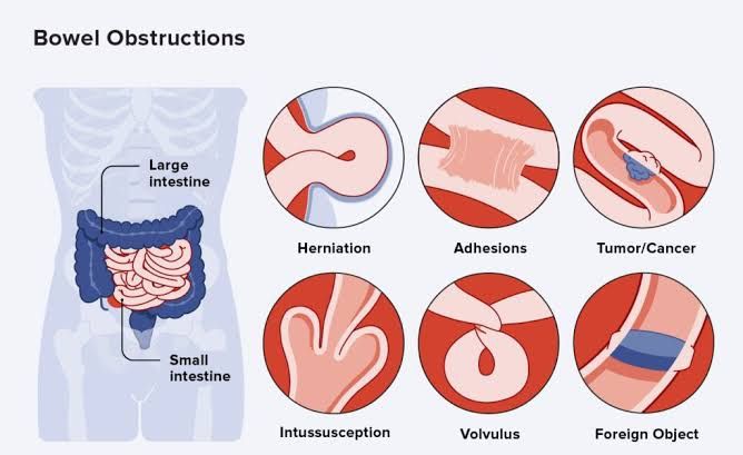
- Colorectal Cancer: This is one of the leading causes of colonic obstruction in adults. Tumors can grow within the lumen of the colon, leading to narrowing or complete blockage. Colorectal cancer accounts for approximately 30-50% of all cases of colonic obstruction.
- Diverticulitis: Inflammation and infection of diverticula (small pouches that can form in the colon wall) can lead to scarring and strictures, causing obstruction. Diverticulitis is responsible for about 20-25% of cases.
- Benign Strictures: These may arise from conditions such as inflammatory bowel disease (IBD), particularly Crohn’s disease, which can cause segmental narrowing due to inflammation and fibrosis. Benign strictures account for around 10-15% of colonic obstructions.
- Volvulus: This occurs when a loop of intestine twists around itself, leading to obstruction and potential ischemia. Sigmoid volvulus is more common in older adults and accounts for approximately 5-10% of cases.
- Intussusception: Although more common in children, intussusception can occur in adults, often associated with a lead point such as a tumor or polyp. It represents about 1-5% of adult colonic obstructions.
2. Functional Obstructions
Functional obstructions are caused by impaired motility rather than a physical blockage:
- Ileus: A temporary cessation of bowel activity often following surgery or due to electrolyte imbalances can lead to functional obstruction. While not strictly a mechanical cause, it is important to recognize ileus as it may mimic obstructive symptoms.
3. Other Causes
Other less common causes include:
- Foreign Bodies: Ingested objects can occasionally cause obstruction.
- Adhesions: Post-surgical adhesions may lead to twisting or kinking of the bowel but are more commonly associated with small bowel obstructions.
Frequency Overview
In summary, while there are several potential causes for colonic obstruction in adults, colorectal cancer remains the most prevalent cause followed by diverticulitis and benign strictures related to inflammatory conditions like IBD. Volvulus and intussusception are less frequent but still significant contributors to this clinical picture.
Conclusion
Understanding these causes helps clinicians develop appropriate diagnostic strategies and treatment plans for patients presenting with symptoms suggestive of colonic obstruction.
Outline for Diagnostic Studies, Preoperative Management, and Treatment of Volvulus, Intussusception, Impaction, and Obstructing Colon Cancer
1. Volvulus
Diagnostic Studies:
- Clinical Evaluation: Assess symptoms such as abdominal pain, distension, constipation, and vomiting.
- Imaging Studies:
- X-ray: Abdominal X-rays may show signs of bowel obstruction or air-fluid levels.
- CT Scan: A CT scan with contrast is the gold standard for diagnosing volvulus; it can reveal the twisted segment of bowel and any associated ischemia.
- Ultrasound: Particularly useful in pediatric cases to visualize the affected bowel.
Preoperative Management:
- Fluid Resuscitation: Initiate IV fluids to correct dehydration and electrolyte imbalances.
- Nasogastric Tube (NGT) Placement: Decompress the stomach to relieve distension.
- Antibiotics: Administer broad-spectrum antibiotics to prevent infection due to potential bowel necrosis.
Treatment:
- Endoscopic Detorsion: In some cases, endoscopy can be used to untwist the volvulus.
- Surgical Intervention: If endoscopic methods fail or if there is evidence of ischemia:
- Resection of necrotic bowel segments may be necessary.
- Surgical fixation (e.g., mesopexy) may be performed to prevent recurrence.
2. Intussusception
Diagnostic Studies:
- Clinical Evaluation: Look for classic symptoms like intermittent abdominal pain, “currant jelly” stools in children, and a palpable abdominal mass.
- Imaging Studies:
- Ultrasound: First-line imaging in children; it can show the target sign indicative of intussusception.
- CT Scan: In adults or when ultrasound is inconclusive; it provides detailed images of the intussuscepted segment.
Preoperative Management:
- Fluid Resuscitation: Similar to volvulus management; ensure hydration and electrolyte balance.
- NGT Placement: To relieve pressure from bowel obstruction.
- Observation for Complications: Monitor closely for signs of perforation or peritonitis.
Treatment:
- Non-surgical Reduction: Air contrast enema can reduce intussusception in children effectively.
- Surgical Intervention:
- If non-surgical methods fail or if there are complications (e.g., perforation), surgical resection of the affected segment may be required.
3. Impaction
Diagnostic Studies:
- Clinical Evaluation: Assess for symptoms like severe constipation, abdominal pain, and rectal bleeding.
- Imaging Studies:
- Abdominal X-ray: Can confirm fecal impaction by showing a large fecal mass in the colon.
- CT Scan or Ultrasound: May be used if complications are suspected.
Preoperative Management:
- Bowel Preparation: Use enemas or oral laxatives to clear impacted stool if surgery is planned due to complications (e.g., perforation).
- Fluid Resuscitation & Electrolyte Monitoring: Important if there’s significant dehydration from prolonged constipation.
Treatment:
- Manual Disimpaction: In cases where conservative measures fail; this may involve digital removal under anesthesia if necessary.
- Surgical Intervention: Rarely needed but may involve resection if there is significant damage or obstruction due to chronic impaction.
4. Obstructing Colon Cancer
Diagnostic Studies:
- Clinical Evaluation: Symptoms include change in bowel habits, weight loss, anemia, and abdominal pain.
- Imaging Studies:
- Colonoscopy with Biopsy: Essential for diagnosis; allows visualization and tissue sampling of lesions.
- CT Scan of Abdomen/Pelvis: To assess extent of disease and identify metastases.
Preoperative Management:
- Nutritional Support & Bowel Preparation: Optimize nutritional status preoperatively; consider total parenteral nutrition (TPN) if unable to tolerate oral intake.
- Stenting Consideration (if applicable): Colonic stenting can relieve obstruction temporarily before definitive surgery.
Treatment:
- Surgical Resection: The primary treatment involves resection of the obstructed segment along with adjacent lymph nodes.
- An anastomosis may be performed unless there are contraindications (e.g., poor perfusion).
- In some cases where resection is not feasible due to advanced disease, palliative care options such as colostomy might be considered.
Potential Complications of Inadequate Treatment for Mechanical Bowel Obstruction
Mechanical bowel obstruction can occur in either the large or small intestine and is characterized by a blockage that prevents the normal passage of intestinal contents. If treatment is inadequate, several serious complications can arise, which can significantly impact patient health and may even be life-threatening. Below, we will discuss these potential complications in detail.
1. Ischemia and Necrosis
One of the most critical complications of untreated bowel obstruction is ischemia, which occurs when blood flow to a section of the intestine is compromised. The obstruction can lead to increased intraluminal pressure, resulting in decreased venous return and ultimately arterial supply to the affected area. This lack of blood flow can cause tissue death (necrosis), leading to perforation of the bowel wall. When necrosis occurs, it creates a risk for peritonitis, an inflammatory response in the abdominal cavity due to leakage of intestinal contents.
2. Perforation
If ischemia progresses without intervention, it can result in bowel perforation. This condition allows intestinal contents, including bacteria and toxins, to spill into the abdominal cavity, leading to peritonitis. Perforation is a surgical emergency that requires immediate intervention; failure to address this complication promptly can lead to sepsis—a systemic inflammatory response that can result in multi-organ failure.
3. Infection and Sepsis
The presence of necrotic tissue or perforated bowel increases the risk of infection within the abdominal cavity. Bacterial overgrowth from stagnant intestinal contents combined with exposure to pathogens from perforated areas can lead to severe infections such as intra-abdominal abscesses or generalized peritonitis. If these infections are not managed effectively, they may progress to sepsis, characterized by fever, tachycardia, hypotension, and altered mental status.
4. Electrolyte Imbalance and Dehydration
Bowel obstructions often lead to significant fluid loss due to vomiting and inability to absorb nutrients or fluids through the obstructed segment of the intestine. This situation can result in dehydration and electrolyte imbalances (e.g., hypokalemia). Severe electrolyte disturbances can have profound effects on cardiac function and muscle contraction.
5. Malnutrition
Chronic bowel obstruction may prevent adequate nutrient absorption over time, leading to malnutrition. Patients may experience weight loss, muscle wasting, and deficiencies in essential vitamins and minerals if they cannot maintain proper nutritional intake due to ongoing obstruction.
6. Short Bowel Syndrome
In cases where surgical intervention becomes necessary after prolonged obstruction—especially if resection of necrotic segments occurs—there is a risk for short bowel syndrome (SBS). SBS results from having insufficient functional bowel length for adequate digestion and absorption of nutrients post-surgery.
7. Adhesions Formation
Following surgical treatment for bowel obstruction (if performed), there is a risk that adhesions—bands of fibrous scar tissue—may form between loops of intestine or other abdominal organs as part of the healing process. These adhesions themselves can lead to future obstructions or chronic pain syndromes.
In summary, inadequate treatment for mechanical large- or small-bowel obstruction poses significant risks including ischemia and necrosis leading to perforation; infection resulting in sepsis; electrolyte imbalances; malnutrition; potential development of short bowel syndrome; and formation of adhesions post-surgery.


