Understanding the Term Hernia
A hernia is a medical condition that occurs when an organ or tissue protrudes through a weak spot in the surrounding muscle or connective tissue. This typically happens in the abdominal area but can also occur in other regions of the body, such as the groin. The most common type of hernia involves abdominal organs pushing through the abdominal wall, leading to a noticeable bulge. While some hernias may not cause symptoms, others can lead to discomfort, pain, and serious complications if left untreated.
Causes of Hernias
Hernias can develop due to various factors including:
- Congenital defects present at birth
- Age-related muscle weakness
- Chronic coughing or sneezing
- Heavy lifting or straining during physical activities
- Obesity, which increases pressure on abdominal walls
- Previous surgeries that weaken muscle structure
Basic Anatomical Knowledge
To understand hernias better, it’s essential to have a grasp of basic anatomy related to this condition:
- Abdominal Cavity: The abdominal cavity houses several vital organs, including the stomach, intestines, liver, and spleen. It is surrounded by layers of muscle and connective tissue that provide structural support.
- Muscle Layers: The abdominal wall consists of several layers:
- Skin: The outermost layer that protects underlying structures.
- Fascia: A connective tissue layer that encases muscles.
- Muscles: The primary muscles involved include the rectus abdominis (the “six-pack” muscles), external obliques, internal obliques, and transversus abdominis. These muscles work together to support the abdomen and maintain intra-abdominal pressure.
- Weakness or Defect: Hernias occur at sites where there is a weakness or defect in these muscle layers or fascia. This can be congenital (present at birth) or acquired due to factors such as aging, injury, surgery, or repetitive strain.
- Types of Hernias:
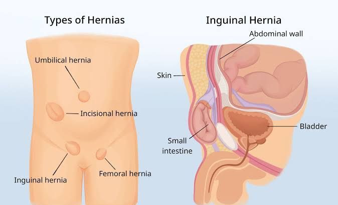
- Inguinal Hernia: Occurs in the inguinal canal in the groin area; more common in males.
- Femoral Hernia: More common in females; occurs just below the inguinal ligament.
- Umbilical Hernia: Occurs near the belly button; often seen in infants but can affect adults.
- Hiatal Hernia: Occurs when part of the stomach pushes through the diaphragm into the chest cavity.
- Symptoms and Complications: Symptoms may include visible bulges during physical activity, discomfort or pain at the site of protrusion, and potential complications like incarceration (where tissue becomes trapped) or strangulation (where blood supply is cut off), leading to tissue death.
Understanding these anatomical aspects helps clarify how hernias develop and why they may require surgical intervention for repair.
Differentiating Hernia Types Based on Physical Exam
The following is a detailed differentiation of the various types of hernias based on physical examination:
- Inguinal Hernia:
- Physical Exam Findings: Inguinal hernias are typically found in the groin area. During a physical exam, the clinician may ask the patient to cough or perform a Valsalva maneuver to increase intra-abdominal pressure, which may cause a bulge to appear in the inguinal canal. The bulge can often be palpated above the pubic bone and may be reducible (able to be pushed back into the abdomen). There are two subtypes:
- Indirect Inguinal Hernia: This type occurs lateral to the inferior epigastric vessels and is often congenital. It may extend into the scrotum in males.
- Direct Inguinal Hernia: This type occurs medial to the inferior epigastric vessels and is usually acquired due to weakness in the abdominal wall.
- Physical Exam Findings: Inguinal hernias are typically found in the groin area. During a physical exam, the clinician may ask the patient to cough or perform a Valsalva maneuver to increase intra-abdominal pressure, which may cause a bulge to appear in the inguinal canal. The bulge can often be palpated above the pubic bone and may be reducible (able to be pushed back into the abdomen). There are two subtypes:
- Femoral Hernia:
- Physical Exam Findings: Femoral hernias present as a bulge below the inguinal ligament, specifically in the femoral canal, which is located medial to the femoral vein and lateral to the pubic tubercle. On examination, it may be more difficult to detect than an inguinal hernia because it can be smaller and less prominent. It is more common in females.
- Umbilical Hernia:
- Physical Exam Findings: Umbilical hernias appear as a bulge at or near the umbilicus (navel). They can be easily identified during a physical exam when patients are asked to perform maneuvers that increase abdominal pressure, such as coughing or straining. These hernias are often reducible and may become more prominent when standing or straining.
- Incisional Hernia:
- Physical Exam Findings: Incisional hernias occur at previous surgical incision sites where there has been a weakness in the abdominal wall. During examination, there will typically be a palpable bulge at or near the site of surgery that becomes more pronounced with increased intra-abdominal pressure (e.g., coughing). These hernias can vary significantly in size depending on factors such as time since surgery and tension on surrounding tissues.
- Hiatal Hernia:
- Physical Exam Findings: Hiatal hernias involve protrusion of part of the stomach through the diaphragm into the thoracic cavity. While they cannot be directly palpated during a physical exam like other types of hernias, signs such as gastroesophageal reflux disease (GERD) symptoms may suggest their presence. A clinician might use auscultation for bowel sounds over areas where they should not normally be present if there is significant displacement.
- Epigastric Hernia:
- Physical Exam Findings: Epigastric hernias occur between the umbilicus and xiphoid process along the midline of the abdomen. On examination, they present as small, palpable masses that may become more prominent with increased abdominal pressure.
In summary, while all types of hernias involve protrusions through weaknesses in muscle layers or fascia, their specific characteristics during physical examination—such as location, reducibility, size, and relation to other anatomical landmarks—allow for differentiation among them.
Common Complications of Hernia: Strangulation, Ischemia, and Bowel Obstruction
Hernias occur when an organ or tissue protrudes through a weak spot in the surrounding muscle or connective tissue. While many hernias are asymptomatic and can be managed conservatively, they can lead to serious complications if not treated appropriately. The most common complications associated with hernias include strangulation, ischemia, and bowel obstruction. Below is a detailed examination of each complication.
1. Strangulation
Strangulation occurs when the blood supply to the herniated tissue is compromised due to constriction by the surrounding tissues. This situation is particularly dangerous because it can lead to tissue necrosis (death) if not promptly addressed.
- Mechanism: In a strangulated hernia, the contents of the hernia sac (often part of the intestine) become trapped and cannot return to their normal position. The pressure from the surrounding tissues can cut off blood flow.
- Symptoms: Patients may experience severe pain at the site of the hernia, swelling, redness, and signs of systemic illness such as fever or tachycardia (increased heart rate). There may also be nausea and vomiting.
- Consequences: If left untreated, strangulation can lead to irreversible damage to the affected tissue within hours, necessitating surgical intervention to remove necrotic sections of bowel or other affected organs.
2. Ischemia
Ischemia refers specifically to a reduction in blood flow that deprives tissues of oxygen and nutrients necessary for cellular metabolism.
- Relation to Hernias: Ischemia often accompanies strangulation but can also occur independently in cases where there is significant tension on blood vessels supplying the herniated area.
- Symptoms: Symptoms may overlap with those of strangulation but are characterized by more gradual onset pain that may worsen over time. Patients might also present with abdominal distension and changes in bowel habits.
- Consequences: Prolonged ischemia can lead to cell death and subsequent perforation of the bowel wall, resulting in peritonitis (inflammation of the abdominal cavity), which is a life-threatening condition requiring immediate medical attention.
3. Bowel Obstruction
Bowel obstruction occurs when there is a blockage that prevents food or liquid from passing through the intestines.
- Types: Obstructions can be classified as either mechanical (due to physical blockage) or functional (due to impaired motility). In hernias, mechanical obstructions are more common.
- Symptoms: Symptoms typically include cramping abdominal pain, inability to pass gas or stool, vomiting (which may contain bile), and abdominal distension.
- Consequences: If not resolved quickly—often through surgical intervention—bowel obstruction can lead to ischemia and strangulation as described above. It may also result in perforation of the bowel leading to peritonitis.
In summary, while hernias themselves may initially present without symptoms or complications, they carry risks that necessitate careful monitoring and timely intervention when complications arise. Strangulation leads directly to ischemia due to compromised blood flow; both conditions can precipitate bowel obstruction if not addressed promptly.
Overview of Hernias and Other Causes of Groin Masses
Understanding Hernias
A hernia occurs when tissue, such as part of the intestine or abdominal fat, protrudes through a weak spot in the abdominal wall. Inguinal hernias are particularly common and occur in the inguinal canal, which is located in the groin area. They can be classified into two main types: indirect and direct inguinal hernias. Indirect inguinal hernias are often congenital and occur due to a failure of the inguinal canal to close properly during fetal development. Direct inguinal hernias typically develop over time due to muscle weakness.
Characteristics of Hernias
- Bulge Appearance: Hernias typically present as a noticeable bulge in the groin that may become more prominent when standing, coughing, or straining.
- Reduction: Many hernias can be pushed back into place (reduced) manually, especially if they are not incarcerated or strangulated.
- Symptoms: Patients may experience discomfort or pain at the site of the bulge, especially during physical activities or prolonged standing.
- Physical Examination Findings: A healthcare provider can often diagnose a hernia through physical examination alone, sometimes requiring maneuvers like coughing to make the hernia more apparent.
Other Causes of Groin Masses
While hernias are a common cause of groin masses, several other conditions can also lead to similar presentations:
- Lymphadenopathy: Enlarged lymph nodes due to infections (like sexually transmitted infections), malignancies (such as lymphoma), or systemic diseases can present as masses in the groin area.
- Characteristics: These masses are usually firm and tender upon palpation and may be associated with systemic symptoms like fever or weight loss.
- Lipomas: These benign tumors consist of fatty tissue and can occur anywhere on the body, including the groin.
- Characteristics: Lipomas are soft, movable under the skin, and generally painless unless they press on surrounding structures.
- Hydroceles: A hydrocele is an accumulation of fluid around a testicle that can cause swelling in the scrotum or groin area.
- Characteristics: Hydroceles typically present as a smooth, non-tender swelling that transilluminates (light passes through it) when examined.
- Spermatic Cord Issues: Conditions affecting the spermatic cord, such as varicoceles (enlarged veins) or torsion (twisting), can also result in groin swelling.
- Characteristics: Varicoceles may feel like a “bag of worms” and are usually asymptomatic but can cause discomfort; torsion is acute and painful.
- Infections/Abscesses: Infections in the skin or deeper tissues can lead to abscess formation in the groin region.
- Characteristics: These masses tend to be red, warm, swollen, and painful; systemic signs such as fever may also be present.
- Tumors/Malignancies: Both benign and malignant tumors can arise in this region.
- Characteristics: Tumors may vary widely in presentation but often require imaging studies for definitive diagnosis.
Diagnostic Approach
To differentiate between these conditions:
- A thorough medical history is essential to identify associated symptoms (e.g., pain, fever).
- Physical examination plays a crucial role; specific characteristics of each type of mass will guide further investigation.
- Imaging studies such as ultrasound may be employed for better visualization—this is particularly useful for distinguishing between solid masses (like tumors) versus fluid-filled structures (like hydroceles).
- Laboratory tests might be necessary if infection or malignancy is suspected based on clinical findings.
Differentiating Hernias from Other Causes of Groin Masses
- Anatomical Location:
- Hernias: The most common types of hernias in the groin are inguinal hernias (which can be direct or indirect) and femoral hernias. Inguinal hernias occur when tissue bulges through a weak spot in the abdominal muscles in the inguinal region. Direct inguinal hernias protrude directly through the abdominal wall, while indirect inguinal hernias pass through the inguinal canal. Femoral hernias occur below the inguinal ligament.
- Other Causes: Other causes of groin masses may include lymphadenopathy (swollen lymph nodes), tumors (benign or malignant), abscesses, and varicoceles. These conditions typically have different anatomical locations and characteristics compared to hernias.
- Clinical Presentation:
- Hernias: Patients with a hernia often present with a palpable mass that may increase in size with activities such as coughing, straining, or lifting heavy objects. The mass may be reducible (able to be pushed back into place) or irreducible (cannot be pushed back). Symptoms may also include discomfort or pain at the site.
- Other Causes: Lymphadenopathy may present as firm, mobile masses that are tender to palpation and often associated with systemic symptoms like fever. Tumors may present as fixed masses that do not change with position or activity and can be associated with weight loss or other systemic symptoms. Abscesses typically present with localized pain, swelling, redness, and warmth over the affected area.
- Pathophysiology:
- Hernias: The primary cause of a hernia is a combination of increased intra-abdominal pressure and weakness in the abdominal wall musculature due to congenital factors, previous surgical incisions, aging, obesity, or chronic cough.
- Other Causes: Lymphadenopathy can result from infections (viral/bacterial), malignancies (lymphoma/metastatic cancer), or inflammatory conditions (sarcoidosis). Tumors arise from abnormal cell growth due to genetic mutations and can vary widely in behavior depending on their nature (benign vs malignant).
- Diagnosis:
- Diagnosis of a hernia is primarily clinical but can be confirmed via imaging studies such as ultrasound or CT scans if necessary.
- For other causes of groin masses, additional diagnostic workup including blood tests, imaging studies (like MRI for soft tissue evaluation), biopsy for histological examination might be required.
- Management:
- Hernias often require surgical intervention for repair if symptomatic; however, asymptomatic small hernias may be monitored.
- Management for other causes varies widely depending on etiology—ranging from antibiotics for infections to oncological treatments for tumors.
Diagnostic Evaluation and Management Plan for a Patient with Hernia
1. Initial Assessment and History Taking
The first step in evaluating a patient with a suspected hernia involves a thorough history and physical examination. Key components include:
- History of Present Illness (HPI): Assess the onset, duration, and characteristics of any bulge or discomfort. Inquire about associated symptoms such as pain, nausea, vomiting, or changes in bowel habits.
- Past Medical History: Document any previous surgeries, particularly abdominal or pelvic surgeries that may predispose to hernia formation.
- Family History: Note any family history of hernias or connective tissue disorders.
- Social History: Evaluate lifestyle factors such as occupation (heavy lifting), exercise habits, and smoking status.
- Review of Systems: Investigate gastrointestinal symptoms that may suggest complications like incarceration or strangulation.
2. Physical Examination
A comprehensive physical examination is crucial:
- Inspection: Look for visible bulges in the abdomen or groin area while the patient is standing and straining (e.g., Valsalva maneuver).
- Palpation: Gently palpate the area to assess the size, reducibility, tenderness, and any signs of incarceration (non-reducible) or strangulation (severe tenderness, discoloration).
- Special Tests: Perform specific maneuvers to elicit symptoms; for example, cough impulse test for inguinal hernias.
3. Diagnostic Imaging
If the diagnosis remains uncertain after clinical evaluation or if there are concerns about complications:
- Ultrasound: This is often the first-line imaging modality for evaluating groin hernias due to its non-invasive nature and ability to visualize soft tissues.
- CT Scan: A CT scan of the abdomen/pelvis can be useful for complex cases or when intra-abdominal contents are involved. It provides detailed images that can help identify the type and extent of the hernia.
4. Differential Diagnosis
Consider other conditions that may mimic hernia symptoms:
- Lipomas
- Lymphadenopathy
- Abscesses
- Tumors
5. Management Options
Management strategies depend on the type of hernia (inguinal, femoral, umbilical, incisional) and whether it is symptomatic:
- Watchful Waiting: For asymptomatic small hernias without complications in elderly patients or those with significant comorbidities.
- Surgical Intervention:
- Elective Surgery: Recommended for symptomatic hernias to prevent complications. Techniques include:
- Open repair: Traditional method involving an incision.
- Laparoscopic repair: Minimally invasive approach using small incisions.
- Emergency Surgery: Required for incarcerated or strangulated hernias presenting with acute pain, nausea/vomiting, or systemic signs like fever.
- Elective Surgery: Recommended for symptomatic hernias to prevent complications. Techniques include:
6. Postoperative Care
After surgical intervention:
- Monitor for complications such as infection, hematoma formation, recurrence of the hernia, and chronic pain.
- Provide instructions on activity restrictions during recovery (typically avoiding heavy lifting for several weeks).
7. Follow-Up
Schedule follow-up appointments to assess healing and address any postoperative concerns. Educate patients about lifestyle modifications that may reduce recurrence risk (e.g., weight management).
In summary, a structured approach involving thorough history taking, physical examination, appropriate imaging studies if needed, differential diagnosis consideration, tailored management strategies based on symptomatology and urgency will ensure effective evaluation and treatment of patients with hernias.
Overview of Hernia Surgery
Surgery is the primary treatment for hernias, which occur when an organ or tissue protrudes through a weak spot in the surrounding muscle or connective tissue. The most common types of hernia surgeries are open surgery, laparoscopic (minimally invasive) surgery, and robotic surgery. The choice of surgical method depends on various factors, including the type and location of the hernia, the patient’s overall health, and the surgeon’s expertise.
Indications for Surgery
Not all hernias require immediate surgical intervention. However, surgery is generally recommended if the hernia causes symptoms such as pain or discomfort, or if there is a risk of complications like strangulation (where blood supply to the herniated tissue is cut off). Symptoms that may necessitate emergency surgery include sudden severe pain, nausea, vomiting, fever, and a bulge that cannot be pushed back into place.
Preoperative Assessment
Before surgery, a thorough evaluation is conducted to determine the patient’s suitability for the procedure. This includes:
- Physical Examination: The surgeon will assess the size and location of the hernia.
- Medical History Review: Any pre-existing conditions such as diabetes or obesity that could complicate recovery are evaluated.
- Diagnostic Tests: Imaging studies like ultrasound or CT scans may be performed to understand better the hernia’s characteristics.
Patients are also advised on how to prepare for surgery, which may include dietary restrictions and arranging transportation home post-surgery.
Types of Hernia Surgery
- Open Surgery (Herniorrhaphy): This traditional approach involves making a larger incision near the hernia site to repair it directly. The surgeon pushes the protruding tissue back into place and reinforces the abdominal wall with stitches or mesh.
- Laparoscopic Surgery: This minimally invasive technique uses several small incisions through which specialized instruments are inserted. A camera provides visualization on a monitor, allowing for precise repairs with less postoperative pain and quicker recovery times compared to open surgery.
- Robotic Surgery: Similar to laparoscopic surgery but utilizes robotic systems that provide enhanced precision and control during the procedure. It can be particularly beneficial for complex cases but typically requires more time than other methods.
Surgical Procedure
The surgical process generally follows these steps:
- Anesthesia Administration: Patients are usually placed under general anesthesia; however, local anesthesia may be used in certain cases.
- Incision Creation: Depending on the type of surgery chosen (open or laparoscopic), appropriate incisions are made.
- Hernia Repair:
- For open repair, tissues are repositioned manually.
- For laparoscopic repair, instruments are used to manipulate tissues through small incisions.
- Mesh may be applied to strengthen the area and reduce recurrence risk.
- Closure of Incisions: After completing the repair, incisions are closed using sutures or staples.
- Recovery Monitoring: Patients are moved to a recovery area where vital signs are monitored until they regain consciousness from anesthesia.
Postoperative Care
Following surgery, patients typically experience some discomfort managed with pain relievers prescribed by their healthcare provider. Recovery protocols often include:
- Resting for 24 hours post-surgery.
- Gradually resuming light activities such as walking within days after surgery.
- Avoiding heavy lifting or strenuous activities for several weeks to prevent complications such as recurrence or strain on healing tissues.
Patients should watch for signs of complications like increased pain, swelling at the incision site, fever, or changes in bowel habits and report these immediately to their healthcare provider.
Risks Associated with Hernia Surgery
While hernia surgeries are generally safe procedures with low complication rates (approximately 16% recurrence rate within ten years), they do carry risks including:
- Infection at incision sites
- Chronic pain
- Fluid accumulation (seroma)
- Injury to surrounding organs
- Complications related to anesthesia
In rare cases, serious complications can occur that may require further medical intervention.
In conclusion, surgical intervention for hernias is a common procedure aimed at relieving symptoms and preventing complications associated with untreated hernias. The choice between open versus minimally invasive techniques depends on individual patient factors and surgeon preference.


