Understanding the Pathophysiology of Pneumothorax and Its Management
(a) Pathophysiology of Pneumothorax
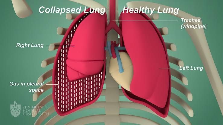
A pneumothorax occurs when air enters the pleural space, which is the area between the lung and the chest wall. This accumulation of air creates pressure that can cause the lung to collapse either partially or fully. The pathophysiological mechanisms behind pneumothorax can be categorized into two main types: spontaneous and traumatic.
- Spontaneous Pneumothorax: This type occurs without any obvious cause or injury. It can be further divided into:
- Primary Spontaneous Pneumothorax (PSP): Typically occurs in healthy individuals, often young males, without underlying lung disease. It is believed to arise from the rupture of small blebs (air-filled sacs) on the surface of the lungs.
- Secondary Spontaneous Pneumothorax (SSP): Occurs in individuals with pre-existing lung conditions such as chronic obstructive pulmonary disease (COPD), cystic fibrosis, or pneumonia. The underlying pathology contributes to weakened lung tissue, making it more susceptible to collapse.
- Traumatic Pneumothorax: This type results from an injury to the chest that allows air to enter the pleural space. Causes include:
- Blunt trauma: Such as rib fractures or blunt force injuries.
- Penetrating trauma: Such as stab wounds or gunshot wounds.
- Iatrogenic causes: Resulting from medical procedures like central line placement or mechanical ventilation.
The presence of air in the pleural space disrupts normal negative pressure, which is essential for lung expansion during inhalation. As a result, affected areas of the lung may collapse, leading to impaired gas exchange and respiratory distress.
(b) Management of Pneumothorax
The management of pneumothorax depends on its size, symptoms, and underlying cause. Treatment options can range from observation to invasive procedures:
- Observation: For small, asymptomatic pneumothoraces (less than 15-20% of lung volume), careful monitoring may be sufficient as many resolve spontaneously over time.
- Supplemental Oxygen: Administering oxygen can enhance reabsorption of air from the pleural space at a rate significantly higher than room air alone.
- Needle Aspiration: For symptomatic patients with small spontaneous pneumothoraces, needle aspiration can be performed to remove air from the pleural space effectively.
- Chest Tube Placement (Thoracostomy): In cases where needle aspiration fails or for larger pneumothoraces, a chest tube may be inserted to continuously drain air and allow for lung re-expansion.
- Surgical Intervention:
- Video-Assisted Thoracoscopic Surgery (VATS): Indicated for recurrent pneumothoraces or those not responding to other treatments; it involves minimally invasive techniques to repair blebs or perform pleurodesis.
- Pleurodesis: A procedure that involves introducing a sclerosing agent into the pleural space to adhere the lung to the chest wall and prevent recurrence.
- Emergency Management for Tension Pneumothorax: This life-threatening condition requires immediate decompression via needle thoracostomy followed by definitive treatment with tube thoracostomy if necessary.
In summary, understanding both the pathophysiology and management strategies for pneumothorax is crucial in providing effective care and preventing complications associated with this condition.
Differential Diagnosis for Fluid in the Pleural Space
The differential diagnosis for fluid in the pleural space, commonly referred to as pleural effusion, can be categorized based on the characteristics of the fluid (transudative vs. exudative) and the underlying causes. Understanding these categories is crucial for determining appropriate management and treatment.
1. Transudative Effusions
Transudative effusions are typically caused by systemic factors that alter hydrostatic or oncotic pressures, leading to an imbalance in fluid movement. Common causes include:
- Congestive Heart Failure (CHF): The most frequent cause of transudative pleural effusions, where increased pulmonary capillary pressure leads to fluid accumulation.
- Cirrhosis: Liver dysfunction can lead to decreased oncotic pressure due to low albumin levels, resulting in fluid leakage into the pleural space.
- Nephrotic Syndrome: Similar to cirrhosis, this condition leads to significant protein loss through urine, decreasing plasma oncotic pressure.
- Atelectasis: Collapse of lung tissue can create a negative intrapleural pressure that draws fluid into the pleural space.
- Peritoneal Dialysis: Fluid from the peritoneal cavity may leak into the pleura.
- Constrictive Pericarditis: Impaired filling of the heart can lead to increased venous pressure and subsequent pleural effusion.
2. Exudative Effusions
Exudative effusions are characterized by high protein content and are typically associated with local inflammatory processes. Common causes include:
- Pneumonia: Parapneumonic effusion occurs when infection spreads from lung tissue into the pleura.
- Malignancy: Cancer can cause malignant pleural effusions either through direct invasion or obstruction of lymphatic drainage.
- Tuberculosis (TB): TB can lead to tuberculous pleuritis, causing lymphocyte-predominant exudates.
- Pulmonary Embolism (PE): Can result in exudative effusion due to infarction or inflammation of lung tissue.
- Pancreatitis: Inflammatory mediators may cause fluid accumulation in the pleura.
- Chylothorax: Leakage of lymphatic fluid into the pleural space due to trauma or malignancy affecting lymphatic vessels.
3. Less Common Causes
In addition to common causes, there are several less frequent conditions that may also result in pleural effusion:
- Hemothorax: Accumulation of blood in the pleural space often due to trauma or malignancy.
- Empyema: Collection of pus within the pleura usually secondary to infection.
- Post-surgical complications: Following thoracic surgery, patients may develop effusions due to inflammation or injury.
In summary, identifying whether a pleural effusion is transudative or exudative is essential for narrowing down potential diagnoses and guiding further investigation and treatment.
Understanding Hemothorax and Chylothorax
(a) Hemothorax: Definition and Causes
A hemothorax is defined as the accumulation of blood in the pleural cavity, which can result from various causes. The most common causes include:
- Trauma: This is the leading cause of hemothorax, often resulting from blunt or penetrating chest injuries. Examples include motor vehicle accidents, falls, or stab wounds.
- Medical Conditions: Certain medical conditions such as malignancies (lung cancer), pulmonary embolism, or vascular malformations can lead to bleeding into the pleural space.
- Procedures: Invasive procedures like thoracentesis or central line placement can inadvertently puncture blood vessels, causing a hemothorax.
Pathophysiology
When blood accumulates in the pleural space, it can lead to increased pressure on the lungs, impairing their ability to expand fully during respiration. This results in respiratory distress and decreased oxygenation.
Symptoms
Patients with hemothorax may present with:
- Chest pain
- Shortness of breath
- Hypotension (in cases of significant blood loss)
- Dullness to percussion on examination
Diagnosis
Diagnosis typically involves imaging studies such as:
- Chest X-ray: Can show fluid levels in the pleural space.
- CT Scan: Provides a more detailed view and helps assess the extent of bleeding.
Treatment Options
The treatment for hemothorax depends on its severity:
- Observation: Small hemothoraces may resolve spontaneously without intervention.
- Thoracostomy (Chest Tube Placement): For larger collections of blood or symptomatic patients, a chest tube is inserted to drain the fluid and allow lung re-expansion.
- Surgical Intervention: In cases where there is ongoing bleeding or if a significant volume of blood is present, surgical options such as thoracotomy may be necessary to control the source of bleeding.
(b) Chylothorax: Definition and Causes
Chylothorax refers to the accumulation of lymphatic fluid (chyle) in the pleural cavity. This condition arises primarily due to disruption of lymphatic drainage pathways.
- Trauma: Similar to hemothorax, trauma can also cause chylothorax by damaging lymphatic vessels.
- Malignancy: Tumors affecting lymph nodes or structures around the thoracic duct can obstruct normal lymphatic flow.
- Surgery: Procedures involving thoracic surgery may inadvertently damage lymphatic vessels leading to chylous leakage.
Pathophysiology
Chyle contains triglycerides and other lipids absorbed from the intestines; when it leaks into the pleural space, it alters fluid dynamics and can lead to respiratory compromise similar to that seen in hemothorax.
Symptoms
Patients may exhibit symptoms including:
- Dyspnea (shortness of breath)
- Chest discomfort
- Weight loss (due to malabsorption)
Physical examination might reveal dullness on percussion over affected areas.
Diagnosis
Diagnosis usually involves:
- Imaging Studies (X-ray/CT): To identify fluid accumulation.
- Fluid Analysis via Thoracentesis: The analysis will show elevated triglyceride levels (>110 mg/dL) confirming chylous effusion.
Treatment Options
Management strategies for chylothorax include:
- Conservative Management:
- Dietary modifications such as a low-fat diet supplemented with medium-chain triglycerides (MCTs) which are easier for absorption without relying heavily on lymphatics.
- Drainage via chest tube if symptomatic or large volumes are present.
- Surgical Intervention:
- If conservative measures fail after several days or if there is significant ongoing leakage, surgical options like ligation of the thoracic duct may be considered.
- Medications:
- Octreotide has been used off-label in some cases to reduce lymphatic flow.
In conclusion, both hemothorax and chylothorax involve fluid accumulation in the pleural space but differ significantly in etiology and management strategies. Understanding these differences is crucial for effective diagnosis and treatment.
Stages of Development of an Empyema
Empyema develops through three distinct stages, each characterized by specific changes in the pleural space and the nature of the fluid present. Understanding these stages is crucial for effective diagnosis and treatment.
Stage 1: Simple Empyema (Exudative Phase)
In this initial stage, excess fluid begins to accumulate in the pleural cavity. This fluid can be sterile or may become infected, leading to the presence of pus. The body responds to the infection by increasing vascular permeability, allowing more fluid to enter the pleural space. Symptoms may start to manifest during this phase, but they are often nonspecific.
Stage 2: Complicated Empyema (Fibrinopurulent Phase)
As the condition progresses, the fluid in the pleural cavity thickens and becomes more viscous due to inflammation. This stage is marked by a buildup of neutrophils and other inflammatory cells, which leads to the formation of “pockets” or loculated effusions within the pleural space. The presence of fibrinous material can also occur, complicating drainage efforts and potentially leading to further lung impairment.
Stage 3: Frank Empyema (Organizing Phase)
In this final stage, the infected fluid causes scarring on the inner layers of the lungs and pleura. The fibrous tissue that forms can restrict lung expansion, resulting in significant difficulty breathing. This stage represents a chronic condition where surgical intervention may be necessary to remove fibrous tissue and pus pockets to restore normal lung function.
Recognizing these stages is essential for healthcare providers as it guides treatment decisions ranging from antibiotic therapy and drainage procedures to more invasive surgical options.
Typical Characteristics of Pleural Tumors
1. Origin and Frequency
Approximately 95% of pleural tumors are metastatic, meaning they originate from cancers that have spread from other parts of the body. Primary pleural tumors are rare, accounting for only 0.3% to 3.5% of cases. Among primary pleural tumors, about 90% are malignant mesotheliomas, while solitary fibrous tumors make up about 5%, with the remaining 5% consisting of other types.
2. Types of Pleural Tumors
- Malignant Mesothelioma: This is the most common type of primary pleural tumor and is strongly associated with asbestos exposure.
- Solitary Fibrous Tumor: These can be benign or malignant and typically arise from the visceral pleura (80%) or other areas like the parietal pleura, mediastinum, or diaphragm (20%). While they often present as asymptomatic masses, about 12% can exhibit malignant characteristics.
3. Imaging Characteristics
Pleural tumors may not always be clearly visible on a PA chest X-ray; however, thoracic CT scans usually reveal well-circumscribed, non-invasive lesions. MRI can also be utilized to assess these masses further.
4. Symptoms and Presentation
Many pleural tumors are asymptomatic in their early stages, but as they grow, they may cause symptoms such as chest pain, dyspnea (difficulty breathing), or cough depending on their size and location.
5. Diagnosis
A definitive diagnosis often requires histopathological examination through biopsy methods such as Tru-cut biopsy or thoracoscopic (VATS) biopsy. Immunohistochemical analysis is essential for diagnosing solitary fibrous tumors.
6. Treatment Options
The treatment approach varies significantly based on whether the tumor is benign or malignant:
- Benign Tumors: Complete surgical removal is typically recommended along with at least a 2 cm margin of normal tissue.
- Malignant Tumors: Surgical options may be limited; oncological treatments are generally preferred depending on the cancer type and stage.
7. Recurrence Rates
Recurrence rates differ between benign and malignant tumors; benign tumors have an approximate long-term recurrence rate of 8%, while malignant tumors tend to recur more frequently.
In summary, pleural tumors exhibit a range of characteristics based on their origin (primary vs metastatic), type (benign vs malignant), imaging features, symptoms, diagnostic methods, treatment approaches, and recurrence rates.


