Understanding and Assigning the Glasgow Coma Score
The Glasgow Coma Scale (GCS) is a clinical tool used to assess a patient’s level of consciousness and neurological functioning, particularly in cases of head injury or other conditions that may impair consciousness. The GCS evaluates three aspects of a patient’s responsiveness: eye opening, verbal response, and motor response. Each component is scored separately, and the total score can range from 3 to 15, with lower scores indicating more severe impairment.
Step 1: Assess Eye Opening
The eye-opening response is scored from 1 to 4:
- 1 point: No eye opening.
- 2 points: Eyes open in response to pressure (e.g., a pinch).
- 3 points: Eyes open in response to voice.
- 4 points: Eyes open spontaneously.
Step 2: Assess Verbal Response
The verbal response is scored from 1 to 5:
- 1 point: No verbal response.
- 2 points: Incomprehensible sounds (moans or groans).
- 3 points: Utters incoherent words.
- 4 points: Confused but coherent speech.
- 5 points: Oriented and converses normally.
Step 3: Assess Motor Response
The motor response is scored from 1 to 6:
- 1 point: No movement.
- 2 points: Extension to pain (decerebrate posture).
- 3 points: Abnormal flexion to pain (decorticate posture).
- 4 points: Normal flexion (bends arm at elbow rapidly).
- 5 points: Localizes pain (brings hand above clavicle to stimulus).
- 6 points: Obeys commands.
Step 4: Calculate Total GCS Score
To calculate the total GCS score, add the scores from each of the three categories:
Total GCS = Eye Opening + Verbal Response + Motor Response
For example, if a patient has an eye score of 3, a verbal score of 4, and a motor score of 5, their total GCS would be:
Total GCS = 3 + 4 + 5 = 12
Interpreting the Total GCS Score
The total GCS score ranges from a minimum of 3 (indicating deep coma or unresponsiveness) to a maximum of 15 (indicating full consciousness). The scores can be interpreted as follows:
- A score of 3–8 indicates severe brain injury or coma.
- A score of 9–12 indicates moderate brain injury.
- A score of 13–15 indicates mild brain injury.
This scoring system allows healthcare providers to quickly assess and communicate the patient’s level of consciousness and track changes over time.
Presentation of Brain Herniation Syndromes in the Setting of Trauma
Brain herniation syndromes occur when brain tissue is displaced due to increased intracranial pressure (ICP), often as a result of trauma, tumors, or other pathological processes. In the context of trauma, these syndromes can arise from contusions, hemorrhages, or swelling that lead to a critical increase in ICP. Understanding the types and presentations of brain herniation is crucial for timely diagnosis and intervention.
Types of Brain Herniation Syndromes
- Subfalcine Herniation: This occurs when the cingulate gyrus is pushed under the falx cerebri due to unilateral mass effect. Clinically, it may present with contralateral leg weakness or changes in consciousness.
- Transtentorial Herniation (Uncal Herniation): This type involves the medial temporal lobe being displaced downward through the tentorial notch. Symptoms include:
- Ipsilateral pupil dilation (due to compression of cranial nerve III)
- Contralateral hemiparesis
- Altered level of consciousness
- Central Herniation: This occurs when there is downward displacement of the brainstem and diencephalon through the tentorial notch. It can lead to:
- Bilateral pupillary changes
- Decerebrate posturing
- Respiratory irregularities
- Tonsillar Herniation: This involves displacement of the cerebellar tonsils through the foramen magnum, which can compress vital centers in the medulla oblongata. Clinical signs may include:
- Severe headache
- Neck stiffness
- Cardiopulmonary instability
Clinical Presentation Following Trauma
In trauma settings, patients may present with a range of symptoms depending on the type and severity of herniation:
- Altered Consciousness: Patients may exhibit confusion, lethargy, or coma.
- Neurological Deficits: Depending on which areas are affected by herniation, deficits such as weakness, sensory loss, or cranial nerve palsies may be observed.
- Pupil Changes: As mentioned earlier, pupil size and reactivity can provide clues about herniation; for instance, an enlarged pupil on one side suggests uncal herniation.
- Posturing Responses: Abnormal posturing (decerebrate or decorticate) can indicate severe brain injury and potential herniation.
Diagnostic Imaging
CT scans are typically used as a first-line imaging modality in trauma cases to identify signs of increased ICP and potential herniation. Key findings might include:
- Midline shift indicating mass effect
- Compression of ventricles
- Presence of hemorrhage or edema
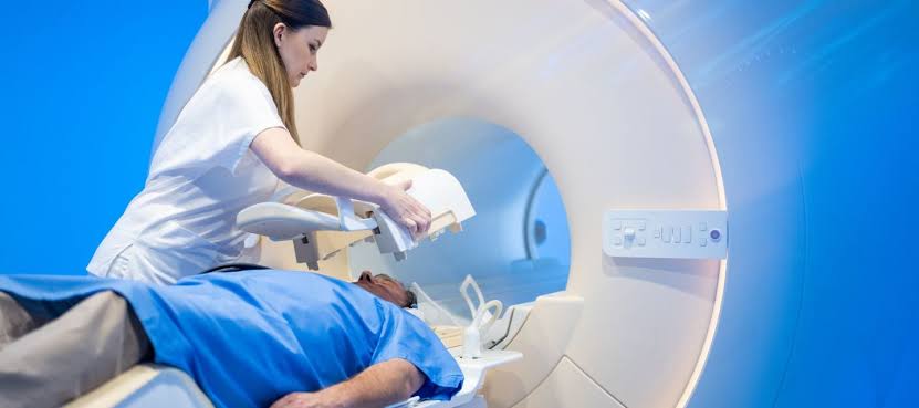
MRI may be utilized for further evaluation but is less common in acute settings due to time constraints.
Management Strategies
The management of brain herniation syndromes focuses on reducing ICP and addressing the underlying cause:
- Medical Management:
- Administration of osmotic agents like mannitol or hypertonic saline.
- Sedatives to reduce metabolic demand.
- Surgical Intervention:
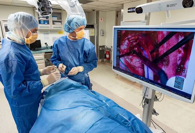
- Decompressive craniectomy may be indicated if medical management fails.
- Evacuation of hematomas if present.
- Monitoring and Supportive Care:
- Continuous monitoring in an intensive care setting.
- Supportive measures including ventilation support if respiratory function is compromised.
In conclusion, prompt recognition and management of brain herniation syndromes following trauma are essential for improving patient outcomes.
Initiating Management of Elevated Intracranial Pressure in Head Trauma
Elevated intracranial pressure (ICP) is a critical condition often encountered in patients with head trauma. The management of elevated ICP is essential to prevent secondary brain injury and improve patient outcomes. The initial approach involves a combination of general prophylactic measures, medical treatments, and potential surgical interventions.
Step 1: General Prophylactic Measures
Immediate actions should focus on stabilizing the patient and preventing further increases in ICP. Key measures include:
- Patient Positioning: Elevate the head of the bed to 30 degrees to facilitate venous drainage from the brain.
- Sedation and Analgesia: Administer adequate sedation to minimize agitation, which can increase ICP through elevated intrathoracic pressure.
- Temperature Control: Maintain normothermia as fever can exacerbate ICP by increasing metabolic demand.
Step 2: Monitoring and Assessment
Continuous monitoring of neurological status is crucial. This includes:
- Neurological Examination: Use tools like the Glasgow Coma Scale (GCS) to assess consciousness level.
- Intracranial Pressure Monitoring: Invasive methods such as intraventricular catheters are considered gold standards for measuring ICP directly.
Step 3: Medical Management
If ICP exceeds 20 mmHg, initiate medical therapies aimed at reducing ICP:
- Hyperosmolar Therapy: Administer mannitol or hypertonic saline. Mannitol draws fluid out of the brain tissue into circulation, while hypertonic saline increases serum osmolality and reduces cerebral edema.
- Hyperventilation: Temporarily reduce PCO2 through controlled ventilation to induce vasoconstriction, thereby lowering CBF and ICP. However, this should be limited due to risks associated with prolonged use.
- Fluid Management: Ensure euvolemia; avoid hypotonic fluids that can worsen cerebral edema.
Step 4: Surgical Interventions
In cases where medical management fails or if there are significant mass lesions contributing to increased ICP:
- Decompressive Craniectomy: Consider surgical intervention such as decompressive craniectomy for refractory elevated ICP or significant brain swelling.
Step 5: Ongoing Assessment and Adjustment of Treatment
Regularly reassess the patient’s neurological status and adjust treatment protocols based on response. Continuous monitoring of vital signs, fluid balance, and laboratory values is essential for effective management.
By following these steps systematically, healthcare providers can effectively manage elevated intracranial pressure in patients suffering from head trauma, aiming to minimize secondary brain injury and optimize outcomes.
Recognition and Management of Concussion, Brain Contusion, and Diffuse Axonal Injury
1. Recognition of Concussion
Concussion is a type of mild traumatic brain injury (TBI) that occurs due to a blow or jolt to the head or body, causing the brain to move rapidly within the skull. Recognizing concussion involves identifying symptoms and signs, which can be immediate or delayed. Common symptoms include:
- Headache
- Confusion or feeling as if in a fog
- Dizziness or balance problems
- Nausea or vomiting
- Sensitivity to light or noise
- Difficulty concentrating or remembering
Healthcare professionals often use standardized assessment tools like the SCAT5 (Sport Concussion Assessment Tool) for evaluation. It is crucial to note that loss of consciousness is not necessary for a concussion diagnosis.
2. Recognition of Brain Contusion
A brain contusion is a bruise on the brain tissue resulting from trauma. Symptoms may overlap with those of concussion but can also include:
- Loss of consciousness (which may last longer than with a concussion)
- Seizures
- Weakness in limbs
- Changes in behavior
Brain imaging techniques such as CT scans or MRIs are essential for diagnosing contusions, as they can reveal bleeding and swelling in the brain.
3. Recognition of Diffuse Axonal Injury (DAI)
Diffuse axonal injury occurs when rotational forces cause widespread damage to the axons in the brain, leading to severe functional impairment. Symptoms can include:
- Prolonged unconsciousness (coma)
- Severe cognitive deficits
- Motor impairments
DAI is typically diagnosed through advanced imaging techniques like MRI, which can show changes in white matter integrity.
4. Management Strategies
- Concussion Management: The management of concussion primarily involves physical and cognitive rest until symptoms resolve. Gradual return-to-play protocols should be followed under medical supervision, ensuring that athletes do not return too soon, which could lead to further injury.
- Brain Contusion Management: Management may require hospitalization for monitoring and supportive care. In cases where there is significant swelling or bleeding, surgical intervention may be necessary to relieve pressure on the brain.
- Diffuse Axonal Injury Management: DAI management focuses on intensive rehabilitation therapies tailored to individual needs since recovery can vary widely among patients. Supportive care in an intensive care unit may be required initially, followed by physical therapy, occupational therapy, and speech therapy as appropriate.
5. Follow-Up Care
Regardless of the type of injury sustained, follow-up care is critical for all patients who have experienced TBI. This includes regular assessments by healthcare providers specializing in brain injuries and neuropsychological evaluations if cognitive deficits are present.
In summary, recognizing these injuries involves understanding their distinct symptoms and utilizing appropriate diagnostic tools while management strategies focus on rest, rehabilitation, and monitoring for complications.
Recognition and Management of Acute Subdural and Epidural Hematoma
Acute subdural hematomas (SDH) and epidural hematomas (EDH) are critical conditions that arise from traumatic brain injury, leading to the accumulation of blood in the subdural or epidural spaces, respectively. Prompt recognition and management are essential to minimize morbidity and mortality associated with these conditions.
Recognition of Acute Subdural Hematoma (SDH)
- Clinical Presentation: Patients may present with altered consciousness, headache, focal neurological deficits, or seizures. The symptoms can vary based on the size of the hematoma and the rate of accumulation.
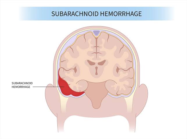
- Imaging Studies: A non-contrast CT scan of the head is the gold standard for diagnosis. An acute SDH appears as a crescent-shaped hyperdense area that conforms to the contour of the brain.
- Risk Factors: Elderly patients, those on anticoagulants, or individuals with a history of falls are at higher risk for developing SDHs.
Recognition of Acute Epidural Hematoma (EDH)
- Clinical Presentation: EDHs often present with a classic “lucid interval,” where patients may initially lose consciousness but then regain it before deteriorating again. Symptoms include headache, confusion, and focal neurological signs.
- Imaging Studies: A non-contrast CT scan shows an EDH as a biconvex (lens-shaped) hyperdense collection that does not cross suture lines due to its location between the skull and dura mater.
- Risk Factors: Commonly associated with skull fractures, particularly in younger individuals due to trauma.
Management of Acute Subdural Hematoma
- Initial Assessment: Assess airway, breathing, circulation (ABCs), and perform a neurological examination using tools like the Glasgow Coma Scale (GCS).
- Surgical Indications:
- Surgical intervention is indicated if there is significant mass effect on imaging or if the patient exhibits clinical deterioration.
- Generally, surgical options include craniotomy or burr hole evacuation depending on hematoma size and patient stability.
- If GCS is less than 9 or there is midline shift greater than 5 mm on CT imaging, surgery should be considered urgently.
- Non-Surgical Management: In cases where SDH is small (<10 mm) without midline shift or significant symptoms, close monitoring may be appropriate.
Management of Acute Epidural Hematoma
- Initial Assessment: Similar to SDH management; focus on ABCs and neurological status assessment.
- Surgical Indications:
- Immediate surgical intervention is typically required for symptomatic EDHs due to their rapid progression.
- Craniotomy is performed to evacuate the hematoma; this is critical as delayed treatment can lead to increased intracranial pressure and potential herniation.
- If there is a significant mass effect or neurological decline observed during monitoring, surgery should be expedited.
- Postoperative Care: Continuous monitoring in an intensive care setting post-surgery for complications such as rebleeding or infection.
Conclusion
The timely recognition and management of acute subdural and epidural hematomas are crucial for improving patient outcomes following traumatic brain injuries. Understanding clinical presentations along with appropriate imaging studies allows for effective decision-making regarding surgical interventions when indicated.
Recognizing and Managing Penetrating Head Trauma Including Gunshot Wounds
Penetrating head trauma refers to injuries where an object breaches the skull and enters the cranial cavity. This can occur due to various mechanisms, with gunshot wounds being one of the most severe forms. The management of such injuries is critical as they can lead to significant morbidity and mortality.
Recognition of Penetrating Head Trauma
- Clinical Presentation:
- Patients may present with altered mental status, loss of consciousness, focal neurological deficits, or seizures.
- External signs may include visible wounds, bleeding from the ears or nose (indicative of cerebrospinal fluid leakage), and signs of increased intracranial pressure (ICP) such as headache, vomiting, or changes in pupil size.
- Mechanism of Injury:
- Understanding the mechanism is crucial; gunshot wounds often cause extensive damage due to high-velocity projectiles that create a temporary cavitation effect in addition to direct tissue destruction.
- Initial Assessment:
- Conduct a thorough primary survey following Advanced Trauma Life Support (ATLS) guidelines.
- Assess airway, breathing, circulation, disability (neurological status), and exposure/environmental control.
Management of Penetrating Head Trauma
- Immediate Care:
- Ensure airway patency; consider intubation if there are signs of compromised airway or altered consciousness.
- Control any external bleeding using direct pressure; avoid applying pressure directly over a penetrating object.
- Neurological Evaluation:
- Perform a rapid neurological assessment using the Glasgow Coma Scale (GCS) to determine the level of consciousness and potential need for urgent intervention.
- Imaging Studies:
- Obtain a CT scan of the head without contrast as it is essential for assessing intracranial hemorrhage, foreign bodies, and overall brain integrity.
- MRI may be considered later if needed but is not typically used in acute settings due to longer acquisition time.
- Surgical Intervention:
- Consult neurosurgery urgently for cases involving significant brain injury or when there is evidence of mass effect or intracranial hemorrhage.
- Surgical options may include debridement of necrotic tissue, removal of foreign bodies, and evacuation of hematomas.
- Postoperative Management:
- Monitor for complications such as infection (e.g., meningitis), seizures, and further neurological decline.
- Manage ICP through appropriate measures including elevation of the head, sedation, osmotic agents like mannitol or hypertonic saline if indicated.
- Rehabilitation:
- Long-term management may involve rehabilitation services including physical therapy, occupational therapy, and neuropsychological support depending on the extent of neurological impairment.
Conclusion
The management of penetrating head trauma requires prompt recognition and immediate action to stabilize the patient while preparing for potential surgical intervention. Continuous monitoring and supportive care are essential components throughout the treatment process.
Principles of Management of Skull Fractures and Related Conditions
1. Overview of Skull Fractures
Skull fractures can be classified into three main categories: open, closed, and basal skull fractures. Each type has distinct characteristics and management principles.
- Open Skull Fractures: These involve a break in the skull that penetrates the skin, exposing the brain to the external environment. This type of fracture is associated with a higher risk of infection and requires immediate surgical intervention to clean the wound and repair any damage.
- Closed Skull Fractures: In these cases, the skull is fractured without any breach in the skin. The management typically involves monitoring for neurological deficits and potential complications such as intracranial hemorrhage.
- Basal Skull Fractures: These occur at the base of the skull and can lead to serious complications due to their proximity to vital structures such as cranial nerves and blood vessels. Symptoms may include raccoon eyes (periorbital ecchymosis), Battle’s sign (postauricular ecchymosis), or cerebrospinal fluid (CSF) leaks.
2. Management Principles
Open Skull Fractures:
- Immediate Care: Control bleeding, assess neurological status, and prevent infection.
- Surgical Intervention: Debridement of contaminated tissue, repair of dura mater if torn, and stabilization of bone fragments.
- Postoperative Care: Monitor for signs of infection or neurological deterioration.
Closed Skull Fractures:
- Observation: Patients are often monitored for changes in consciousness or neurological function.
- Imaging Studies: CT scans are commonly used to assess for intracranial bleeding or other complications.
- Symptomatic Treatment: Pain management and monitoring for potential complications like seizures or hematomas.
Basal Skull Fractures:
- Assessment for CSF Leak: A clear fluid leaking from the nose or ears indicates a CSF leak, which can increase infection risk (e.g., meningitis).
- If a CSF leak is suspected, bed rest, head elevation, and hydration are recommended.
- Surgical repair may be necessary if conservative measures fail.
3. Cerebrospinal Fluid Leak Management
Cerebrospinal fluid leaks can occur with both open and basal skull fractures. The management includes:
- Conservative Measures: Bed rest, hydration, caffeine intake (to promote CSF production), and avoiding activities that increase intracranial pressure (e.g., straining).
- Surgical Options: If conservative treatment does not resolve the leak within several days or if there are recurrent leaks or infections, surgical intervention may be required to repair the dura mater.
4. Chronic Subdural Hematoma Management
A chronic subdural hematoma is a collection of blood that forms between the brain’s surface and its outer protective covering (the dura mater) over a period of time, typically several weeks after an initial bleeding event. This condition often arises from the tearing of bridging veins due to head injuries, particularly in older adults whose brains have shrunk with age, making them more susceptible to such injuries. Symptoms can vary widely and may include headaches, confusion, weakness, and in severe cases, loss of consciousness or coma.
The management includes:
- Diagnosis via Imaging: CT or MRI scans help confirm the presence of a hematoma.
- Observation vs. Surgery: Small asymptomatic hematomas may be observed; however, larger ones causing symptoms typically require surgical intervention through burr hole drainage or craniotomy.
In children specifically:
- They may present differently than adults; thus careful monitoring is essential after head trauma.
In summary, effective management of skull fractures—whether open, closed, or basal—requires a thorough understanding of their implications on brain health and appropriate interventions based on individual patient needs.


