Extracellular, Intracellular, and Intravascular Volume in a 70-kg Man
In a 70-kg man, the distribution of body fluid is typically categorized into three main compartments: extracellular volume (ECV), intracellular volume (ICV), and intravascular volume. Understanding these compartments is crucial for comprehending physiological processes, fluid balance, and the effects of various medical conditions.
(a) Extracellular Volume (ECV)
Extracellular volume refers to the fluid outside of cells and constitutes about one-third of total body water. In a 70-kg adult male, the total body water is approximately 60% of body weight, which translates to about 42 liters (L). Therefore, the extracellular volume would be roughly:
- ECV = Total Body Water × Proportion of ECV
- ECV ≈ 42 L × 1/3 ≈ 14 L
The extracellular fluid can be further divided into two main components: interstitial fluid and plasma. Interstitial fluid accounts for about three-quarters of the ECV, while plasma makes up the remaining quarter. Thus:
- Interstitial Fluid ≈ 14 L × 0.75 ≈ 10.5 L
- Plasma Volume ≈ 14 L × 0.25 ≈ 3.5 L
(b) Intracellular Volume (ICV)
Intracellular volume refers to the fluid contained within cells and constitutes about two-thirds of total body water. For a typical adult male weighing 70 kg, this can be calculated as follows:
- ICV = Total Body Water × Proportion of ICV
- ICV ≈ 42 L × (2/3) ≈ 28 L
This large compartment is critical for cellular function as it contains cytoplasm and organelles necessary for metabolic processes.
(c) Intravascular Volume
Intravascular volume specifically refers to the blood plasma within the circulatory system. As mentioned earlier, this is part of the extracellular volume but is often considered separately due to its importance in hemodynamics and circulation. The intravascular volume can be estimated based on plasma volume:
- Intravascular Volume ≈ Plasma Volume ≈ 3.5 L
To summarize:
- Total Body Water: Approximately 42 L
- Extracellular Volume: Approximately 14 L
- Interstitial Fluid: Approximately 10.5 L
- Plasma Volume (Intravascular): Approximately 3.5 L
- Intracellular Volume: Approximately 28 L
Thus, in summary, for a typical 70-kg man:
- The intracellular volume is approximately 28 liters.
- The extracellular volume is approximately 14 liters.
- The intravascular volume is approximately 3.5 liters.
This distribution highlights how fluids are compartmentalized in the human body and underscores their importance in maintaining homeostasis.
Endogenous Factors Affecting Renal Control of Sodium and Water Excretion
The renal control of sodium and water excretion is a complex process influenced by various endogenous factors. These factors can be broadly categorized into hormonal, neural, and physiological mechanisms. Below, we will explore each category in detail.
1. Hormonal Factors
Hormones play a crucial role in regulating sodium and water balance in the body. The primary hormones involved include:
- Aldosterone: This steroid hormone is produced by the adrenal cortex and promotes sodium reabsorption in the distal convoluted tubule and collecting duct of the nephron. Increased levels of aldosterone lead to enhanced sodium retention, which subsequently affects water retention due to osmotic forces.
- Antidiuretic Hormone (ADH): Also known as vasopressin, ADH is secreted by the posterior pituitary gland in response to increased plasma osmolality or decreased blood volume. It acts on the collecting ducts to increase water permeability, allowing for greater water reabsorption and concentrating urine.
- Natriuretic Peptides (ANP and BNP): Atrial natriuretic peptide (ANP) and brain natriuretic peptide (BNP) are released from cardiac atria and ventricles, respectively, in response to increased blood volume or pressure. These peptides promote natriuresis (sodium excretion) by inhibiting aldosterone secretion and increasing glomerular filtration rate (GFR), leading to increased sodium and water excretion.
- Renin-Angiotensin-Aldosterone System (RAAS): This system is activated when there is a decrease in renal perfusion pressure or blood volume. Renin is released from the juxtaglomerular cells of the kidneys, converting angiotensinogen from the liver into angiotensin I, which is then converted to angiotensin II by ACE (angiotensin-converting enzyme). Angiotensin II stimulates aldosterone release, increases thirst sensation, promotes vasoconstriction, and enhances sodium reabsorption.
2. Neural Factors
The autonomic nervous system also influences renal function:
- Sympathetic Nervous System Activation: Increased sympathetic activity can lead to reduced renal blood flow through vasoconstriction of afferent arterioles. This results in decreased GFR and enhanced sodium reabsorption as a compensatory mechanism to maintain blood pressure during stress or low blood volume states.
- Parasympathetic Nervous System: While less influential than sympathetic activation on renal function directly, parasympathetic stimulation can affect overall fluid balance indirectly through its effects on heart rate and vascular tone.
3. Physiological Factors
Several physiological conditions can impact renal control of sodium and water excretion:
- Blood Volume Status: Changes in blood volume significantly influence renal function. For instance, hypovolemia triggers mechanisms such as RAAS activation leading to increased sodium retention while hypervolemia leads to increased excretion via natriuretic peptides.
- Plasma Osmolality: The concentration of solutes in plasma affects ADH secretion; higher osmolality stimulates ADH release leading to more water reabsorption while lower osmolality suppresses it resulting in dilute urine production.
- Age: Aging can affect kidney function through structural changes that reduce nephron number and glomerular filtration rate over time, impacting both sodium handling and fluid balance.
In summary, endogenous factors affecting renal control of sodium and water excretion include hormonal influences such as aldosterone, ADH, natriuretic peptides, and components of the RAAS; neural inputs primarily from sympathetic activation; as well as physiological conditions including blood volume status, plasma osmolality, and age-related changes.
24-Hour Sensible and Insensible Fluid and Electrolyte Losses in the Routine Postoperative Patient
Postoperative patients experience various physiological changes that can significantly affect their fluid and electrolyte balance. Understanding these losses is crucial for effective postoperative management, as it helps guide fluid replacement therapy and prevent complications such as dehydration or electrolyte imbalances.
(a) Sensible Fluid Losses
Sensible losses refer to fluid losses that can be measured accurately. In a postoperative setting, these typically include:
- Urinary Output:
- The kidneys play a vital role in regulating fluid balance. After surgery, urinary output may vary depending on factors such as the type of surgery, anesthesia used, and patient hydration status. A typical urinary output for adults is about 0.5 to 1 mL/kg/hour. For an average adult weighing 70 kg, this translates to approximately 1,680 mL over 24 hours.
- Drainage from Surgical Sites:
- Many postoperative patients have drains placed at surgical sites to remove excess fluids or blood. The amount of drainage can vary widely based on the procedure performed but may range from 50 to 200 mL per day.
- Gastrointestinal Losses:
In summary, sensible fluid losses in a routine postoperative patient can total anywhere from approximately 2,000 to 3,000 mL within a 24-hour period when considering urinary output, surgical drainage, and gastrointestinal losses.
(b) Insensible Fluid Losses
Insensible losses are those that cannot be easily measured but still contribute significantly to overall fluid loss. These include:
- Respiratory Losses:
- During normal respiration, water vapor is lost through exhalation. The average insensible water loss via respiration is estimated at about 300-400 mL per day under normal conditions; however, this can increase in postoperative patients due to factors like increased respiratory rate or depth of breathing.
- Cutaneous Losses:
- Skin also contributes to insensible fluid loss through perspiration and transcutaneous evaporation. In a resting state, insensible losses from the skin are generally around 500-700 mL per day but can increase with fever or increased activity levels.
In total, insensible fluid losses for a routine postoperative patient may range from approximately 800 to 1,100 mL over a 24-hour period.
(c) Electrolyte Losses
Fluid loss is often accompanied by electrolyte loss; thus understanding these changes is essential for proper management:
- Sodium (Na+):
- Sodium is primarily lost through urine and gastrointestinal secretions postoperatively. Typical sodium loss through urine can be around 100-200 mmol/day depending on hydration status and renal function.
- Potassium (K+):
- Potassium loss occurs mainly through urine as well; average potassium excretion ranges from about 40-80 mmol/day in healthy individuals but may vary based on dietary intake and renal function post-surgery.
- Chloride (Cl-) and Bicarbonate (HCO3-):
- Chloride typically follows sodium in terms of excretion patterns while bicarbonate levels might fluctuate based on acid-base status during recovery.
In conclusion, both sensible and insensible fluid losses must be carefully monitored in postoperative patients to ensure adequate hydration and electrolyte balance for optimal recovery outcomes.
The total estimated daily fluid loss for a routine postoperative patient could therefore range between approximately 2,800 to 4,100 mL when combining both sensible (2,000-3,000 mL) and insensible (800-1,100 mL) losses along with associated electrolyte considerations.
Signs and Symptoms of Dehydration
Dehydration occurs when the body loses more fluids than it takes in, leading to an insufficient amount of water for normal bodily functions. The signs and symptoms can vary based on the severity of dehydration and can be categorized into mild, moderate, and severe.
Mild to Moderate Dehydration Symptoms:
- Thirst: A common early indicator that the body needs more fluids.
- Dry or Sticky Mouth: A lack of saliva can lead to a dry sensation in the mouth.
- Less Urination: Decreased frequency of urination or dark yellow urine indicates concentrated waste due to low fluid intake.
- Dry, Cool Skin: Skin may feel less moist than usual.
- Headache: Often caused by reduced fluid levels affecting brain function.
- Muscle Cramps: Loss of electrolytes can lead to muscle spasms.
- Flushed Skin: Increased blood flow to the skin may cause a reddish appearance.
- Low Blood Pressure: Can occur as blood volume decreases.
Severe Dehydration Symptoms:
- Extreme Thirst: An intense desire for fluids is present.
- Very Dry Mouth and Mucous Membranes: The mouth may feel parched with little moisture.
- Rapid Heart Rate and Breathing: The heart works harder to maintain blood flow with lower fluid levels.
- Sunken Eyes: Eyes may appear hollow or sunken due to loss of fluid around them.
- Sleepiness or Lack of Energy: Severe fatigue can result from inadequate hydration.
- Confusion or Irritability: Mental confusion may arise as the brain is affected by dehydration.
- Fainting or Dizziness: Particularly when standing up quickly, indicating low blood pressure.
Symptoms in Infants and Young Children: Infants and young children may exhibit different signs:
- Dry mouth and tongue
- No tears when crying
- Fewer wet diapers (less than six per day)
- Sunken eyes or cheeks
- Sunken soft spot on top of the skull (fontanelle)
- Listlessness or irritability
Recognizing these signs early is crucial for effective treatment. If severe symptoms are present, immediate medical attention is necessary.
Objective Ways of Measuring Fluid Balance
1. Fluid Input Measurement Fluid input refers to all the fluids a patient consumes or receives through various routes. This includes:
- Oral Intake: All liquids consumed, including water, soups, and beverages.
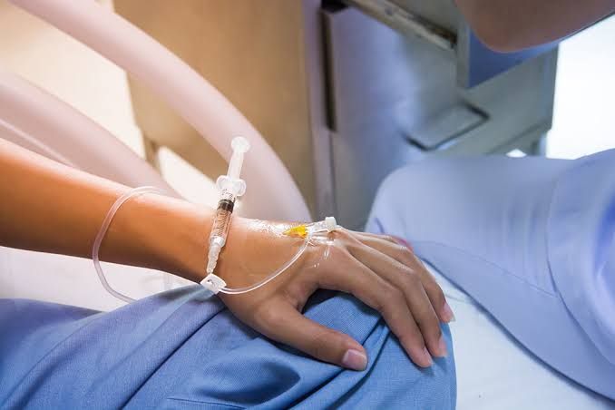
- Intravenous (IV) Fluids: Any fluids administered through an IV line, which are often recorded in milliliters (ml).
- Enteral Feeding: Fluids given via nasogastric tubes or percutaneous endoscopic gastrostomy (PEG) tubes.
Accurate documentation of fluid intake is essential for assessing overall fluid balance.
2. Fluid Output Measurement Fluid output encompasses all fluids lost from the body and can be measured through:
- Urine Output: The volume of urine produced, typically measured in ml over specific time intervals. Urine color and concentration can also provide insights into hydration status.
- Gastrointestinal Losses: This includes any fluid lost through vomiting, diarrhea, or stoma output.
- Insensible Losses: These are losses that occur without visible signs, such as perspiration and respiratory losses. While harder to quantify precisely, estimates can be made based on factors like fever or increased respiratory rate.
3. Daily Weight Monitoring Daily weight measurements can provide a practical assessment of fluid balance. A sudden increase in weight may indicate fluid retention (overload), while a decrease could suggest dehydration or effective diuresis. It is important to measure weight at the same time each day under similar conditions (e.g., after voiding and before breakfast) for consistency.
4. Laboratory Tests Certain laboratory tests can help assess fluid balance indirectly:
- Serum Electrolytes: Measurements of sodium, potassium, and other electrolytes can indicate hydration status and kidney function.
- Blood Urea Nitrogen (BUN) and Creatinine Levels: Elevated levels may suggest dehydration or impaired kidney function.
- Urine Specific Gravity: This test measures the concentration of solutes in urine; higher values indicate concentrated urine often associated with dehydration.
5. Fluid Balance Charts Healthcare providers often use fluid balance charts to document both input and output systematically over 24 hours. These charts allow for quick calculations of net fluid balance by summing total inputs and outputs, helping clinicians make informed decisions regarding patient management.
By employing these objective methods for measuring fluid balance, healthcare professionals can effectively monitor patients’ hydration status and adjust treatment plans accordingly.
Normal Electrolyte Values in Body Secretions
Electrolytes are essential minerals that carry an electric charge and are crucial for various bodily functions. They exist in different body fluids, including blood, urine, and other secretions. The normal values of electrolytes can vary slightly depending on the specific fluid being measured. Below are the typical normal electrolyte values found in common body secretions:
1. Blood Electrolyte Values:
- Sodium: 136-144 mEq/L
- Potassium: 3.7-5.1 mEq/L (serum)
- Magnesium: 1.4-1.9 mEq/L (serum)
- Calcium: 2.16-2.6 mEq/L
- Phosphate: 0.87-1.55 mEq/L
- Chloride: 97-105 mEq/L
- Bicarbonate: 22-30 mEq/L
These values represent the concentration of electrolytes in the bloodstream, which is critical for maintaining fluid balance, nerve function, and muscle contractions.
2. Urine Electrolyte Values: Urine electrolyte levels can vary significantly based on hydration status, diet, and overall health but generally fall within these ranges:
- Sodium: 40-220 mEq/day (varies widely based on dietary intake)
- Potassium: 25-125 mEq/day (also varies with diet)
- Chloride: 110–250 mEq/day
- Calcium: <300 mg/day (varies with dietary intake)
Urinary electrolyte levels provide insight into kidney function and hydration status.
3. Saliva Electrolyte Values: Saliva contains lower concentrations of electrolytes compared to blood and urine but still plays a role in oral health:
- Sodium: Approximately 10–20 mmol/L
- Potassium: Approximately 5–15 mmol/L
- Chloride: Approximately 5–15 mmol/L
Salivary electrolytes help maintain oral pH balance and contribute to digestion.
4. Sweat Electrolyte Values: Sweat is another important secretion that contains electrolytes, particularly during physical exertion:
- Sodium: Approximately 40–60 mmol/L (can be higher in individuals with high salt diets or during intense exercise)
- Potassium: Approximately 4–8 mmol/L
- Chloride: Approximately similar to sodium levels
Sweat composition can vary widely among individuals based on genetics, acclimatization to heat, and hydration status.
In summary, normal electrolyte values differ across various body secretions but are vital for maintaining homeostasis within the body.
Possible Causes (Differential Diagnosis) of Electrolyte and fluid disorders
Electrolyte and fluid disorders can arise from a variety of causes, which can be broadly categorized into several groups:
- Dehydration: This can result from inadequate fluid intake, excessive fluid loss (e.g., vomiting, diarrhea, sweating), or conditions such as diabetes insipidus.
- Renal Disorders: Kidney diseases can lead to imbalances in electrolytes due to impaired filtration and excretion. Conditions like acute kidney injury (AKI) or chronic kidney disease (CKD) are common culprits.
- Hormonal Imbalances: Disorders affecting hormones that regulate fluid and electrolyte balance, such as adrenal insufficiency (Addison’s disease) or hyperaldosteronism (Conn’s syndrome), can cause significant disturbances.
- Medications: Certain drugs, including diuretics, ACE inhibitors, and lithium, can disrupt normal electrolyte levels.
- Acid-Base Disorders: Conditions that alter the acid-base balance in the body (e.g., metabolic acidosis or alkalosis) often accompany electrolyte imbalances.
- Malnutrition: Deficiencies in dietary intake of essential electrolytes like potassium, magnesium, and calcium can lead to imbalances.
Appropriate Laboratory Studies Needed
To diagnose electrolyte and fluid disorders accurately, several laboratory studies are essential:
- Basic Metabolic Panel (BMP): This test measures key electrolytes including sodium, potassium, chloride, bicarbonate, glucose, and creatinine levels.
- Comprehensive Metabolic Panel (CMP): In addition to BMP components, this panel includes liver function tests and total protein levels which may provide additional context for electrolyte abnormalities.
- Urine Electrolyte Tests: These tests help assess renal handling of electrolytes by measuring concentrations of sodium, potassium, calcium, and magnesium in urine.
- Arterial Blood Gas (ABG): This test evaluates acid-base status and oxygenation; it is particularly useful in cases of suspected respiratory or metabolic acidosis/alkalosis.
- Thyroid Function Tests: Since thyroid hormones influence metabolism and fluid balance, these tests may be indicated if thyroid dysfunction is suspected.
- Hormonal Assays: Tests for aldosterone and cortisol levels may be necessary if an adrenal disorder is suspected.
Treatment of Common Electrolyte and Fluid Disorders
The treatment approach varies depending on the specific disorder identified but generally includes:
- Fluid Replacement Therapy:
- For dehydration or hypovolemia due to losses from vomiting or diarrhea, oral rehydration solutions containing electrolytes may be sufficient for mild cases.
- Severe cases may require intravenous fluids with isotonic solutions like Normal Saline or Lactated Ringer’s solution.
- Electrolyte Replacement:
- Hypokalemia (low potassium): Potassium supplements orally or intravenously depending on severity; monitoring is crucial due to potential cardiac effects.
- Hyponatremia (low sodium): Treatment depends on the cause; hypertonic saline may be used cautiously in severe cases while addressing underlying issues.
- Hypercalcemia (high calcium): Hydration with saline followed by diuretics to promote calcium excretion; bisphosphonates may also be indicated.
- Addressing Underlying Causes:
- If medications are causing imbalances, adjusting dosages or switching medications might be necessary.
- Hormonal therapies may be required for endocrine disorders affecting electrolyte balance.
- Monitoring and Follow-Up:
- Regular monitoring of serum electrolytes during treatment is critical to avoid overcorrection which could lead to further complications.
In summary, understanding the differential diagnosis for electrolyte and fluid disorders involves recognizing various underlying causes ranging from dehydration to hormonal imbalances. Appropriate laboratory studies are vital for accurate diagnosis while treatment focuses on correcting the specific electrolyte imbalance along with addressing any underlying conditions.


